This protocol describes an approach for biologically fine-tuning platelet-rich fibrin through enrichment with cobalamin and alpha-tocopherol prior to centrifugation, enabling controlled modulation of growth factor and cytokine release under in vitro conditions.
A subscription to JoVE is required to view this content. Sign in or start your free trial.
Research Article
Enrichment of Platelet-Rich Fibrin Using Cobalamin and Alpha-Tocopherol to Modulate Growth Factor Release
In This Article
Summary
Abstract
Platelet-rich fibrin (PRF) is an autologous biomaterial widely used in regenerative dentistry due to its biocompatibility and sustained release of growth factors. Recent research has shifted toward biologically modulating PRF to enhance its regenerative performance without altering its fundamental preparation principles. This study presents a protocol for biologically fine-tuning PRF through enrichment with cobalamin and alpha-tocopherol prior to centrifugation and evaluates their effects on growth factor and proinflammatory cytokine release under in vitro conditions. Peripheral blood samples obtained from healthy volunteers were processed using a fixed centrifugation protocol. Cobalamin or alpha-tocopherol was added to whole blood before centrifugation to obtain bioactively modified PRF matrices. The release profiles of platelet-derived growth factor (PDGF), transforming growth factor-β1 (TGF-β1), vascular endothelial growth factor (VEGF), insulin-like growth factor-1 (IGF-1), fibroblast growth factor-2 (FGF-2), interleukin-1β (IL-1β), and tumor necrosis factor-α (TNF-α) were quantified at 24 h and 72 h using an enzyme-linked immunosorbent assay. Cobalamin-enriched PRF was associated with significant intergroup differences in TGF-β1 levels and reduced release of proinflammatory cytokines, indicating a shift toward a more favorable regenerative microenvironment. In contrast, increases observed in FGF-2 and IGF-1 were time-dependent and detected across all groups, without demonstrating a cobalamin-specific effect. Alpha-tocopherol-enriched PRF exhibited a selective increase in VEGF release, suggesting a potential proangiogenic influence. These findings demonstrate that targeted vitamin enrichment enables controlled modulation of PRF bioactivity while preserving its autologous and anticoagulant-free nature. This protocol provides a practical framework for biologically fine-tuning PRF and supports its use as a tunable regenerative platform for future translational and clinical applications.
Introduction
Platelet-rich fibrin (PRF) is a second-generation autologous platelet concentrate that has been widely adopted in periodontology, implant dentistry, and oral surgery because of its biocompatibility, ease of preparation, and capacity for sustained release of growth factors1,2. PRF is obtained by centrifugation of whole blood without the use of anticoagulants or exogenous activators, resulting in a three-dimensional fibrin matrix that entraps platelets and leukocytes. These cellular components serve as endogenous reservoirs of growth factors, including platelet-derived growth factor (PDGF), transforming growth factor-β1 (TGF-β1), vascular endothelial growth factor (VEGF), insulin-like growth factor-1 (IGF-1), and fibroblast growth factor-2 (FGF-2), which collectively regulate angiogenesis, cell migration, proliferation, and extracellular matrix remodeling during tissue regeneration3,4.
Beyond its biological composition, the regenerative performance of PRF is strongly influenced by the architecture of its fibrin network and the biological activity of its cellular constituents. For this reason, recent investigations have moved beyond conventional preparation protocols and have begun to conceptualize PRF as a biologically tunable carrier system rather than a passive scaffold5,6. Structural modification strategies, such as the incorporation of biomaterials including hyaluronic acid and collagen, have demonstrated that the physical and mechanical properties of PRF can be altered, leading to changes in fibrin organization, mechanical stability, and growth factor release kinetics7. These findings have contributed to a growing body of evidence supporting the adaptability of PRF to specific regenerative demands.
In parallel with biomaterial-based approaches, vitamin-based enrichment has emerged as a biologically relevant strategy for modulating PRF bioactivity. Vitamins are known to regulate fundamental cellular processes such as metabolism, redox balance, collagen synthesis, angiogenesis, and inflammatory signaling. In particular, vitamins such as ascorbic acid, cholecalciferol, cobalamin, and alpha-tocopherol have been shown to influence cellular behavior and tissue healing through distinct molecular pathways8. Previous in vitro studies have demonstrated that vitamin-enriched PRF formulations can modulate growth factor release profiles, inflammatory cytokine expression, and biomechanical characteristics of the fibrin matrix, indicating that vitamin supplementation represents an effective means of biologically modifying PRF without altering its autologous nature9.
Cobalamin (Vitamin B12) and alpha-tocopherol (vitamin E) are essential micronutrients involved in key biological processes relevant to tissue homeostasis and repair. Vitamin B12 plays a critical role in nucleic acid synthesis, cellular metabolism, and erythropoiesis, and its deficiency has been associated with oral mucosal alterations and periodontal tissue breakdown10,11. Vitamin E functions as a potent antioxidant that limits lipid peroxidation, reduces oxidative stress, and contributes to collagen stabilization and modulation of inflammatory responses12,13. Importantly, although the present work builds upon previous investigations exploring different biological and physiological modulators of PRF, the current study introduces a distinct protocol specifically for enrichment with cobalamin and alpha-tocopherol prior to centrifugation. Accordingly, the aim of this study is to describe a reproducible protocol for preparing cobalamin- and alpha-tocopherol-enriched PRF and to evaluate the effects of this bioactive fine-tuning approach on growth factor and proinflammatory cytokine release under in vitro conditions.
Access restricted. Please log in or start a trial to view this content.
Protocol
This experimental study was conducted in accordance with the ethical principles outlined in the Declaration of Helsinki and received approval from the Clinical Research Ethics Committee of Çukurova University (Approval No: 139/25; Date: 8th December, 2023). Prior to participation, all individuals were informed about the study procedures, and written informed consent was obtained from each participant. All procedures involving human blood samples were performed in compliance with institutional biosafety regulations. Appropriate personal protective equipment (PPE), including gloves, masks, and laboratory coats, was used throughout the experimental process. Biological materials were handled using aseptic techniques, and all waste was disposed of in accordance with established biohazard management protocols. All reagents, consumables, and equipment used in this study are listed in the Table of Materials, including manufacturer details and relevant specifications to ensure reproducibility.
1. Participant selection
Participants were recruited from systemically healthy adults aged between 20 years and 30 years who demonstrated adequate compliance with the study procedures. Individuals with a history of systemic disease, hematological disorders, or conditions known to affect platelet function or wound healing were not considered eligible. Additional exclusion criteria included pregnancy or lactation, current smoking, recent infection, known hypersensitivity to cobalamin or alpha-tocopherol, and the use of vitamin supplements or medications that could interfere with platelet activity within the preceding six months. To reduce biological variability, only volunteers with normal baseline hematological findings were enrolled, and all participants were evaluated prior to inclusion to ensure suitability for PRF preparation. As a preliminary exploratory clinical investigation, this study used a sample size based on feasibility and the availability of eligible participants during the study period, and samples from twelve participants were analyzed.
2. Blood collection and group allocation
Peripheral venous blood was collected from each participant using additive-free 10 mL glass tubes under aseptic conditions. A total of five tubes were obtained per participant. One tube was allocated for complete blood count analysis, and one tube was used to determine baseline serum cobalamin and alpha-tocopherol levels. The remaining three tubes were assigned for platelet-rich fibrin (PRF) preparation. Among these, one tube served as the control PRF group without any additives, one tube received cobalamin (1 mL) prior to centrifugation to obtain cobalamin-enriched PRF, and one tube received alpha-tocopherol (0.5 mL) prior to centrifugation to obtain alpha-tocopherol-enriched PRF. Cobalamin was administered as a sterile injectable solution (1000 µg/mL), and alpha-tocopherol was used in ampoule form (2 mL). All samples were processed immediately after blood collection to avoid premature coagulation.
3. PRF preparation
Peripheral venous blood samples were collected from each participant using sterile, anticoagulant-free glass tubes. Immediately following collection, the tubes were placed into a centrifuge and processed without delay to prevent premature coagulation. Centrifugation was performed at 2700 rpm (708 × g) for 12 min (at room temperature) using a fixed-angle rotor system. Upon completion of centrifugation, three distinct layers were observed: a lower red blood cell fraction, an upper platelet-poor plasma layer, and an intermediate fibrin clot representing platelet-rich fibrin (PRF). The PRF clot was carefully separated from the red blood cell layer using sterile instruments and isolated for further processing. For the preparation of enriched PRF groups, the designated additives were introduced into the blood samples immediately prior to centrifugation under standardized conditions. This approach enabled the incorporation of bioactive compounds into the developing fibrin matrix during clot formation (Figure 1).
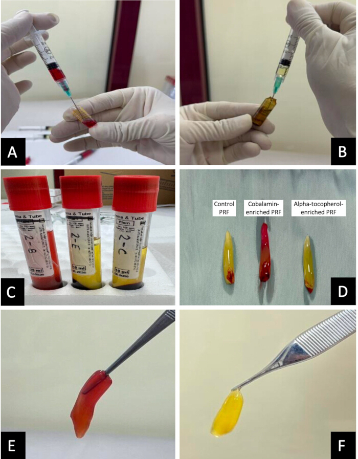
Figure 1: Flowchart demonstrating the methodological protocol for obtaining the PRF samples. 1 mL of cobalamin (A) and 0.5 mL of alpha-tocopherol (B) were added to blood samples prior to centrifugation. The resultant PRF samples in the tubes (C) and after being separated from the red blood cell layer (D). Final cobalamin-enriched PRF (E) and alpha-tocopherol-enriched PRF (F). Please click here to view a larger version of this figure.
4. In vitro incubation of PRF samples
Each PRF clot was transferred into a sterile tube, and cell culture medium was added at a standardized ratio of 1 mL per 1 g of clot (1:1, v/w) to ensure a consistent diffusion environment across all samples. The tubes were placed on an orbital shaker and incubated at 37 °C for a total duration of 72 h to facilitate the gradual release of growth factors and cytokines from the PRF matrix. Supernatants were collected at 24 h and 72 h, transferred into sterile microtubes, and stored at −80 °C until further analysis. Cell culture medium was used without serum supplementation to avoid interference from exogenous growth factors and to ensure that all measured biomolecules originated solely from the PRF matrix.
5. Growth factor ELISA analysis
The concentrations of platelet-derived growth factor (PDGF), transforming growth factor-β1 (TGF-β1), vascular endothelial growth factor (VEGF), insulin-like growth factor-1 (IGF-1), fibroblast growth factor-2 (FGF-2), interleukin-1β (IL-1β), and tumor necrosis factor-α (TNF-α) in the collected supernatants were quantified using enzyme-linked immunosorbent assay kits according to the manufacturers’ instructions. Absorbance values were measured at 450 nm using a microplate reader, and analyte concentrations were calculated based on standard calibration curves.
6. Statistical analysis
Statistical analyses were performed using IBM SPSS Statistics for Windows (Version 20.0). Data were expressed as mean ± standard deviation and median (minimum–maximum), as applicable. Normality of data distribution was assessed using the Shapiro–Wilk test. Intra-group comparisons between time points were conducted using the paired t-test or Wilcoxon signed-rank test, while inter-group comparisons were performed using repeated-measures analysis of variance or the Friedman test, as appropriate. A p-value of <0.05 was considered statistically significant.
Access restricted. Please log in or start a trial to view this content.
Results
Successful preparation of control, cobalamin-enriched, and alpha-tocopherol-enriched platelet-rich fibrin (PRF) was achieved in all participants. Macroscopic examination confirmed the formation of well-structured PRF clots with preserved fibrin integrity and sufficient mechanical stability, allowing standardized handling, incubation, and subsequent biochemical analyses (Table 1).
Access restricted. Please log in or start a trial to view this content.
Discussion
The present study demonstrates that platelet-rich fibrin (PRF) can be biologically fine-tuned through targeted enrichment with cobalamin and alpha-tocopherol prior to centrifugation, resulting in distinct and reproducible modulation of growth factor and cytokine release profiles. These findings reinforce the evolving concept that PRF is not merely a passive autologous scaffold but a biologically responsive matrix whose regenerative behavior can be adjusted through carefully selected bioactive interventions without alteri...
Access restricted. Please log in or start a trial to view this content.
Disclosures
The authors have nothing to disclose.
Materials
| Name | Company | Catalog Number | Comments |
|---|---|---|---|
| Alpha-Tocopherol (Vitamin E) | AKSU FARMA, Istanbul, Turkey | A11HA03 | Ampule EVIGEN 2 mL |
| Centrifuge (40° fixed-angle rotor) | Process for PRF, Nice, France | 913023410238 | Duo Centrifuge |
| Cobalamin (Vitamin B12) | DEVA HOLDING, Istanbul, Turkey | B03BA01 | Ampule DODEX 1000 mcg/mL |
| Eppendorf Tubes | Axygen | PCR-02-C | 200 piece |
| Glass Blood Collection Tubes | Hema & Tube (Istanbul, Turkey) | HNM8080 | (10 mL) No anticoagulant |
| HRP Solution | Included in ELISA kit | ||
| Human ELISA Kit - FGF | Wuhan Fine Biotech | EH0541 | Fine Test |
| Human ELISA Kit - IGF-1 | Wuhan Fine Biotech | ER0030 | Fine Test |
| Human ELISA Kit - PDGF | Wuhan Fine Biotech | ER1240 | Fine Test |
| Human ELISA Kit - TGFβ-1 | Wuhan Fine Biotech | EH0287 | Fine Test |
| Human ELISA Kit - VEGF | Wuhan Fine Biotech | EH0327 | Fine Test |
| Human IL-1β ELISA Kit | Wuhan Fine Biotech | EH0185 | Fine Test |
| Human TNF-α ELISA Kit | Wuhan Fine Biotech | EH0302 | Fine Test |
| Micropipettes & Tips | Eppendorf | 3123000063 | 1 Set |
| Microplate Reader | Biochrome | EZ Read 400 | 450 nm |
| Orbital Shaker | Stuart | SSM3 | Mini Gyro-Rocke |
| Precision Scale | Sartorius | H51-D | 1 piece |
| RPMI-1640 Cell Culture Medium | HyClone, Cytiva, USA | SH30027.01 | 500 mL |
| Sterile Scissors | ASA Dental (Istanbul, Turkey) | 0305-1 | 1 piece |
| Stop Solution | Included in ELISA kit | ||
| Substrate Solution | Included in ELISA kit | ||
| Wash Buffer | Included in ELISA kit |
References
- Dohan, D. M., Choukroun, J., Diss, A., Dohan, S. L., Dohan, A. J. J., et al. Platelet-rich fibrin (PRF): a second-generation platelet concentrate. Part I: technological concepts and evolution. Oral Surg Oral Med Oral Pathol Oral Radiol Endod. 101 (3), e37-e44 (2006).
- Miron, R. J., Zucchelli, G., Pikos, M. A., Salama, M., Lee, S., et al. Use of platelet-rich fibrin in regenerative dentistry: a systematic review. Clin Oral Investig. 21 (6), 1913-1927 (2017).
- Kobayashi, E., Flückiger, L., Fujioka-Kobayashi, M., Sawada, K., Sculean, A., et al. Comparative release of growth factors from PRP, PRF, and advanced-PRF. Clin Oral Investig. 20 (9), 2353-2360 (2016).
- Choukroun, J., Diss, A., Simonpieri, A. Platelet-rich fibrin (PRF): a second-generation platelet concentrate. Part IV: clinical effects on tissue healing. Oral Surg Oral Med Oral Pathol Oral Radiol Endod. 101 (3), e56-e60 (2006).
- Miron, R. J., Fujioka-Kobayashi, M., Bishara, M., Zhang, Y., Hernandez, M., Choukroun, J. Platelet-rich fibrin and soft tissue wound healing: a systematic review. Tissue Eng Part B Rev. 23 (1), 83-99 (2017).
- Ghanaati, S., Booms, P., Orlowska, A., Kubesch, A., Lorenz, J., et al. Advanced platelet-rich fibrin: a new concept for cell-based tissue engineering by means of inflammatory cells. J Oral Implantol. 40 (6), 679-689 (2014).
- Demirbilek, F., Alkaya, B., Ucak Turer, O., Yilmaz, M. B., Haytac, M. C., Ozcan, M. Enhancing platelet-rich fibrin: influence of hyaluronic acid and collagen on growth factor release and biomechanical properties in vitro. J Vis Exp. (222), e68693 (2025).
- Fawzy El-Sayed, K. M., Cosgarea, R., Sculean, A., Doerfer, C. Can vitamins improve periodontal wound healing/regeneration?. Periodontol 2000. 94 (1), 539-602 (2024).
- Ozcan, M., Topcu, B. E., Alkaya, B., Yilmaz, M. B., Ucak Turer, O., Haytac, M. C. Effects of ascorbic acid and cholecalciferol on growth factor release, cytokines, and biomechanical properties of platelet rich fibrin: in vitro. study. BMC Oral Health. 26 (1), 262 (2026).
- Kozlak, S. T., Walsh, S. J., Lalla, R. V. Reduced dietary intake of vitamin B12 and folate in patients with recurrent aphthous stomatitis. J Oral Pathol Med. 39 (5), 420-423 (2010).
- Alekhya, K., Muppirala, S. Oral manifestations of vitamin B12 (cobalamin) deficiency: a review. Int Healthc Res J. 3 (1), 9-15 (2019).
- Pavithra, R., Ramaprabha, G., Rajasekar, S., Lakshmi Sree, S. Vitamin deficiency and periodontal disease: a tie-in relationship. Sch J Appl Med Sci. 5 (1A), 74-81 (2017).
- Singh, N., Chander Narula, S., Kumar Sharma, R., Tewari, S., Kumar Sehgal, P. Vitamin E supplementation, superoxide dismutase status, and outcome of scaling and root planing in patients with chronic periodontitis: a randomized clinical trial. J Periodontol. 85 (2), 242-249 (2014).
- Miron, R. J., Fujioka-Kobayashi, M., Sculean, A., Zhang, Y. Optimization of platelet-rich fibrin. Periodontol 2000. 94 (1), 79-91 (2024).
- Miron, R. J., Estrin, N. E., Ahmad, P., Farshidfar, N., Fujioka-Kobayashi, M., et al. Thirty years of autologous platelet concentrates: from platelet-rich plasma to platelet-rich fibrin. J Periodontal Res. , (2025).
- Caruana, A., Savina, D., Macedo, J. P., Soares, S. C. From platelet-rich plasma to advanced platelet-rich fibrin: biological achievements and clinical advances in modern surgery. Eur J Dent. 13 (2), 280-286 (2019).
- Pietruszka, P., Chruścicka, I., Duś-Ilnicka, I., Paradowska-Stolarz, A. PRP and PRF-subgroups and divisions when used in dentistry. J Pers Med. 11 (10), 944 (2021).
- Dohan Ehrenfest, D. M., Bielecki, T., Jimbo, R., Barbé, G., Del Corso, M., et al. Do the fibrin architecture and leukocyte content influence the growth factor release of platelet concentrates? An evidence-based answer comparing a pure platelet-rich plasma (P-PRP) gel and a leukocyte- and platelet-rich fibrin (L-PRF). Curr Pharm Biotechnol. 13 (7), 1145-1152 (2012).
- Schär, M. O., Diaz-Romero, J., Kohl, S., Zumstein, M. A., Nesic, D. Platelet-rich concentrates differentially release growth factors and induce cell migration in vitro. Clin Orthop Relat Res. 473 (5), 1635-1643 (2015).
- Isobe, K., Watanebe, T., Kawabata, H., Kitamura, Y., Okudera, T., et al. Mechanical and degradation properties of advanced platelet-rich fibrin (A-PRF), concentrated growth factors (CGF), and platelet-poor plasma-derived fibrin (PPTF). Int J Implant Dent. 3 (1), 17 (2017).
- El Bagdadi, K., Kubesch, A., Yu, X., Al-Maawi, S., Orlowska, A., et al. Reduction of relative centrifugal forces increases growth factor release within solid platelet-rich-fibrin (PRF)-based matrices: a proof of concept of LSCC (low speed centrifugation concept). Eur J Trauma Emerg Surg. 45 (3), 467-479 (2019).
- Yaşar, N. &. #. 1. 9. 9. ;., Ünsal, G., Kocaelli, H. A., Orhan, K., Sağ, F. B. Comparative analysis of local application of titanium-platelet rich fibrin (T-PRF) and leukocyte-platelet rich fibrin (L-PRF) in bone defect healing: a micro-CT and histopathological study in rabbit models. Microsc Res Tech. 88 (7), 2073-2084 (2025).
- O'Leary, F., Samman, S. Vitamin B12 in health and disease. Nutrients. 2 (3), 299-316 (2010).
- Traber, M. G., Atkinson, J. Vitamin E, antioxidant and nothing more. Free Radic Biol Med. 43 (1), 4-15 (2007).
- Ozcan, M., Arçay Polat, G., Alkaya, B., Uçak Türer, O., Yilmaz, M. B., Haytac, M. C. Evaluation of circadian variation in growth factors of platelet-rich fibrin: a pilot study. J Vis Exp. (224), e68902 (2025).
- Ozcan, M., Ciritci Kabaklı, S., Alkaya, B., Isler, S. C., Ucak Turer, O., Oksuz, H., et al. The impact of local and systemic penicillin on antimicrobial properties and growth factor release in platelet-rich fibrin: in vitro. study. Clin Oral Investig. 28 (1), 61 (2024).
Access restricted. Please log in or start a trial to view this content.