The purpose of this study was to identify the key active components of Coptis chinensis and elucidate its action in the intervention of cerebral palsy (CP) by means of affinity ultrafiltration and experimental verification.
Research Article
Identification of AChE-Targeting Anti-Cerebral Palsy Constituents From Coptis chinensis Using Affinity Ultrafiltration And Experimental Validation
In This Article
Summary
Abstract
The purpose of this study was to identify the key active components of Coptis chinensis and elucidate its mechanisms of action in the intervention of cerebral palsy (CP) by means of affinity ultrafiltration. Using acetylcholinesterase (AChE) as the target, coptisine was screened out from the extract of C. chinensis as the component with the strongest binding capacity, which exhibited a binding rate of 21.9% and significant in vitro inhibitory activity (IC₅₀ = 3.24 µg/mL). Molecular docking and molecular dynamics simulations demonstrated that coptisine could bind stably to the active pocket of AChE. Subsequently, verification experiments were carried out on a neonatal rat model of hypoxic-ischemic brain damage (HIBD). The results showed that intervention with coptisine (10 mg/kg) significantly improved spatial learning and memory deficits in the model animals, increased cerebral blood flow in the injured hemisphere, alleviated pathological damage of brain tissues, and inhibited the abnormal activation of AChE in the brain. In summary, this study combined affinity ultrafiltration technology and confirmed that coptisine, identified from C. chinensis extract, can inhibit AChE and exert neuroprotective effects and cerebral blood flow-improving effects. It serves as the key material basis for C. chinensis to ameliorate CP-like neurological deficits, thereby providing candidate molecules and scientific evidence for the development of related drugs.
Introduction
Cerebral palsy (CP) is a permanent syndrome caused by non-progressive damage to the developing brain of fetuses or infants, characterized primarily by motor dysfunction and postural abnormalities. Although multiple clinical treatment approaches are currently available for CP, there remains a lack of ideal drugs that can fundamentally repair neurological deficits with minimal side effects. In contrast to chemically synthesized drugs, which usually act on single targets and carry relatively high potential toxicity, traditional Chinese medicines (TCMs), with their advantages of multi-component, multi-target, and holistic regulation as well as the favorable safety profiles demonstrated in long-term clinical practice, provide new insights and a reservoir of drug candidates for the treatment of complex brain disorders1,2,3,4,5,6. Therefore, exploring highly effective and low-toxicity chemical components from TCMs holds significant scientific significance and clinical value7.
Among the complex pathological mechanisms of cerebral palsy, cholinergic nervous system dysfunction plays a pivotal role8,9. Acetylcholinesterase (AChE) is a key enzyme that hydrolyzes the neurotransmitter acetylcholine (ACh). Excessively high activity of this enzyme leads to ACh depletion, impairs cholinergic signal transmission, and consequently exacerbates motor control and cognitive dysfunctions. Therefore, AChE has emerged as an important and well-established drug target for the development of neuroprotective agents. Inhibiting AChE activity to elevate ACh levels in the synaptic cleft is recognized as one of the effective strategies for ameliorating CP-related neurological deficits. In the context of hypoxic-ischemic brain injury, disruption of cholinergic neurotransmission may further aggravate neuronal dysfunction, thereby contributing to motor and cognitive impairments relevant to CP. Therefore, AChE was selected in this study not only as a classical neurofunctional target, but also as a mechanistically relevant entry point for screening bioactive compounds with potential anti-CP effects.
Modern research has shown that medicinal plants have a wide range of pharmacological activity values. As a classic traditional Chinese medicine for clearing heat and drying dampness, Coptis chinensis constitutes a complex system abundant in various isoquinoline alkaloids. In addition to the well-known berberine, it also contains a series of components with similar structures and diverse pharmacological activities, such as coptisine, epiberberine, palmatine, and jatrorrhizine10. These alkaloids collectively form the material basis for the pharmacological effects of C. chinensis. Previous studies have demonstrated that C. chinensis and its alkaloids possess anti-inflammatory, antioxidant, and potential neuroprotective properties. However, given such a complex component system, traditional research methods are difficult to rapidly and accurately identify the key active components that directly target AChE, which greatly limits the in-depth elucidation of the mechanisms underlying the effects of C. chinensis on brain injury11.
Compared with conventional activity-guided isolation, which usually requires repeated fractionation, bioactivity testing, and purification and may lead to the loss or neglect of low-abundance but high-affinity compounds, affinity ultrafiltration enables rapid screening of target-binding ligands directly from complex mixtures. This technique enables efficient and rapid "fishing" of active ligands that bind to specific targets (e.g., AChE) directly from the multi-component crude extracts of TCMs. It is particularly suitable for systems with multiple alkaloid components, such as C. chinensis, allowing for the accurate identification of effective components that directly interact with the target from the complex mixture12. Combined with computational approaches including molecular docking and molecular dynamics simulations, it can intuitively predict the binding mode, interaction types, and complex stability between components and target proteins at the atomic level, thereby providing robust theoretical evidence and mechanistic explanations for the results of experimental screening13.
Based on the above background, this study proposes the following scientific hypothesis: among the various alkaloids present in C. chinensis, there exist active components that can specifically inhibit AChE, and these components are the material basis for its potential anti-cerebral palsy effects. To verify this hypothesis, we integrated modern screening techniques with pharmacological validation methods: first, using AChE as the target, affinity ultrafiltration technology was employed to rapidly screen potential inhibitors from the multi-alkaloid extract of C. chinensis; further, molecular docking and molecular dynamics simulations were utilized to clarify the binding mechanism at the atomic level; finally, in vivo animal experiments were performed to verify the improving effects of the screened components on the behavior, brain tissue pathology, and AChE activity of rats with CP.
Protocol
All animal welfare and experimental procedures were approved by the Laboratory Animal Welfare Ethics Committee of The First People's Hospital of Zunyi (Ethics No. 2025-2-362). During the modeling and treatment period, animal body condition and survival were recorded daily.
Preparation of C. chinensis extract
In this study, an Accelerated Solvent Extractor was used for the accelerated extraction of the n-butanol fraction from C. chinensis medicinal materials. A total of 1 g of dried powder of C. chinensis was accurately weighed and placed into 40 mL extraction cells, respectively. Metal gaskets and glass fiber filter membranes were sequentially placed above and below the extraction cell to filter the sample, and the sample was wrapped with quartz sand. During the extraction process, the temperature and pressure were set to 90 °C and 100 bar, respectively. The accelerated extract was collected, and the extraction solvent was evaporated to dryness using a rotary evaporator to obtain dried extracts of different polar fractions. Methanol:water (50:50, v/v) was selected as the reconstitution solvent. The extract was filtered through a 0.45 µm microporous membrane and stored at 4 °C. Before sample detection, the inhibitory activity of the blank reconstitution solvent on AChE was investigated. The results showed that its inhibitory effect was negligible, indicating that the selection of the reconstitution solvent in this experiment would not affect the experimental results.
Acetylcholinesterase inhibition assay
The experiment was performed in a 96-well plate. First, PBS was added, followed by AChE (0.1 U/mL) and C. chinensis samples sequentially. After incubation at 37 °C for 10 min, 5,5′-Dithiobis (2-nitrobenzoic acid) (DTNB) chromogenic solution (2.5 mmol/mL) and ATChI substrate (10 mmol/mL) were added. The mixture was incubated at 37 °C for another 10 min, and the reaction was terminated by adding 60 µL of 1% sodium dodecyl sulfate (SDS). The absorbance was measured at 405 nm. Each C. chinensis sample was set up with 4 groups: Control group a: PBS (120 µL), AChE solution (20 µL), DTNB solution (40 µL), ATChI solution (40 µL), and SDS solution (60 µL) were added sequentially; Blank control group b: PBS (120 µL), DTNB solution (40 µL), ATChI solution (40 µL), and SDS solution (80 µL); Sample group c: PBS (80 µL), C. chinensis extract (20 µL), AChE solution (20 µL), DTNB solution (40 µL), ATChI solution (40 µL), and SDS solution (60 µL); Sample blank group d: PBS (100 µL), C. chinensis extract (20 µL), DTNB solution (40 µL), ATChI solution (40 µL), and SDS solution (60 µL). Huperzine A was used as the positive control with an initial concentration of approximately 2.5 mg/mL. The initial concentration of the n-butanol fraction extract was about 1 mg/mL. Both huperzine A and the samples were diluted with a 60% gradient. The inhibition rate was calculated using the following formula: Inhibition rate (%) = [1 - (c - d)/(a - b)] × 100%, where a, b, c, and d represent the absorbance values of control group a, blank control group b, sample group c, and sample blank group d, respectively.
The inhibitory rate at each tested concentration was calculated using the formula described above. Concentration–inhibition curves were then constructed by plotting the inhibitory rate against the logarithm of compound concentration. IC20, IC50, and IC80 values were calculated by nonlinear regression fitting using GraphPad Prism software based on the fitted concentration–response curves, and were defined as the concentrations required to produce 20%, 50%, and 80% inhibition of AChE activity, respectively.
Affinity ultrafiltration assay
Affinity ultrafiltration was performed to screen AChE-binding compounds from the C. chinensis extract. Briefly, AChE solution (0.1 U/mL in PBS, pH 7.4) was used as the active enzyme. The inactivated enzyme control was prepared by heating the same AChE solution in a boiling water bath for 10 min. For each reaction, 200 µL of C. chinensis extract solution was mixed with 200 µL of active or inactivated AChE solution and incubated at 37 °C for 30 min. After incubation, the mixture was transferred to a 30 kDa ultrafiltration centrifuge tube and centrifuged at 12,000 × g for 10 min at 4 °C. The retained enzyme-ligand complex was washed three times with 200 µL of PBS under the same centrifugation conditions to remove unbound components. Subsequently, 200 µL of 90% methanol was added to dissociate and elute the ligands bound to AChE, followed by incubation for 10 min and centrifugation at 12,000 × g for 10 min. The eluate was collected, filtered, and analyzed by high-performance liquid chromatography (HPLC). Compounds enriched in the active enzyme group relative to the inactivated enzyme control were considered potential AChE-binding components14.
HPLC analysis of major alkaloids metabolites
Chromatographic analysis was performed using an HPLC system equipped with a quaternary pump, an autosampler, a column oven, and a ultraviolet–visible (UV-Vis) detector. Separation was achieved on a C18 chromatographic column (250 × 4.6 mm, 5 µm) maintained at 25 °C. The mobile phase consisted of acetonitrile (A) and 30 mmol/L ammonium bicarbonate solution containing 0.7% ammonia and 0.25% triethylamine (B), delivered at a flow rate of 1.0 mL/min. The gradient elution program was as follows: 0–15 min, 90%–75% B; 15–25 min, 75%–70% B; 25–50 min, 70%–55% B. The injection volume was 5 µL, and UV detection was performed at 270 nm.
For quantitative analysis of the major alkaloids in C. chinensis, each sample (0.02 g) was accurately weighed and ultrasonically extracted with 10 mL of hydrochloric acid–methanol solution (1:100, v/v) for 30 min. After filtration, the weight loss was compensated with the same extraction solvent to the original weight, and the final solution was filtered through a 0.45 µm membrane before HPLC analysis. Eight authentic reference standards, including magnoflorine, glaucine, jatrorrhizine, columbamine, epiberberine, coptisine, palmatine, and berberine, were analyzed under the same chromatographic conditions. Calibration curves were constructed for these analytes using reference solutions. Because the abundance levels and detector responses of the eight alkaloids differed considerably, the calibration range for each analyte was individually optimized based on literature reports and preliminary experiments, rather than applying an identical fold-dilution scheme to all compounds. Therefore, the high-to-low concentration ratios of the linear ranges were not expected to be identical among the eight analytes. The limits of detection (LOD) and limits of quantification (LOQ) were estimated at signal-to-noise ratios of approximately 3 and 10, respectively.
For analysis of affinity ultrafiltration eluates, the ligands dissociated from AChE after ultrafiltration were collected from repeated experiments and concentrated prior to HPLC analysis to ensure adequate signal intensity for detection. The concentrated eluates were then analyzed using the same HPLC conditions as described above. The chromatographic peaks in the eluates were assigned by targeted comparison with the eight authentic reference standards and with the chromatographic profile of the C. chinensis extract obtained under identical analytical conditions. Therefore, the present analysis was not intended for de novo structural elucidation of unknown compounds, but for targeted determination of whether the known major alkaloids in C. chinensis were enriched in the active-enzyme eluate relative to the inactivated-enzyme control.
Molecular docking assay
In the present study, docking analysis was focused on coptisine because it was identified as the top-ranked AChE-binding constituent by affinity ultrafiltration and also showed the strongest in vitro inhibitory activity among the major alkaloids. Protein crystals were retrieved from the Protein Data Bank (PDB). Protein crystals with co-crystallized ligands were selected as the final docking receptors, and all target protein crystals were derived from humans. The receptor file was opened using PyMOL software, and the receptor and ligand files were separated and saved; the receptor was opened with MGLTools software, followed by removing water molecules, adding hydrogen atoms and charges, merging non-polar hydrogen atoms, calculating local atomic charges, etc., and then saved in pdbqt format; the ligand file was opened with MGLTools software, hydrogen atoms and charges were added, and saved as a pdbqt format file; grid box was opened, spacing was set to 1.000, the number of grid points was set, the central point coordinates of the box were selected according to the co-crystallized ligand, and saved as a GPF file15,16.
Chemical component structures were first collected from the SciFinder database and saved in cdx format; each molecular three-dimensional structure was displayed using the ChemBio3D Ultra module, energy optimization was performed with the MM2 force field, saved in mol2 format, and charge parameters were removed. Using AutoDock, the mol2 format file was opened with MGLTools software, hydrogen atoms and charges were added to the ligand, rotatable chemical bonds were observed, and saved as a pdbqt format file. The AutoDock Vina (version 1.2.5) model was applied to perform molecular docking evaluation.
Molecular dynamics simulation assay
Gaussian 16 and GaussView 6 software were used for ligand structure optimization. After importing the mol2 file of the ligand structure into GaussView 6, the task type was set to Optimization, the state of the target molecule was selected as Ground State, the calculation method was density functional theory (DFT), the spin multiplicity was defaulted to Default Spin, the functional used was B3LYP, the solvent model was IEFPCM, and the solvent was water. After the completion of molecular structure optimization, Gaussian 16 was used to convert the generated chk file into an fch file, which was then imported into Multiwfn software to calculate the RESP charge of the ligand.
After the calculation of RESP charge, the generated chg file was imported into sobtop_1.0 (dev5) software to generate the ligand topology file required for molecular dynamics simulation. The molecular dynamics simulation was performed using GROMACS 2019.6 software with the AMBER99 force field. The structure files of the protein molecule and small molecule were imported into the software, a triclinic box with a periodic boundary of 1.0 nm was established, and the TIP3P water model was used for filling. The Particle Mesh Ewald (PME) method was employed to calculate long-range electrostatic interactions, and the gmx geion command was used to introduce an appropriate number of sodium ions and chloride ions to neutralize the charge of the entire system. The simulation was carried out at a constant temperature of 298 K and a standard atmospheric pressure of 100 kPa. For energy minimization, the steepest descent method (steep) was implemented. The gmx grompp and gmx mdrun commands were used to perform 10,000 steps of NVT (isothermal-isochoric ensemble) equilibrium and NPT (isothermal-isobaric ensemble) equilibrium, with the temperature coupling constant and pressure coupling constant set to 0.1 ps and 0.5 ps, respectively, and the duration of the equilibrium simulation was 100 ps. After the system reached equilibrium, the time step was set to 2 fs, and a 10 ns molecular dynamics simulation was performed on the complex, with trajectory data saved every 2 ps. The 10 ns production run was selected as an exploratory simulation length to provide a short-timescale dynamic assessment of the docked coptisine–AChE complex, rather than exhaustive conformational sampling. This setting was used to evaluate whether the docked complex could maintain a relatively stable binding mode over time. The gmx rms, gmx rmsf, gmx gyrate, and gmx hbonds commands were further used to calculate the root mean square deviation (RMSD), root mean square fluctuation (RMSF), radius of gyration of the protein, hydrogen bonds, and solvent-accessible surface area. QTgrace was used for result visualization.
Animal experiment verification
Eighteen healthy neonatal Sprague-Dawley (SD) rat pups (7 days postnatal) of either sex were selected. All experimental animals were housed in a specific pathogen-free (SPF) animal experimental facility. This facility was equipped with a strict barrier system, which could effectively prevent the invasion of microorganisms and parasites. The environmental parameters of the housing room were automatically monitored and regulated by a central control system: temperature 22 ± 2 °C, relative humidity 55% ± 10%, and light cycle 12-h light/12-h dark cycle. A neonatal rat model of hypoxic-ischemic brain damage (HIBD) modified by the classic Rice-Vannucci method was used to simulate the pathological and behavioral characteristics of CP.
Briefly, postnatal day 7 rat pups were anesthetized with 2–3% isoflurane for induction and 1–2% for maintenance. After adequate anesthesia, a midline cervical incision was made, and the left common carotid artery was carefully isolated and permanently ligated. The incision was then sutured, and the pups were allowed to recover on a warming pad for 1 h. Subsequently, the pups in the model and coptisine groups were exposed to hypoxia in a chamber containing 8% O₂ balanced with 92% N₂ for 2 h at 37 °C to induce hypoxic-ischemic brain injury. Sham-operated animals underwent the same anesthesia and surgical exposure without carotid ligation or hypoxic treatment. After hypoxia, the pups were returned to their dams and monitored daily for general condition, survival, and feeding behavior throughout the experimental period.
The experimental rats were randomly divided into 3 groups: Sham group: only the left common carotid artery was separated without ligation or hypoxia treatment, and an equal volume of solvent was administered postoperatively; Model group: hypoxic-ischemic brain damage modeling surgery was performed, and an equal volume of solvent was administered postoperatively; Coptisine group: after modeling, coptisine (10 mg/kg) was administered by gavage. The dose of coptisine (10 mg/kg) was selected as an in vivo dose based on previously reported in vivo/pharmacokinetic studies of coptisine17. Drugs were administered once a day at a fixed time for 14 consecutive days of intervention.
Detection of animal indicators
The Morris water maze (MWM) test equipment consisted of a large circular water pool (diameter approximately 150 cm) with obvious visual cues on the pool wall. The pool water was stratified into opaque milky white, and the water temperature was controlled at 23 ± 1 °C. A transparent platform was placed in the target quadrant, submerged 1–2 cm below the water surface. Training was conducted 4 times a day: the animals were placed into the water facing the pool wall from four different entry points on the pool wall, and the time required for the animals to find and climb onto the hidden platform from entry into the water, namely the "escape latency", was recorded. Each training session was limited to 60 s. If the animal found the platform within 60 s, it was allowed to stay on it for 15 s to consolidate memory. If not found, the animal was gently guided to the platform and allowed to stay for 15 s. After 7 days of training, the hidden platform was removed. The animals were placed into the water from the entry point opposite the original platform and allowed to swim freely for 60 s, and the swimming trajectory of the animals was recorded by a video system. Through the MWM test, indicators such as average escape latency, time spent in the target quadrant, and the number of crossings over the original platform position were recorded.
For laser speckle contrast imaging (LSCI) detection: After anesthesia, the rats were fixed on a stereotaxic instrument, and the skull was exposed and kept moist. A laser speckle blood flow imaging system was used to continuously collect raw speckle images for 30 s directly above the skull. Professional software was used to convert the spatiotemporally varying speckle contrast into a cerebral blood flow perfusion unit (PU) distribution map. For quantitative analysis, a manually delineated cortical ROI was selected within the exposed ipsilateral hemisphere in each image according to the lesion-relevant cortical perfusion area and visible anatomical landmarks, and the average perfusion value within this ROI was used for intergroup comparison.
Brain tissues were collected, and pre-cooled PBS was added according to the weight-volume ratio. Mechanical homogenization was performed on ice, followed by centrifugation at 8000 × g at 4 °C for 10–15 min, and the supernatant was collected for testing. Brain tissues were quickly put into 4% paraformaldehyde for fixation for more than 24 h to maintain tissue morphology. Hematoxylin-eosin (HE) staining was performed through operations such as dehydration, clearing, and paraffin embedding, and the degree of brain injury was observed under an optical microscope.
For AChE activity measurement, the cerebral cortex from the injured (ipsilateral) hemisphere was collected and homogenized according to the manufacturer's instructions. Tissue samples were homogenized in extraction buffer and centrifuged at 8000 × g for 10 min at 4 °C, and the supernatant was collected for analysis. AChE activity was determined using a commercial acetylcholinesterase activity assay kit (microplate/colorimetric method). This assay is based on the hydrolysis of acetylcholine by AChE, followed by reaction with DTNB to generate a chromogenic product, and the absorbance was measured at 412 nm for the calculation of enzyme activity. Because this kit is an enzyme activity-based colorimetric assay rather than a conventional antigen-quantifying enzyme-linked immunosorbent assay (ELISA) kit, a standard detection range and analytical sensitivity were not provided by the manufacturer. In the present study, the measured AChE activity values in cortical tissue were approximately within the range of 25–80 nmol/min/mg protein.
Statistical analysis
All experimental data were expressed as mean ± standard deviation (SD) and analyzed using GraphPad Prism software. Comparisons among groups were performed using one-way analysis of variance (ANOVA) followed by Tukey's multiple comparisons test. For the Morris water maze test, the final training result and probe trial parameters were analyzed using one-way ANOVA followed by Tukey's multiple comparisons test. A value of P < 0.05 was considered statistically significant18. The correlation between cerebral blood flow perfusion in the ROI and AChE activity in the ipsilateral cerebral cortex was analyzed using Pearson correlation analysis. A value of P < 0.05 was considered statistically significant.
Results
Analysis of chemical components in C. chinensis extract
Alkaloids are the main accumulated components and also the major active components of C. chinensis medicinal materials. In this study, chromatographic technology was used to analyze the main chemical components in C. chinensis (Figure 1A). As shown in Table 1, all eight analytes exhibited good linearity within their respective calibration ranges. Because the contents and detector responses of these alkaloids differed considerably, the calibration ranges were individually optimized for each analyte. (Figure 1B). As shown in Table 1, the linear relationship, linear range, and other parameters of each chemical component were good, which could be simultaneously used for the determination of chemical components in C. chinensis.
| Metabolites | Calibration Equation | Linear Range (μg/mL) | Correlation Coefficient | LOD (μg/mL) | LOQ (μg/mL) |
| Magnoflorine | y = 4,773,370.49x + 191.05 | 2.29–39.49 | 0.9994 | 0.25 | 0.82 |
| Glaucine | y = 13,875,850.99x - 5,362.91 | 0.45–45.65 | 0.9995 | 0.37 | 1.22 |
| Jatrorrhizine | y = 14,793,030.49x + 697.38 | 2.08–51.3 | 0.9992 | 0.49 | 1.63 |
| Columbamine | y = 31,009,479.81x - 1,095.99 | 1.79–42.01 | 0.9994 | 0.38 | 1.24 |
| Epiberberine | y = 19,062,766.74x + 2,287.64 | 1.22–86.81 | 0.9996 | 1.01 | 3.33 |
| Coptisine | y = 19,040,819.50x - 273.45 | 3.96–98.84 | 0.9991 | 0.88 | 1.54 |
| Palmatine | y = 19,209,155.06x + 6,732.96 | 3.15–77.62 | 0.9994 | 0.98 | 3.23 |
| Berberine | y = 18,878,819.98x - 8,645.71 | 38.73–470.04 | 0.9996 | 6.94 | 23.7 |
Table 1: The linear relationship, linear range, and other parameters of each chemical component in HPLC analysis.
Previous study results have shown that the main active components of C. chinensis are concentrated in the n-butanol fraction13. Based on the accelerated solvent extractor, the extract of the n-butanol fraction of C. chinensis was obtained. The results showed that berberine was the main chemical component in C. chinensis, with a content as high as 62.23 ± 4.45 mg/g. Secondly, epiberberine, coptisine, and palmatine were the components with relatively high accumulation in C. chinensis, and their contents were 10.03 ± 2.72 mg/g, 19.45 ± 2.18 mg/g, and 16.39 ± 1.84 mg/g, respectively. Magnoflorine, glaucine, columbamine, and jatrorrhizine were the chemical components with low accumulation in C. chinensis, and their contents were 4.65 ± 1.50 mg/g, 2.63 ± 0.98 mg/g, 3.78 ± 0.78 mg/g, and 3.98 ± 0.65 mg/g, respectively (Figure 1C). The metabolic information of these chemical components provides a basis for subsequent AChE inhibition assay and affinity ultrafiltration analysis.
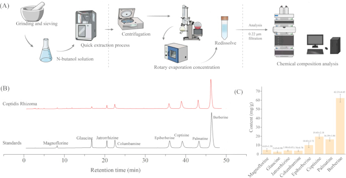
Figure 1: Analysis of chemical components in C. chinensis extract. (A) Extraction protocol of C. chinensis extract. (B) Chromatogram of main chemical components in extract. (C) Quantitative analysis of major alkaloids, expressed in milligrams per gram (mg/g). Please click here to view a larger version of this figure.
Analysis of the inhibitory effect of C. chinensis extract on AChE
By successfully constructing an in vitro AChE inhibition model, we detected the inhibitory effects of C. chinensis extract and its positive control, huperzine A, on AChE. The changes in the inhibition rate of C. chinensis extract on AChE activity at different concentrations are shown in Figure 2. C. chinensis extract also exhibited a concentration-dependent inhibitory trend, and the increase in the inhibition rate was relatively gentle within the entire test concentration range. This indicates that the inhibition of AChE by C. chinensis extract may not be caused by a single component, but by the combined effect of multiple active components (Figure 2A). These components have varying inhibitory abilities, which together constitute a comprehensive and gradual inhibitory effect. Figure 2B shows the inhibitory effect of the positive control huperzine A. In the low concentration range, the inhibition rate increased sharply with the increase in concentration, and the curve was very steep, indicating that huperzine A has a very high affinity for AChE. Using the logistic regression algorithm, we calculated the IC₂₀, IC₅₀, and IC₈₀ values of C. chinensis extract and huperzine A for AChE inhibition. The results showed that the IC₂₀, IC₅₀, and IC₈₀ of C. chinensis extract for AChE inhibition were 7.99 ± 0.42 µg/mL, 28.07 ± 0.88 µg/mL, and 98.72 ± 1.36 µg/mL, respectively (Figure 2C). The IC₂₀, IC₅₀, and IC₈₀ of huperzine A for AChE inhibition were 22.63 ± 1.52 µg/mL, 74.05 ± 15.26 µg/mL, and 177.25 ± 8.47 µg/mL, respectively. The results indicated that the inhibitory activity of C. chinensis extract on AChE was superior to that of the positive drug huperzine A (Figure 2D). This result provides a basis for the subsequent use of affinity ultrafiltration technology to fish out AChE-inhibitory components from C. chinensis extract.
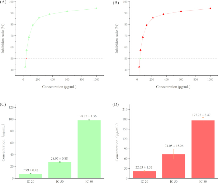
Figure 2: Analysis of the inhibitory effect of C. chinensis extract on AchE. (A) Concentration-dependent inhibitory curve of C. chinensis extract on AChE. (B) Concentration-dependent inhibitory curve of huperzine A on AChE. (C) IC₂₀, IC₅₀, and IC₈₀ values of C. chinensis extract for AChE inhibition, expressed in micrograms per milliliter (µg/mL). (D) IC₂₀, IC₅₀, and IC₈₀ values of huperzine A for AChE inhibition, expressed in micrograms per milliliter (µg/mL). Please click here to view a larger version of this figure.
Affinity ultrafiltration analysis of C. chinensis extract with AChE
Affinity ultrafiltration is a technology for rapidly screening active ligands that bind to specific targets from complex mixtures. Its core principle is to separate small molecule ligand-biomacromolecule target complexes from unbound free small molecules by utilizing the molecular weight cutoff property of ultrafiltration membranes. The pre-prepared AChE and C. chinensis extract were co-incubated at 37 °C for a period of time. During this period, the active small molecules (ligands) in the extract that can bind to the active site of the protein will form ligand-protein complexes with the protein, while other molecules without binding ability remain in a free state. Free small-molecule components were screened out by ultrafiltration. A dissociation solution was added to the ultrafiltered centrifuge tube to inactivate the protein, thereby releasing the bound active small molecules. Finally, the concentrated eluates obtained from the active AChE group and the inactivated-enzyme control group were analyzed by HPLC under the same chromatographic conditions, and the major bound components were assigned by comparison with the eight authentic reference standards. (Figure 3A).
After multiple ultrafiltration operations, HPLC analysis showed that berberine was the most bound active component, followed by palmatine and coptisine, which may be related to the high content of these components in C. chinensis extract (Figure 3B). Through comparative analysis of ultrafiltration experiments with inactive enzyme and active enzyme, it was found that coptisine may be the active component with the highest binding rate to AChE, and its binding rate was the highest among all alkaloid components, reaching 21.9% (Figure 3C). It is inferred that coptisine may be the most critical active component in C. chinensis acting on the AChE target. To further verify this result, we analyzed the main alkaloid components for in vitro AChE inhibition. The results showed that coptisine had the lowest IC₅₀ for AChE inhibition, which was 3.24 µg/mL (Figure 3D). The conclusion of single-component verification confirmed that coptisine may be the optimal active component for AChE inhibition in C. chinensis.
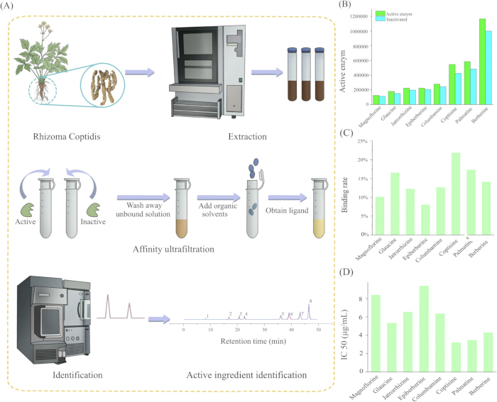
Figure 3: Affinity ultrafiltration analysis of C. chinensis extract binding to AChE. (A) Schematic illustration of the workflow for screening AChE-binding components by affinity ultrafiltration. (B) HPLC analysis of AChE-bound components in C. chinensis extract. (C) Binding rates of the major alkaloids in C. chinensis extract to AChE. (D) IC50 values of the major alkaloids for AChE inhibition, expressed in micrograms per milliliter (µg/mL). Please click here to view a larger version of this figure.
Molecular docking and molecular dynamics analysis of coptisine with AChE
Because coptisine showed both the highest binding rate in the affinity ultrafiltration assay and the lowest IC₅₀ value in the in vitro AChE inhibition assay, it was selected as the representative lead compound for subsequent molecular docking and molecular dynamics analyses. After removing redundant ligands and water molecule solvents from the human acetylcholinesterase crystal (PDB ID: 6O4W; Resolution: 2.35 Å; Source: Human; Co-crystallized ligand: Donepezil) using PyMOL software, the docking site was identified based on the position of donepezil, and molecular docking was performed. Figure 4A shows the schematic diagram of the binding position of coptisine with AChE. It can be seen from the figure that the binding pocket of coptisine with the AChE crystal is mainly located in the narrow and long hydrophobic cavity. The cavity of AChE is narrower and longer, leading to tighter binding with metabolites, which is the reason why this type of compound exerts a better effect on AChE.
According to the literature, a docking energy < -4.25 kcal/mol indicates that the ligand and the target have binding activity; a docking energy < -5.0 kcal/mol indicates good binding activity; and a docking energy < -7.0 kcal/mol indicates strong docking activity between the two19 (Figure 4B). The analysis results showed that the docking energy between coptisine and AChE was < -7.85 kcal/mol, indicating that the two have good binding activity. The two-dimensional results showed that coptisine interacts with AChE mainly through amino acid residues such as PHE338, TYR341, TYR337, ARG526, TRP86, and TRP286 (Figure 4C). In this study, a short-timescale molecular dynamics simulation was further used to preliminarily assess the dynamic stability of the coptisine–AChE complex. The RMSD curve gradually stabilized after the initial rise, and the fluctuation remained within a limited range, suggesting that the docked complex maintained a relatively stable binding mode during the simulation period. (Figures 4D,E). These results emphasize the high affinity between coptisine and AChE, confirming that coptisine is an effective AChE inhibitor.
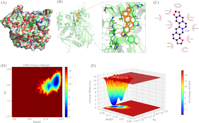
Figure 4: Molecular docking and molecular dynamics analysis of coptisine with AChE. (A) Surface representation showing the predicted binding pocket of coptisine in AChE. (B) Three-dimensional binding mode of coptisine within the active site of AChE. (C) Two-dimensional interaction diagram between coptisine and key amino acid residues of AChE. (D) Two-dimensional Gibbs free energy landscape of the coptisine-AChE complex using RMSD and Rg as reaction coordinates. (E) Three-dimensional Gibbs free energy landscape of the coptisine-AChE complex based on RMSD and Rg. RMSD reflects the overall conformational deviation of the complex, Rg indicates structural compactness, RMSF represents residue-level flexibility, and SASA refers to the solvent-accessible surface area. Please click here to view a larger version of this figure.
Effect of coptisine on AChE activity in CP rat models
Based on the previous screening via affinity ultrafiltration technology, which found that coptisine is the component with the strongest binding ability to AChE in C. chinensis, this study further evaluated the therapeutic effect and potential mechanism of coptisine on the HIBD model of neonatal rats (simulating human CP) in vivo.
The results of the MWM test showed that HIBD model rats exhibited significant spatial learning and memory impairment. In the final training session of the MWM test, HIBD model rats showed a significantly prolonged escape latency compared with the sham group, whereas coptisine treatment markedly shortened the escape latency (Figure 5A); during the spatial exploration period, the swimming trajectory showed a random scattered pattern (Figure 5B); the number of crossings over the original platform position (Figure 5C) and the time spent in the target quadrant (Figure 5D) were significantly reduced. However, coptisine intervention effectively improved these deficits, restoring the above indicators to near-normal levels. There was no significant difference in the total movement distance among the groups (Figure 5E), excluding the interference of motor ability on cognitive results. This indicates that coptisine can specifically improve cognitive dysfunction related to the CP model.
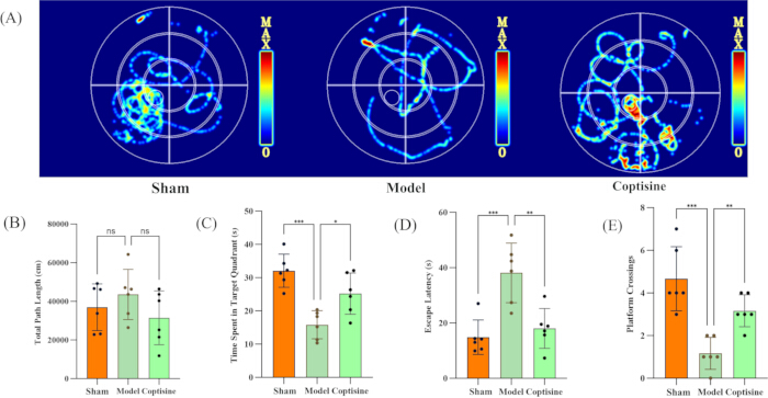
Figure 5: Effect of coptisine on spatial learning and memory ability in HIBD model rats. (A) Representative swimming trajectories of each group during the probe test. (B) total path length of each group, expressed in cm. (C) time spent in the target quadrant of each group, expressed in seconds. (D) escape latency of each group, expressed in seconds. (E) number of platform crossings in each group. *P < 0.05, **P < 0.01, and ***P < 0.001 for the indicated comparisons. Please click here to view a larger version of this figure.
LSCI detection showed that HIBD injury led to a significant decrease in cerebral blood flow within the ipsilateral cortical ROI of model rats. Coptisine treatment effectively increased cerebral blood flow in this lesion-relevant cortical region, restoring it toward near-normal levels (Figures 6A,B). This indicates that coptisine has the effect of improving cerebral microcirculation disorders in the cerebral palsy model. HE staining results showed that the brain tissue structure of the model group was disorganized, with widened intercellular spaces and obvious interstitial edema; a large number of neurons were lost, arranged sparsely and disorderly, most neurons were shrunk and deformed, with pyknotic and deeply stained nuclei, disappeared nucleoli, and some neurons were necrotic and lysed; glial cell proliferation and inflammatory cell infiltration were observed. In contrast, the coptisine intervention group had relatively clear brain tissue layers and reduced edema; neurons were arranged more neatly with reduced loss, most neurons had a near-normal morphology, and only a small number of neurons were slightly shrunk; glial proliferation and inflammatory infiltration were reduced (Figure 6C). These results indicate that coptisine can significantly improve brain injury and insufficient cerebral blood flow perfusion in the HIBD model rats.
To further confirm that the improvement of cerebral palsy-like characteristics in HIBD model rats by coptisine was associated with inhibition of AChE activity, AChE activity in the ipsilateral cerebral cortex was determined using a commercial colorimetric activity assay kit. The results showed that AChE activity in the model group was significantly higher than that in the sham group, whereas coptisine intervention significantly reduced the excessive activation of AChE (Figure 6D). Moreover, correlation analysis showed that cerebral blood flow perfusion in the ROI was significantly negatively correlated with cortical AChE activity (R = -0.71, p = 0.00097), suggesting that improved local cerebral perfusion was associated with reduced AChE activity (Figure 6E). These findings further support the mechanistic association between improved cerebral blood flow and inhibition of abnormal AChE activation in this model.
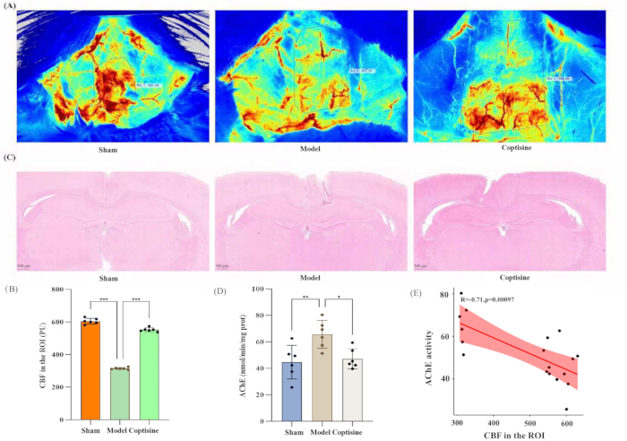
Figure 6. Effect of coptisine on cerebral blood flow, brain histopathology, and cortical AChE activity in HIBD model rats. (A) Representative CBF maps of each group. (B) Quantification of CBF in the cortical ROI, expressed as PU. (C) Representative HE-stained brain sections; scale bar = 500 µm. (D) cortical AChE activity, expressed as nmol/min/mg protein. (E) Correlation analysis between CBF in the ROI and cortical AChE activity. *P < 0.05, **P < 0.01, and ***P < 0.001 for the indicated comparisons. Pearson correlation analysis: R = −0.71, P = 0.00097. Please click here to view a larger version of this figure.
DATA AVAILABILITY:
The datasets supporting the conclusions of this article are included within the article, with all data publicly available.
Discussion
Currently, various methods and technologies have been developed for the prevention and treatment of brain diseases20,21,22. In this study, an integrated strategy of in vitro screening, computational simulation, and in vivo verification was adopted to systematically clarify the material basis and multi-target mechanism of C. chinensis in improving cerebral palsy-like symptoms induced by HIBD. The core finding is that coptisine, a characteristic alkaloid in C. chinensis, rather than berberine with the highest content, is the key active component that potently inhibits AChE and thereby exerts neuroprotective and functional improvement effects. This finding not only provides a modern pharmacological annotation for the traditional efficacy of C. chinensis in "clearing heat and purging fire" by acting on cholinergic targets in the central nervous system but also offers a strong candidate molecule and scientific basis for the development of natural product-based therapeutic strategies for CP.
Firstly, the combined technology of affinity ultrafiltration and liquid chromatography used in this study provides a successful example for efficiently and accurately identifying active components targeting AChE from the complex alkaloid system of C. chinensis. This method directly captures the dynamic binding between components and target proteins in solution, effectively avoiding the loss or neglect of active components in traditional activity-guided separation23. The results showed that coptisine had the highest binding rate (21.9%) and the optimal in vitro inhibitory activity (IC₅₀ = 3.24 µg/mL), which highlights that "quality" (affinity and efficacy for specific targets) is more critical than mere "content" in the study of effective substances of traditional Chinese medicine. On this basis, molecular docking and molecular dynamics simulations were specifically performed for coptisine as the experimentally prioritized lead compound, with the aim of providing mechanistic support for its interaction with AChE. The results revealed the structural basis for coptisine to form a stable complex with the AChE active pocket (key residues including PHE338, TYR341, etc.) at the atomic level, with a binding free energy as low as -7.85 kcal/mol and a stable equilibrium of the simulation system24. This computational biology evidence mutually confirms with the experimental screening results, jointly establishing coptisine as a high-efficiency AChE inhibitor and laying a solid theoretical foundation for subsequent in vivo experiments.
Secondly, animal experiments confirmed the multi-dimensional therapeutic benefits of coptisine in the HIBD model, and its mechanism of action goes beyond single cholinergic regulation. Behaviorally, coptisine significantly reversed the spatial learning and memory deficits in model rats, which directly confirms that increasing acetylcholine levels in the brain by inhibiting AChE is an effective way to improve cognitive dysfunction25. More importantly, this study found additional benefits of coptisine: it significantly increased local cerebral blood flow in the injured brain area and reduced neuronal apoptosis and histopathological damage. These effects suggest that the action of coptisine may be a multi-target synergistic process. Previous studies have reported that coptisine possesses anti-inflammatory and antioxidant activities26,27. However, these effects were not directly examined in the present study. In the current work, the beneficial effects of coptisine were mainly reflected by AChE inhibition, improvement of cholinergic dysfunction, restoration of cerebral blood flow, and attenuation of histopathological damage. Further studies are needed to determine whether anti-inflammatory and antioxidant pathways also participate in its neuroprotective effects in HIBD/CP models.
Despite the encouraging findings, this study still has several limitations. Only a single dose of coptisine (10 mg/kg) was evaluated in vivo, and all measurements were performed at a single experimental endpoint after 14 days of treatment, without dose–response analysis or multi-time-point dynamic observation. In addition, no pharmacokinetic or brain exposure assessment was conducted. Moreover, because the animal experiment was primarily designed as a proof-of-concept validation of the screened candidate compound, no in vivo positive control group was included. Although the current data support an association between coptisine treatment and AChE inhibition, improved cerebral blood flow, and histopathological protection, other potential mechanisms were not directly examined. Another limitation is that the molecular dynamics analysis was based on a single 10 ns exploratory simulation without independent replicate runs. Future studies should further include dose optimization, dynamic time-course evaluation, pharmacokinetic and brain distribution analyses, and direct validation of other potential protective pathways28,29,30,31.
This study comprehensively used modern scientific technologies to reveal that coptisine is the key AChE inhibitor in C. chinensis for improving HIBD. Its therapeutic effect is achieved through a multi-target mechanism of enzyme inhibition, neuroprotection, and improved blood flow. This not only provides new scientific evidence for the neuropharmacological application of C. chinensis but also lays an important research foundation for the development of new multi-modal therapeutic drugs for CP with coptisine as the lead compound.
Disclosures
The authors declare that they have no known competing financial interests or personal relationships that could have appeared to influence the work reported in this paper.
Acknowledgements
This work was supported by the Zunyi Science and Technology Plan Project (Zunshi Kehe Support HZ (2020) No. 110).
Materials
| Name | Company | Catalog Number | Comments |
|---|---|---|---|
| 5,5'-dithiobis(2-nitrobenzoic acid) (DTNB) | CATO Research Chemicals Inc. | 416046844 | Acetylcholinesterase inhibition assay |
| Acetylcholinesterase (AChE) | Sigma-Aldrich Trading Co., Ltd | C1682 | Acetylcholinesterase inhibition assay |
| Acetylthiocholine iodide (ATChI) | Shanghai Adamas Reagent Co., Ltd | 1037408 | Acetylcholinesterase inhibition assay |
| Aetonitrile | Thermo Fisher Scientific Inc | 1162577 | Liquid chromatography mobile phase |
| AutoDock Vina | AutoDock | Version 1.2.5 | |
| Berberine standard | Chengdu Keloma Reagent Co., Ltd | 110713-201814 | Chemical composition analysis |
| Columbamine standard | Chengdu Keloma Reagent Co., Ltd | CHB180712 | Chemical composition analysis |
| Coptisine standard | Chengdu Keloma Reagent Co., Ltd | CHB180309 | Chemical composition analysis |
| E-916 accelerated solvent extractor | Büchi Labortechnik AG, Switzerland | E916-008 | Extraction of n-butanol fraction |
| Epiberberine standard | Chengdu Keloma Reagent Co., Ltd | CHB180309 | Chemical composition analysis |
| Formic acid | Thermo Fisher Scientific Inc | 1270563 | Liquid chromatography mobile phase |
| GaussView 6 | Gaussian | https://gaussian.com/gaussview6/ | |
| Glaucine standard | Chengdu Keloma Reagent Co., Ltd | CHB180615 | Chemical composition analysis |
| Jatrorrhizine standard | Chengdu Keloma Reagent Co., Ltd | CHB180607 | Chemical composition analysis |
| Magnoflorine standard | Chengdu Keloma Reagent Co., Ltd | CHB180205 | Chemical composition analysis |
| Methanol | Thermo Fisher Scientific Inc | 1162578 | Liquid chromatography mobile phase |
| Microporous membrane | Millipore Filter Company | 2139547 | Filtration of extracts |
| Palmatine standard | Chengdu Keloma Reagent Co., Ltd | CHB180226 | Chemical composition analysis |
| Prism | GraphPad | https://www.graphpad.com/ | |
| PyMOL | PyMOL | https://pymol.org/ | |
| Sodium carbonate | Shanghai Adamas Reagent Co., Ltd | 15138805 | Acetylcholinesterase inhibition assay |
| Sodium dodecyl sulfate (SDS) | Shanghai Adamas Reagent Co., Ltd | 11019349 | Acetylcholinesterase inhibition assay |