Two novel candidate PCNT variants (c.5675A>G and c.9734G>T) were identified in a 7-year-old boy with severe growth failure, developmental delay, and dysmorphic features. The findings are consistent with an atypical, suspected PCNT-related primordial dwarfism phenotype. Both variants remain classified as variants of uncertain significance pending further validation
A subscription to JoVE is required to view this content. Sign in or start your free trial.
Case Report
Whole-exome Sequencing Identifies Novel Candidate PCNT Variants in a Child With Overlapping MOPD II Features: A Case Report
* These authors contributed equally
In This Article
Summary
Abstract
A 7-year-old Chinese boy presented with severe postnatal growth failure (height <3rd percentile at age 7 years), global developmental delay, moderate intellectual disability, and characteristic dysmorphic features including hypertelorism, short palpebral fissures, low-set ears, and a broad nasal bridge. A single electrocardiogram demonstrated a borderline corrected QT interval (QTc = 450 ms). No arrhythmias, QT-prolonging medications, electrolyte abnormalities, or relevant family cardiac history were identified. This finding warrants longitudinal cardiology follow-up and should not be interpreted as definitive Long QT syndrome. Whole-exome sequencing identified two novel missense variants in the PCNT gene (NM_006031.5): c.5675A>G (p.Glu1892Gly) in exon 28 and c.9734G>T (p.Arg3245Ile) in exon 45. Both variants were absent from gnomAD, ExAC, the 1000 Genomes Project database, and Chinese population databases, fulfilling ACMG criterion PM2. Although classified as variants of uncertain significance (VUS) because of limited functional evidence and conflicting in silico predictions, the variants occur in a gene associated with primordial dwarfism and are accompanied by partial phenotypic overlap with Microcephalic Osteodysplastic Primordial Dwarfism Type II (MOPD II). However, parental segregation analysis was unavailable; therefore, the variant phase could not be confirmed, and a recessive disease mechanism could not be established. These findings support the presence of candidate PCNT variants in an atypical primordial dwarfism phenotype and illustrate the utility of whole-exome sequencing for generating testable molecular hypotheses in genetically heterogeneous growth disorders. A definitive molecular diagnosis cannot be established at present, and the isolated borderline QTc finding requires further clinical evaluation.
Introduction
Microcephalic Osteodysplastic Primordial Dwarfism Type II (MOPD II) is an exceptionally rare autosomal recessive disorder (estimated prevalence <1:1,000,000) characterized by severe intrauterine and postnatal growth failure, progressive microcephaly, skeletal dysplasia, and distinctive craniofacial features1,2. MOPD II was molecularly linked to biallelic pathogenic variants in the PCNT gene (pericentrin) in 20083. Despite this genetic characterization, clinical diagnosis remains challenging because of significant phenotypic heterogeneity, age-dependent expressivity, and substantial overlap with other primordial dwarfism syndromes, including Seckel syndrome and Meier-Gorlin syndrome4. Conventional diagnostic approaches that rely solely on clinical criteria often yield inconclusive results, particularly in young patients who have not yet developed the complete phenotypic spectrum5.
Whole-exome sequencing (WES) has emerged as a valuable diagnostic approach for genetically heterogeneous disorders, offering distinct advantages over sequential single-gene testing. WES enables the simultaneous interrogation of more than 20,000 protein-coding genes. This approach provides definitive molecular diagnoses in approximately 25%–40% of previously undiagnosed growth disorders, thereby reducing diagnostic delays6. Unlike karyotyping or chromosomal microarray analysis, which primarily detect large structural variants, WES efficiently identifies single-nucleotide variants (SNVs) and small insertions/deletions (InDels) that may underlie monogenic disorders7. In clinically ambiguous cases, WES prioritizes candidate variants for subsequent segregation studies and functional validation rather than providing a standalone definitive molecular diagnosis. This approach is particularly relevant given the broad mutational spectrum of the PCNT gene, which encompasses numerous variants distributed across its 47 exons and exhibits incomplete genotype–phenotype correlations3.
This case report describes a 7-year-old Chinese boy in whom WES identified two novel candidate PCNT missense variants (c.5675A>G and c.9734G>T). The patient presented with profound postnatal growth failure, global developmental delay, and characteristic craniofacial dysmorphism, yet notably lacked overt microcephaly at the initial evaluation, a feature that typically characterizes classic MOPD II. Nevertheless, MOPD II remained a leading diagnostic consideration because microcephaly in PCNT-related disorders often demonstrates age-dependent progression, with some affected individuals developing significant head growth deceleration only during late childhood or adolescence4. Furthermore, the combination of severe short stature, characteristic craniofacial features, intellectual disability, and delayed skeletal maturation closely aligns with the core phenotypic spectrum of PCNT-related primordial dwarfism. This case highlights the utility of WES in generating testable molecular hypotheses in atypical clinical presentations and underscores the importance of longitudinal phenotypic monitoring in resolving diagnostic uncertainty.
Case Presentation:
A 7-year-old boy was referred to the Department of Genetics at Changsha Maternal and Child Health Care Hospital in April 2019 because of profound growth failure and global developmental delay. He was born to healthy, non-consanguineous parents (father's height: 173 cm; mother's height: 160 cm). There was no family history of similar conditions or consanguinity, although the maternal grandmother had a history of diabetes mellitus.
The perinatal history was notable for delivery at 38 weeks of gestation with a birth weight of 2.5 kg. Although the initial clinical record categorized this birth weight as normal, it corresponds to approximately the 10th percentile for gestational age, consistent with small for gestational age (SGA) rather than the severe intrauterine growth restriction typically observed in classic MOPD II. From early childhood, the patient exhibited significant postnatal growth failure with a growth velocity of only 2–3 cm/year. At 7 years of age (April 2019), height was 103.5 cm (–4.04 SD), weight was 16.7 kg (<3rd percentile), and occipitofrontal circumference (OFC) was 51.5 cm (Z-score: –1.2), according to the 2009 Chinese National Growth Standards and age- and sex-specific OFC growth charts.
Skeletal survey radiographs, including anteroposterior views of the left hand and wrist, were obtained in April 2019. Radiographic interpretation by a pediatric radiologist using the Greulich–Pyle atlas demonstrated a bone age of 6 years at a chronological age of 7 years and 1 month. The skeletal survey revealed no evidence of characteristic MOPD II skeletal abnormalities, such as femoral neck dysplasia, metaphyseal flaring, or advanced carpal bone age. Brain magnetic resonance imaging (MRI) demonstrated normal brain structure and myelination without overt malformations, helping to exclude other syndromic causes of severe growth failure and neurodevelopmental delay.
The patient demonstrated delayed motor and language milestones. Formal cognitive assessment using the Wechsler Intelligence Scale for Children (WISC), performed at a chronological age of 7 years and 1 month, yielded a Full-Scale Intelligence Quotient (FSIQ) of 50. According to the Diagnostic and Statistical Manual of Mental Disorders, Fifth Edition (DSM-5), this score is consistent with mild-to-moderate intellectual disability (mild: 50–69; moderate: 35–49). Physical examination revealed hypertelorism, short palpebral fissures, low-set ears, and a broad nasal bridge. Overt microcephaly was not documented at the initial evaluation. A baseline 12-lead electrocardiogram demonstrated a borderline corrected QT interval (QTc) of 450 ms (Figure 1A, B). The patient had no history of syncope, palpitations, or documented arrhythmias. There was no exposure to QT-prolonging medications, serum electrolyte concentrations were within normal limits, and the family history was negative for sudden cardiac death or inherited arrhythmias. Repeat electrocardiography and a formal pediatric cardiology consultation were recommended.
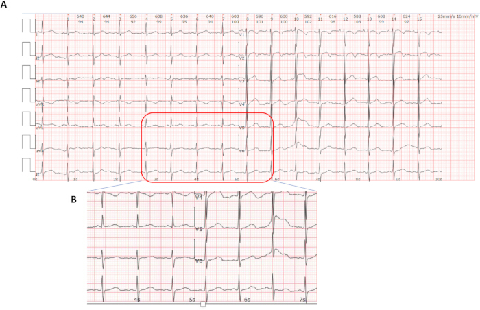
Figure 1: Representative electrocardiogram (ECG) obtained from the patient at the initial clinical evaluation. (A) Standard 12-lead ECG recorded at a paper speed of 25 mm/s and a calibration of 10 mm/mV. The tracing demonstrates a normal sinus rhythm with a heart rate of 97 beats/min. The PR interval measures 116 ms, the QRS duration measures 78 ms, and the corrected QT interval (QTc), calculated using Bazett's formula, measures 450 ms. (B) Magnified view of leads II, V5, and V6, highlighting the T-wave morphology and QT interval. Note: The ECG represents a single baseline recording obtained during the initial clinical evaluation. Please click here to view a larger version of this figure.
Endocrine evaluation confirmed delayed bone age. A growth hormone provocation test demonstrated a normal peak growth hormone response (12.39 ng/mL), excluding growth hormone deficiency as the primary cause of short stature. Thyroid function was normal (TSH: 1.76 mIU/L; FT4: 21.93 pmol/L), and pituitary MRI demonstrated normal morphology (4.5 × 9.1 × 3.1 mm). Following this evaluation, recombinant human growth hormone (rhGH) therapy was initiated on July 28, 2019, along with lifestyle interventions, including dietary protein optimization, regular jumping exercises (e.g., rope skipping), and improved sleep hygiene.
Whole-exome sequencing (WES) performed using peripheral blood DNA identified two novel missense variants in the PCNT gene (NM_006031.5): c.5675A>G (p.Glu1892Gly) in exon 28 and c.9734G>T (p.Arg3245Ile) in exon 45. Both variants were absent from major population databases (gnomAD, ExAC, the 1000 Genomes Project database, and the Chinese-specific Shenzhou Genome Database). In silico prediction tools yielded divergent results. The c.5675A>G variant was predicted to be deleterious by SIFT and possibly damaging by PolyPhen-2 (CADD PHRED score: 18.82), whereas c.9734G>T was predicted to be tolerated or benign (CADD score: 7.87). CNV-seq analysis identified no pathogenic copy number variants (Figure 2). According to the ACMG/AMP guidelines, both variants were classified as Variants of Uncertain Significance (VUS). Parental DNA samples were unavailable; therefore, segregation analysis to determine the phase of the two variants could not be performed.
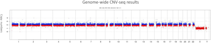
Figure 2: Copy number variant sequencing (CNV-seq) analysis. Genome-wide CNV-seq plot generated from peripheral blood DNA obtained from the patient. The x-axis represents genomic coordinates (chromosomes 1–22, X, and Y), and the y-axis represents the log2 read-depth ratio. Horizontal dashed lines indicate the thresholds for copy number gain (>0.3) and copy number loss (<–0.3). No chromosomal aneuploidies or pathogenic copy number variants above the 100 kb detection threshold were identified. Please click here to view a larger version of this figure.
Diagnosis, assessment, and plan:
Comprehensive clinical, radiological, endocrine, cardiac, and genomic evaluation supported a diagnosis of suspected PCNT-related primordial dwarfism with overlapping features of Microcephalic Osteodysplastic Primordial Dwarfism Type II (MOPD II). The diagnostic assessment included standardized anthropometric measurements, a neurodevelopmental evaluation demonstrating global developmental delay and moderate intellectual disability, baseline 12-lead electrocardiography, a skeletal survey, an endocrine evaluation, brain magnetic resonance imaging (MRI), copy number variant sequencing (CNV-seq), and whole-exome sequencing (WES) (Figure 3). CNV-seq excluded pathogenic copy number variants and large chromosomal abnormalities, whereas WES identified two candidate missense variants in the PCNT gene. According to the ACMG/AMP guidelines, both variants were classified as Variants of Uncertain Significance (VUS). Because parental DNA samples were unavailable, segregation analysis could not be performed to determine whether the variants were present in trans, precluding confirmation of a recessive molecular diagnosis.
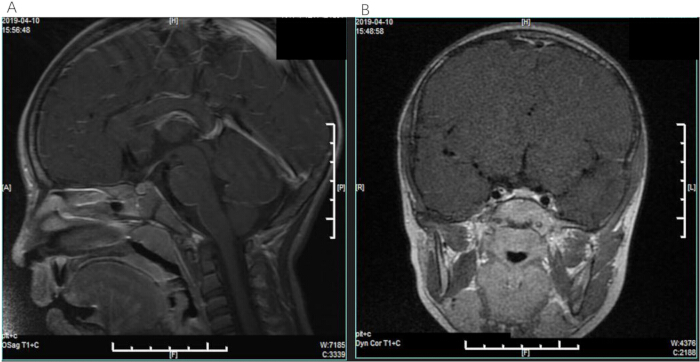
Figure 3: Brain magnetic resonance imaging (MRI) demonstrating normal intracranial findings. (A) Sagittal T1-weighted contrast-enhanced image demonstrating normal midline intracranial structures and brainstem without abnormal enhancement. (B) Coronal T1-weighted contrast-enhanced image demonstrating normal brain parenchyma and sellar region without evidence of an intracranial mass lesion. Please click here to view a larger version of this figure.
Following the initial evaluation, recombinant human growth hormone (rhGH) therapy was initiated on July 28, 2019, along with lifestyle interventions, including dietary protein optimization, regular jumping exercises (e.g., rope skipping), and improved sleep hygiene. Genetic counseling was provided to the family regarding the current VUS classification, the theoretical recurrence risk associated with an autosomal recessive disorder, and available reproductive options, including prenatal diagnosis and preimplantation genetic testing.
Because no curative therapy is currently available for PCNT-related disorders, long-term management focuses on multidisciplinary surveillance. Annual magnetic resonance angiography (MRA) of the brain, beginning at 5 years of age, was recommended for patients with MOPD II because of the recognized risk of cerebrovascular complications, including moyamoya disease, in accordance with published vascular surveillance recommendations. Repeat 12-lead electrocardiography and a formal pediatric cardiology evaluation were recommended to determine whether the borderline QTc interval was reproducible. Pharmacological intervention was not indicated at the time of evaluation but would be reconsidered if persistent QTc prolongation (>460 ms) or symptomatic arrhythmias developed. Continued endocrine follow-up, developmental assessment, speech therapy, occupational therapy, and individualized educational support were also recommended. Growth hormone therapy should continue to be evaluated cautiously because of its uncertain efficacy and the theoretical vascular risks reported in PCNT-related disorders.
Longitudinal follow-up data were unavailable because the patient was lost to follow-up after the initial 2019 evaluation. Consequently, subsequent endocrine monitoring, including assessment of rhGH efficacy and insulin-like growth factor 1 (IGF-1) concentrations, developmental progress, repeat cardiac evaluation, and additional genetic counseling, could not be documented. The long-term clinical course and response to treatment, therefore, remain unknown.
The diagnostic assessment was limited by the unavailability of parental DNA samples for segregation analysis and the absence of functional studies to determine the biological effects of the identified PCNT variants. Consequently, the phase of the two variants (cis versus trans) could not be established, compound heterozygosity could not be confirmed, and both variants remain classified as Variants of Uncertain Significance (VUS). In addition, whole-exome sequencing cannot exclude pathogenic variants located within deep intronic or regulatory regions; therefore, whole-genome sequencing may be considered in future investigations. Given the recognized risk of cerebrovascular and metabolic complications associated with PCNT-related disorders, long-term multidisciplinary surveillance remains appropriate.
Access restricted. Please log in or start a trial to view this content.
Protocol
This study was conducted in strict accordance with the Declaration of Helsinki and was approved by the Ethics Committee of Changsha Hospital for Maternal & Child Health Care (Approval No.CSSFYBJYEC-SOP-005-F07V2.0). Written informed consent was obtained from the patient’s parents for both participation in this study and the publication of clinical data and accompanying images. All patient data and images presented in this manuscript have been strictly de-identified to protect patient privacy. The research tools used in this protocol are listed in the Table of Materials.
1. Initial clinical assessment and phenotypic characterization
- Anthropometric measurements were obtained with the patient standing barefoot in an upright position using calibrated digital stadiometers and electronic scales.
- Height and weight were plotted against the 2009 Chinese National Growth Standards to assess growth status. Occipitofrontal circumference (OFC) was measured using a non-stretchable measuring tape and plotted on age- and sex-specific growth charts to assess for microcephaly, defined as an OFC below –2 SD or below the 3rd percentile.
- A comprehensive dysmorphology examination was performed by a board-certified clinical geneticist to document craniofacial features. Neurodevelopmental assessment was conducted using the Wechsler Intelligence Scale for Children (WISC).
- The Full-Scale Intelligence Quotient (FSIQ) was calculated and interpreted according to the Diagnostic and Statistical Manual of Mental Disorders, Fifth Edition (DSM-5), diagnostic criteria for intellectual disability (mild: 50–69; moderate: 35–49; severe: 20–34; profound: <20).
2. Cardiovascular and skeletal evaluation
- A standard 12-lead electrocardiogram (ECG) was obtained using a paper speed of 25 mm/s and a calibration of 10 mm/mV. The QT interval was measured manually in leads II and V5, and the corrected QT interval (QTc) was calculated using Bazett’s formula (QTc = QT/√RR). QTc prolongation was defined according to pediatric reference values as >440 ms in males.
- Skeletal survey radiographs, including anteroposterior and lateral views of the left hand and wrist, were obtained. Bone age was assessed by a pediatric radiologist using the Greulich–Pyle atlas to evaluate skeletal maturation and assess for skeletal dysplasia.
3. Genetic diagnostic workup
- Chromosomal copy number variant sequencing (CNV-seq) was performed using peripheral blood DNA. Low-pass whole-genome sequencing was performed to assess for chromosomal aneuploidies and pathogenic copy-number variants at a detection resolution greater than 100 kb.
- Whole-exome sequencing (WES) was performed on genomic DNA extracted from peripheral blood leukocytes using a commercial DNA extraction kit. Exome capture was performed using a commercial exome capture kit, followed by sequencing on a next-generation sequencing platform, generating 2 × 150 bp paired-end reads.
- Sequence reads were aligned to the GRCh38/hg38 reference genome using a validated alignment algorithm, and variant calling was performed using a validated bioinformatics pipeline.
- Quality-control metrics demonstrated a mean sequencing depth greater than 100×, with more than 98% of targeted exons achieving at least 20× coverage and PCNT exon coverage exceeding 99%.
- Sanger sequencing was performed to confirm the presence and zygosity of the identified PCNT variants in the proband. Because of the extended interval since the initial clinical evaluation, the original Sanger sequencing chromatograms were no longer available from the institutional archives.
- Parental DNA samples were unavailable; therefore, segregation analysis could not be performed to determine the phase of the identified variants.
- Variant interpretation was performed according to the 2015 ACMG/AMP guidelines. The bioinformatics workflow included variant annotation, population frequency assessment using gnomAD (v2.1.1), ExAC, and the Chinese-specific Shenzhou Genome Database, in silico pathogenicity prediction using SIFT, PolyPhen-2, and CADD (v1.6), evolutionary conservation analysis using PhyloP and multiz alignment (UCSC Genome Browser), and protein domain mapping using UniProt and the AlphaFold Protein Structure Database.
- Published literature and the ClinVar database were also reviewed to assign final variant classifications.
4. Differential diagnosis framework
- Microcephalic Osteodysplastic Primordial Dwarfism Type II (MOPD II; OMIM #210720) was prioritized because of severe prenatal-onset growth failure, intellectual disability, and characteristic craniofacial features despite the absence of overt microcephaly at the initial evaluation (age- and sex-adjusted OFC Z-score > –2 SD).
- Seckel syndrome (OMIM #210600) was considered but excluded because of the absence of a bird-headed facial profile and the absence of pathogenic or likely pathogenic variants in associated genes (e.g., ATR and CEP152) identified through analysis of the WES dataset.
- Meier–Gorlin syndrome (OMIM #224690) was excluded because patellar aplasia or hypoplasia was absent, and no pathogenic or likely pathogenic variants were identified in pre-replication complex genes (e.g., ORC1, ORC4, ORC6, CDC6, CDC45, GMNN, and MCM5) during analysis of the WES dataset.
Access restricted. Please log in or start a trial to view this content.
Results
Comprehensive clinical evaluation and genomic analysis identified a molecular and phenotypic profile consistent with a suspected PCNT-related primordial dwarfism. Whole-exome sequencing (WES) identified two novel missense variants in the PCNT gene (NM_006031.5): c.5675A>G (p.Glu1892Gly) in exon 28 and c.9734G>T (p.Arg3245Ile) in exon 45. Copy number variant sequencing (CNV-seq) identified no pathogenic copy number variants or chromosomal aneuploidies above the 100 kb detection threshold (
Access restricted. Please log in or start a trial to view this content.
Discussion
This case report describes a 7-year-old Chinese boy harboring two novel missense variants in the PCNT gene (c.5675A>G/p.Glu1892Gly and c.9734G>T/p.Arg3245Ile) who presented with profound postnatal growth failure (height 103.5 cm, –4.04 SD at age 7 years), global developmental delay, intellectual disability (Full-Scale IQ = 50 on the Wechsler Intelligence Scale), and characteristic craniofacial dysmorphism but lacked several canonical features of classic MOPD II. Compared with the classic diagnostic...
Access restricted. Please log in or start a trial to view this content.
Disclosures
The authors have nothing to disclose.
Acknowledgements
The authors thank the patient and his family for their participation in this study and for providing consent for publication of this case report. The authors also acknowledge the clinical genetics team at Changsha Maternal and Child Health Care Hospital for their contributions to patient care and phenotypic characterization, as well as the laboratory staff at the Medical Genetics Center for their technical support with whole-exome sequencing. This work was supported by the National Natural Science Foundation of China (Grant No. 82301845) and the Hunan Provincial Natural Science Foundation (Grant No. 2024JJ40187). The funding agencies had no role in the study design, data collection, data analysis, decision to publish, or preparation of the manuscript.
Access restricted. Please log in or start a trial to view this content.
Materials
| Name | Company | Catalog Number | Comments |
|---|---|---|---|
| 12-lead electrocardiograph | GE Healthcare | MAC 5500 HD (Software v2.0) | Cardiac evaluation and QT interval measurement |
| ABI 3730xl Genetic Analyzer | Applied Biosystems | Firmware v3.0 | Sanger sequencing validation |
| ANNOVAR | Children's Hospital of Philadelphia (CHOP) | v2021-02-01 | Variant annotation |
| BigDye Terminator v3.1 Cycle Sequencing Kit | Thermo Fisher Scientific | Cat. No. 4337456 | Sanger sequencing reactions |
| BWA-MEM | Broad Institute | v0.7.17 | Sequence alignment |
| ClinVar | National Center for Biotechnology Information (NCBI) | Accessed June 2026 | Clinical variant database |
| Combined Annotation Dependent Depletion (CADD) | University of Washington | v1.6 | In silico pathogenicity prediction |
| Digital radiography (DR) system | Siemens Healthineers | Ysio Max (syngo.via v4.0) | Skeletal survey and bone age assessment |
| Ensembl Variant Effect Predictor (VEP) | EMBL-EBI | v104 | Variant annotation |
| GATK HaplotypeCaller | Broad Institute | v4.2.6.1 | Variant calling |
| Genome Aggregation Database (gnomAD) | Broad Institute | v2.1.1 / v3.1.2 | Population frequency database |
| GRCh38/hg38 reference genome | Genome Reference Consortium | Build 38, Patch 13 | Sequence alignment reference |
| Low-pass whole-genome sequencing library preparation kit | Berry Genomics | Commercially provided | CNV-seq library preparation |
| NovaSeq 6000 Sequencing System | Illumina | Reagent Kit v1.5 (300 cycles) | Whole-exome sequencing |
| PolyPhen-2 | Harvard Medical School | v2.3.10 | In silico pathogenicity prediction |
| QIAamp DNA Blood Mini Kit | QIAGEN | Cat. No. 51104 | Genomic DNA extraction |
| SIFT | J. Craig Venter Institute | v5.2.2 | In silico pathogenicity prediction |
| SureSelect Human All Exon V6 Kit | Agilent Technologies | Cat. No. G9904B | Exome capture and library preparation |
References
- Gharehdaghi EE, et al. Novel mutation in patients with microcephalic osteodysplastic primordial dwarfism type II (MOPD II). Metab Brain Dis. 2024;40(1):18.
- Bober MB, Jackson AP. Microcephalic osteodysplastic primordial dwarfism, type II: a clinical review. Curr Osteoporos Rep. 2017;15(2):61-69.
- Rauch A, et al. Mutations in the pericentrin (PCNT) gene cause primordial dwarfism. Science. 2008;319(5864):816-19.
- Willems M, et al. Molecular analysis of pericentrin gene (PCNT) in a series of 24 Seckel/microcephalic osteodysplastic primordial dwarfism type II (MOPD II) families. J Med Genet. 2010;47(12):797-802.
- Jurca AD, et al. Clinical challenges in diagnosing primordial dwarfism: insights from a MOPD II case study. Medicina (Kaunas). 2024;60(11):1906.
- Retterer K, et al. Clinical application of whole-exome sequencing across clinical indications. Genet Med. 2016;18(7):696-704.
- Miller DT, et al. Consensus statement: chromosomal microarray is a first-tier clinical diagnostic test for individuals with developmental disabilities or congenital anomalies. Am J Hum Genet. 2010;86(5):749-64.
- Watanabe S, et al. Centriole-independent mitotic spindle assembly relies on the PCNT-CDK5RAP2 pericentriolar matrix. J Cell Biol. 2020;219(12):e202006010.
- Kim J, Kim J, Rhee K. PCNT is critical for the association and conversion of centrioles to centrosomes during mitosis. J Cell Sci. 2019;132(6):jcs225789.
- Duker AL, et al. Microcephalic osteodysplastic primordial dwarfism type II is associated with global vascular disease. Orphanet J Rare Dis. 2021;16(1):231.
- Majumder S, et al. Pericentrin deficiency in smooth muscle cells augments atherosclerosis through HSF1-driven cholesterol biosynthesis and PERK activation. JCI Insight. 2023;8(21):e173247.
- Steinfeldt J, Becker R, Vergarajauregui S, Engel FB. Alternative splicing of pericentrin contributes to cell-cycle control in cardiomyocytes. J Cardiovasc Dev Dis. 2021;8(8):87.
- Waich S, et al. Novel PCNT variants in MOPD II with attenuated growth restriction and pachygyria. Clin Genet. 2020;98(3):282-7.
Access restricted. Please log in or start a trial to view this content.