Here, we present a protocol to standardize the preparation of isolated rabbit duodenal smooth muscle for in vitro pharmacological assessment, utilizing a Horizontal Constant-Temperature Smooth Muscle Experiment System for real-time data recording of contractile activity.
Method Article
Standardized Preparation of Isolated Rabbit Duodenal Smooth Muscle For In Vitro Pharmacological Assessment of Contractile Activity
Xi Peng1,2,3 , Zhizhong Luo1,2,3 , Mulan Li4 , Ruoqi Zhang1,2,3 , Yiwen Bao1,2,3 , Nan Zeng1,2,3 , Rong Liu1,2,3
1School of Pharmacy, Chengdu University of Traditional Chinese Medicine, 2Chinese Medicine Germplasm Resources Innovation and Effective Uses Key Laboratory of Sichuan Province, 3Key Laboratory of Standardization of Chinese Medicine, Ministry of Education, Chengdu University of Traditional Chinese Medicine, 4Department of Laboratory Medicine, Southwest Medical University
Opens in a new tab
In This Article
Summary
Loading...
$$\rightleftharpoonup{xx}$$
$$\longleftharp{xx}$$,
$$\longrightharp{xx}$$,
Abstract
Loading...
$$\rightleftharpoonup{xx}$$
$$\longleftharp{xx}$$,
$$\longrightharp{xx}$$,
The coordinated contractile activity of gastrointestinal smooth muscle forms the physiological basis for maintaining normal digestion, absorption, and transport functions, with its dysfunction closely associated with motility disorders. The isolated organ perfusion technique eliminates complex in vivo interferences—such as neural, endocrine, and hemodynamic factors—serving as a classic model for pharmacological studies. To this end, this study details a stepwise protocol for the standardized preparation of rabbit isolated duodenal smooth muscle. The procedural workflow highlights the critical steps of preparing solutions, euthanizing the rabbit, performing a precise aseptic mid-abdominal laparotomy for rapid tissue excision, suspending the smooth muscle strips, and calibrating transducers for real-time tension recording. Based on this technical platform, we further validated the method by evaluating the effects of Huoxiang Zhengqi Oral Liquid (HXZQ-OL) on spontaneous contractions and acetylcholine/barium chloride-induced tetanic contractions. In conclusion, this protocol yields a robust and highly reproducible methodological framework, providing a broadly applicable in vitro screening platform for the pharmacological assessment of therapeutic agents targeting gastrointestinal motility.
Introduction
Loading...
$$\rightleftharpoonup{xx}$$
$$\longleftharp{xx}$$,
$$\longrightharp{xx}$$,
The coordinated contractile activity of gastrointestinal smooth muscle serves as the physiological foundation for maintaining normal digestive, absorptive, and transport functions1. Its dysfunction is closely associated with various common clinical disorders, including irritable bowel syndrome2, functional dyspepsia3, and postoperative gastrointestinal motility disorders4. Elucidating the molecular and pharmacological mechanisms regulating smooth muscle activity is crucial for developing novel prokinetic or antispasmodic therapies. In this research field, the isolated organ perfusion technique serves as a classic model. It offers practical value by eliminating complex in vivo interferences such as neural, endocrine, and hemodynamic factors, thereby enabling direct and precise assessment of the effects of drugs or active substances on smooth muscle tone5,6.
Establishing stable and reliable protocols for in vitro tissue preparation and data recording is essential for obtaining reproducible, high-quality results. Existing in vitro intestinal muscle experimental methods require further optimization and standardization in several areas: standardized management of experimental animals, meticulous surgical techniques for tissue harvesting, consistent environmental control of perfusion systems, and automated data acquisition coupled with standardized analysis. For instance, perfusion fluid temperature and pH levels, mechanical damage during tissue harvesting, and tissue preload settings can all significantly impact tissue viability and introduce experimental errors. These factors compromise the reliability of results and hinder comparability across different studies7,8.
Therefore, this study aims to establish and elaborate a standardized protocol for preparing isolated duodenal smooth muscle specimens from rabbits and performing real-time tension recording. This protocol systematically describes the entire process, from experimental animal ethics and welfare, aseptic laparotomy, and precise intestinal segment isolation, to achieving environmental control, standardized tension preload settings, and high-fidelity data acquisition using an integrated horizontal constant-temperature perfusion system and a multi-channel physiological signal acquisition system. Based on this standardized technical platform, we further applied cumulative dosing methods to quantitatively evaluate the effects of Huoxiang Zhengqi Oral Liquid (HXZQ-OL), meeting the quality standards of the Chinese Pharmacopoeia (2025 edition)9, on spontaneous contractions and acetylcholine- and barium chloride-induced tetanic contractions in isolated duodenal smooth muscle. Huoxiang Zhengqi Oral Liquid (HXZQ-OL), an oral dosage form of the classic Huoxiang Zhengqi traditional Chinese medicine formula, is commonly used in China for gastrointestinal disorders, providing the rationale for evaluating its direct effects on isolated intestinal smooth muscle. This study not only provides direct experimental pharmacological evidence elucidating the gastrointestinal smooth muscle regulatory effects of HXZQ-OL, but the experimental protocol also serves as a standardized, reproducible methodological model for pharmacological screening and in vitro tissue studies of gastrointestinal smooth muscle.
Protocol
Loading...
$$\rightleftharpoonup{xx}$$
$$\longleftharp{xx}$$,
$$\longrightharp{xx}$$,
Healthy adult New Zealand White rabbits of either sex (weighing 2.0–2.5 kg) were used in this work. All animal experimental procedures were conducted in accordance with the guidelines for the care and use of laboratory animals and were approved by the Laboratory Animal Ethics Committee of Chengdu University of Traditional Chinese Medicine (Approval No.: [2026070]). All efforts were made to minimize animal suffering and to reduce the number of animals used. The workflow diagram is illustrated in Figure 1.
1. Solution preparation
NOTE: All working solutions, including Tyrode's solution, acetylcholine (ACh), barium chloride, atropine, and verapamil, must be prepared fresh immediately before the experiment.
- Prepare 1 L of Tyrode's solution by dissolving 137 mM NaCl, 5.4 mM KCl, 1.8 mM CaCl₂, 0.4 mM NaH₂PO₄, 1.0 mM MgCl₂, 12 mM NaHCO₃, and 5.6 mM D-glucose. Before use, saturate with a 95% O₂ / 5% CO₂ mixture for 15–30 min, adjusting the pH to 7.3–7.4 (see the Table of Materials).
- Prepare a 1 mM ACh stock solution. Take 20 µL and add to 20 mL of the Tyrode's solution. Mix thoroughly to obtain a 1 µM ACh solution.
- Prepare a 1 M barium chloride (BaCl₂) stock solution. Take 20 µL and add to 20 mL of Tyrode's solution. Mix thoroughly to obtain a 1 mM BaCl₂ solution.
NOTE: BaCl₂ is highly toxic and irritating. When handling, wear personal protective equipment in a fume hood and dispose of waste liquid in accordance with regulations. - Prepare a 10 mM atropine sulfate stock solution. Take 20 µL and add to 20 mL of Tyrode's solution. Mix thoroughly to obtain a 10 µM atropine sulfate solution.
- Prepare a 1 mM verapamil hydrochloride stock solution. Take 2 µL and add to 20 mL of Tyrode's solution. Mix thoroughly to obtain a 0.1 µM verapamil hydrochloride solution.
NOTE: The concentrations of the prepared solutions were determined by referencing previous studies6,10,11 and were further optimized through our preliminary experiments to ensure optimal experimental conditions.
2. Preparation of in vitro rabbit duodenal smooth muscle specimens
- Rabbit euthanasia
- Secure the rabbit in the rabbit restraint device (Figure 2A,B and Figure 3A). Perform euthanasia by administering an overdose of sodium pentobarbital (120 mg/kg) via the auricular vein in rabbits (Figure 2C and Figure 3B).
NOTE: Complete death must be confirmed by the following criteria: dilated pupils, absence of pupillary light reflex, cessation of respiration and heartbeat, and loss of plantar reflex.
- Secure the rabbit in the rabbit restraint device (Figure 2A,B and Figure 3A). Perform euthanasia by administering an overdose of sodium pentobarbital (120 mg/kg) via the auricular vein in rabbits (Figure 2C and Figure 3B).
- Preparation and disinfection of the surgical site
- Secure the rabbit in the supine position on the operating table (Figure 2D and Figure 3C). Using surgical scissors (Figure 2E) or an electric shaver, thoroughly remove all abdominal hair to expose the skin in the designated surgical area (Figure 3D).
- Using sponge forceps (Figure 2E), grasp an iodine-soaked cotton ball (Figure 2F,G). Starting at the center of the planned surgical incision, apply the antiseptic solution in a concentric, circular pattern from the inside out (Figure 3E).
NOTE: The disinfection area should extend beyond the incision edges to ensure complete coverage of the entire surgical field skin. After disinfection, wait at least 3 min to allow the antiseptic to dry naturally on the skin surface. - Place a sterile drape (Figure 2H) over the disinfected abdominal area to expose the surgical field (Figure 3F).
- Positioning and incision of the abdominal incision
- Using a scalpel (blade #23) (Figure 2E), make an 8 cm longitudinal incision along the midline of the abdomen from below the xiphoid process to above the symphysis pubis.
- Dissect the skin and subcutaneous tissue layer by layer. Following the linea alba, dissect the anterior sheath of the rectus abdominis muscle, muscle fibers, and peritoneum layer by layer to enter the abdominal cavity (Figure 3G).
- NOTE: During the procedure, avoid damaging abdominal organs while ensuring hemostasis to prevent blood contamination of the surgical field and abdominal organs.
- Duodenal separation
- Open the abdominal cavity along the incision (Figure 3H). Gently retract the hepatic lobes cephalad using moist saline gauze (Figure 2I) to fully expose the stomach and duodenum (Figure 3I).
- Select the initial segment of the duodenum (bulbar or upper descending portion) located 2–3 cm from the pylorus as the tissue harvesting site.
- NOTE: Avoid direct traction or clamping of the target intestinal segment whenever possible to minimize mechanical injury. Instead, use blunt forceps to gently grasp the adjacent mesentery, or use saline-moistened cotton swabs to delicately manipulate and position the tissue.
- Using sterile ophthalmic scissors (Figure 2E), quickly and accurately resect a 1–2 cm (Figure 2J) segment of the duodenal tube (Figure 3J,K).
- Immediately transfer the excised intestinal segment to a culture dish (Figure 2K) containing prechilled (4 °C) Tyrode's solution continuously perfused with a mixed gas (95% O₂ / 5% CO₂).
- Intestinal segment trimming, cleaning, and temporary storage
- Using ophthalmic forceps (Figure 2E), gently dissect the mesentery and surrounding adipose tissue adhering to the intestinal wall.
- Using a syringe filled with cold Tyrode's solution (equipped with a blunt-tip needle) (Figure 2L), gently inject the enema solution into the intestinal cavity. Slowly flush the interior of the intestinal cavity 2–3x to thoroughly clear its contents.
- Place the cleaned intestinal segments in fresh oxygenated cold Tyrode's solution for temporary storage until subsequent experiments.
3. Horizontal constant-temperature smooth muscle experimental system startup and solution infusion
- Add Tyrode's solution to the preheating dish of the DSQG-1 Horizontal Constant-Temperature Smooth Muscle Experiment System (Figure 4B; see the Table of Materials).
- Turn on the system power, touch the operation panel to set the bath temperature to 37 °C, and begin preheating (Figure 4C).
- Press the Infusion button, and the system will automatically dispense 20 mL of Tyrode's solution into the bath tank.
- Press the Drainage button to drain the liquid into the waste collection container completely.
- Press the Rinse button, and the system will automatically complete the process of first draining the contents and then refilling with liquid.
- Press the Stop button to immediately terminate all ongoing infusion, drainage, and rinse processes.
- Press the Inflation button to continuously introduce the mixed gas (95% O₂ / 5% CO₂) into the bath. Adjust the gas flow rate to scale 5 to ensure subsequent experimental tissue oxygenation.
NOTE: Scale 5 indicates a gas flow rate of 100 mL/min. Excessively high gas flow rates may cause increased solution turbulence, affecting the stability of tension recordings.
4. Connecting multi-channel physiological recording and processing system with software operation
- Connect the data cable of the Horizontal Constant-Temperature Smooth Muscle Experiment System transducer to Channel 1 of the Multi-Channel Physiological Recording and Processing System (Figure 4D).
- Turn on the external power supply for the Multi-Channel Physiological Recording and Processing System and start the computer. Double-click the RM6240XC Multi-Channel Physiological Recording and Processing System icon on the desktop to launch the experimental software system (see the Table of Materials).
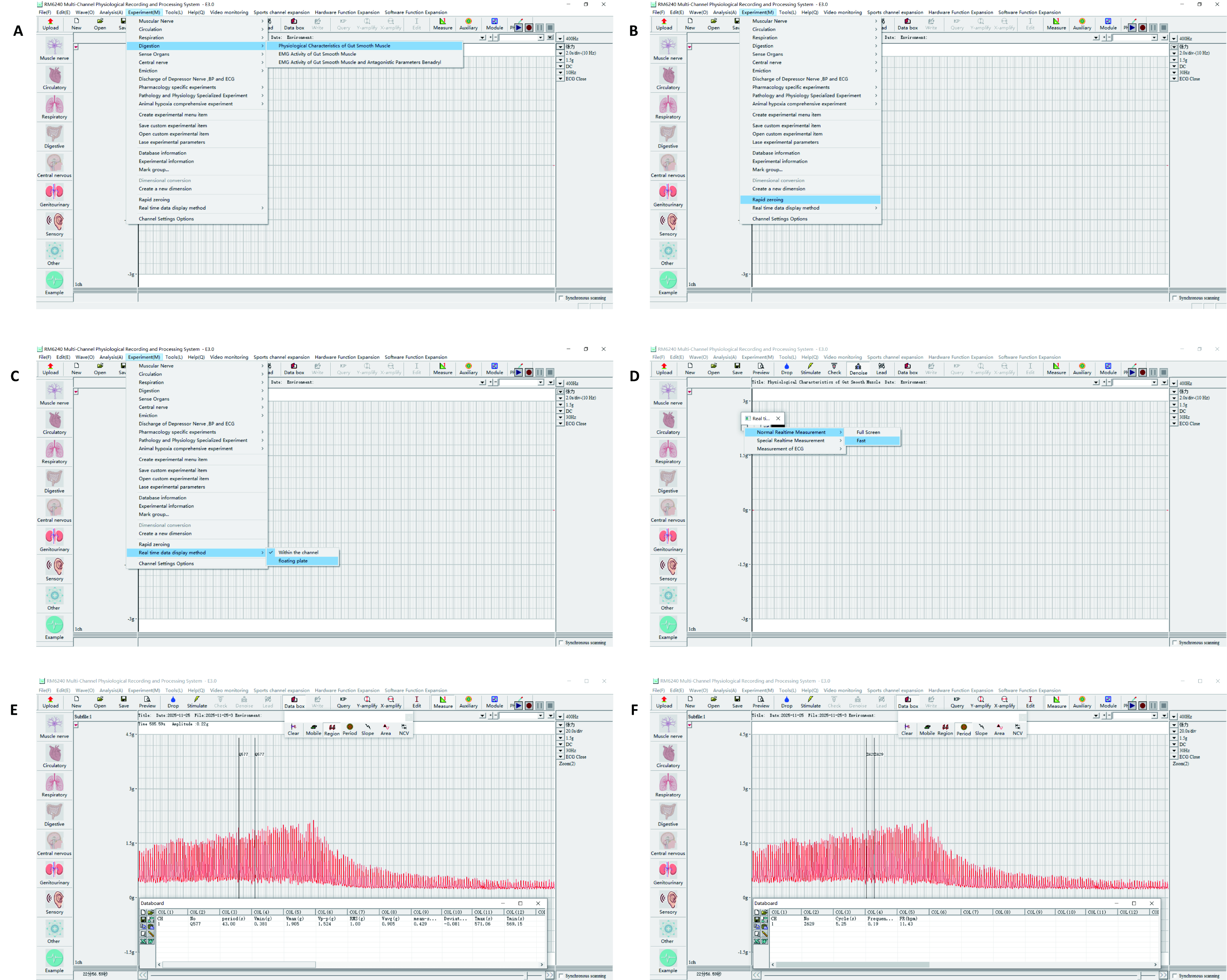
- In the menu bar, select Experiment | Digestion | Physiological Characteristics of Gut Smooth Muscle to load the corresponding experimental module.
- Set the following parameters: Channel mode to Tension, sampling frequency to 400 Hz, sensitivity to 1.5 g, time constant to DC, and filter frequency to 30 Hz.
- Click the Start button in the upper-right corner of the software interface to begin real-time signal acquisition and display. Click the Record button to simultaneously display the oscilloscope and save the acquired signal to the hard drive in real time.
- In the menu bar, select Experiment | Rapid Zeroing to adjust the tension signal to the baseline level.
NOTE: If the signal deviates from the baseline by more than one vertical coordinate unit, manually adjust the zero point of the tension transducer using a screwdriver. - In the menu bar, select Experiment | Real time data display method | floating plate. In the dialog box that appears, choose Normal Realtime Measurement-Fast to display real-time tension data of the intestinal muscle in the floating window.
5. Suspension and tension preload setting of duodenal muscle strip
- Adjust the front-to-back spacing of the tissue hooks by rotating the horizontal screw knob according to the length of the duodenal muscle strip, ensuring it matches the strip's length.
- Maintain the intestinal segment in its natural cylindrical configuration, and align the line of sight with the circular cross-sectional opening of the intestinal lumen.
- Using ophthalmic forceps, gently retract the intestinal opening to maintain patency.
- Gently insert the tip of the tissue hook into the intestinal lumen.
- At a distance of 2–3 mm from the intestinal margin, pierce the upper single-layer intestinal wall from the inside out using the tip of the tissue hook.
NOTE: The tissue hook must penetrate the muscle layer to ensure secure anchoring, avoiding superficial attachment only to the serosal layer, so as to prevent mechanical cutting and tearing of the intestinal wall by the hook when preload is applied. - The other side of the intestinal wall is identical to the first side. After identifying the parallel corresponding point, penetrate only the single-layer intestinal wall at that point (Figure 3L).
- Rotate the horizontal screw knob again to adjust the hook spacing, ensuring the duodenal muscle strip remains naturally relaxed.
- Gradually apply tension by adjusting the one-dimensional fine adjuster until the average tension of the duodenal muscle strip stabilizes at 0.8 ± 0.1 g (Figure 4A).
NOTE: Visually inspect the entire length of the intestine to ensure it remains straight and cylindrical, free of twisting, rotation, or spiral folds. If this is not the case, immediately reduce the tension and readjust the mounting point at one end. - Before commencing the experiment, equilibrate the duodenal muscle strip in the perfusion chamber for 60 min. During this equilibration period, replace the Tyrode's solution with fresh buffer every 15–20 min until its spontaneous contractile rhythm stabilizes.
NOTE: The 60 min equilibration period was considered complete only when the isolated intestinal segments exhibited stable spontaneous contractile activity for at least 15 consecutive min. The objective criteria for stabilization were defined quantitatively as follows: (1) baseline resting tension drift of < ±5%, (2) variance in mean contractile amplitude of < ±10% across consecutive 5 min intervals, and (3) frequency fluctuation of < ± 1 cycle/min. - Record the spontaneous contraction activity curve of duodenal muscle strip as baseline reference data for subsequent experiments.
6. Experimental data acquisition
- For ongoing experiments, in the menu bar, select Tools | Split view to perform real-time statistical analysis on captured waveform data.
- For stored data files, click the Open button in the toolbar, select a saved data file with the .lsd extension, and load historical experiment records.
- Click the Measure icon in the toolbar, select Region mode. Move the mouse cursor over the waveform area, hold down the left mouse button to select any two points, and the system will display a data panel showing the average tension value within that segment.
- Click the Measure icon in the toolbar, select Period mode. Using the left mouse button, click on five consecutive distinct wave peaks (peak points) along the waveform. Right-click the mouse, and the data panel will calculate and display the contraction frequency (in bpm) (Supplemental Figure S1).
Results
Loading...
$$\rightleftharpoonup{xx}$$
$$\longleftharp{xx}$$,
$$\longrightharp{xx}$$,
This experimental protocol describes the standardized preparation method for isolated rabbit duodenal muscle strips and the process of recording their dynamic tension and frequency changes in real time using an isolated organ perfusion system. Following this protocol, HXZQ-OL was sequentially added via cumulative dosing into a 20 mL Tyrode's solution bath, achieving final volume concentrations of 10, 20, 30, 40, and 50 µL/mL in the bath. Referencing the standards of the Chinese Pharmacopoeia (2025 edition), these concentrations correspond to crude drug slices at 6.6, 13.2, 19.8, 26.4, and 33.0 mg/mL, respectively. At the highest HXZQ-OL concentration tested (33.0 mg/mL), the pH (7.37 ± 0.08) and osmolarity (297 ± 4 mOsm/L) of Tyrode's solution were comparable to those of control Tyrode's solution (pH 7.38 ± 0.05; osmolarity 295 ± 3 mOsm/L; n = 3 independent preparations; P > 0.05, Student's t-test). These results indicate that the concentration-dependent reduction in contraction tension was not attributable to nonspecific pH or osmolarity changes (Figure 5A–C). Following administration of each concentration, the next concentration was added only after the intestinal muscle contraction response reached a stable plateau. To further validate the relaxing effect of HXZQ-OL on precontracted intestinal muscle, ACh (1 µM) and BaCl₂ (1 mM) were used to induce stable contraction. In this model, administration of HXZQ-OL (33.0 mg/mL) significantly induced intestinal muscle relaxation, with a relaxation magnitude comparable to that of the positive control drugs atropine sulfate (10 µM) (Figure 5D–G) and verapamil hydrochloride (0.1 µM) (Figure 5H–K). These results indicate that HXZQ-OL produces a concentration-dependent relaxing effect on isolated rabbit duodenal smooth muscle and is functionally consistent with inhibition of cholinergic- and calcium-dependent contractile responses.
In conclusion, the precise concentration-dependent response induced by HXZQ-OL and its relaxing effect on ACh/BaCl₂ precontracted tissue confirm the physiological activity of the specimen and the sensitivity of the recording equipment. This method is capable of accurately capturing subtle changes in smooth muscle tension, thereby validating the stability and reliability of the standardized in vitro tension recording protocol.
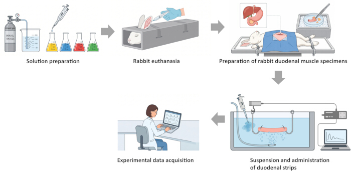
Figure 1: Schematic workflow of the ex vivo rabbit duodenal experiment. Please click here to view a larger version of this figure.
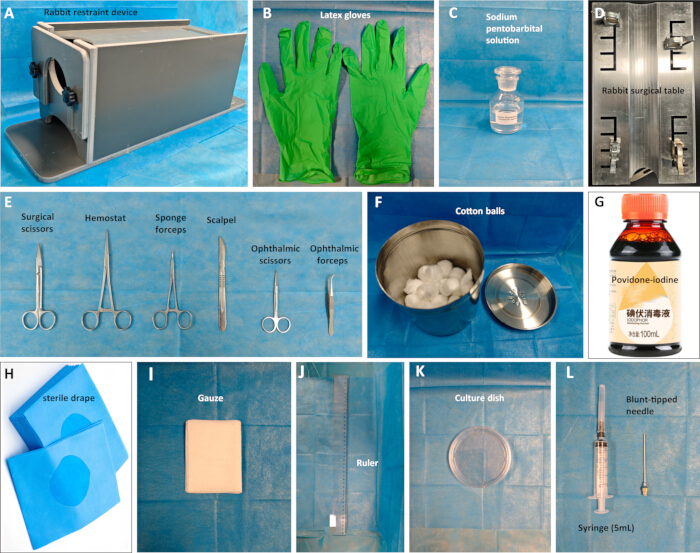
Figure 2: Key experimental materials. (A) Rabbit restraint device (B) Latex gloves (C) Sodium pentobarbital solution (D) Rabbit surgical table (E) Surgical instruments (F) Cotton balls (G) Povidone-iodine (H) Sterile drape (I) Gauze (J) Ruler (K) Culture dish (L) Syringe, Blunt-tipped needle. Please click here to view a larger version of this figure.
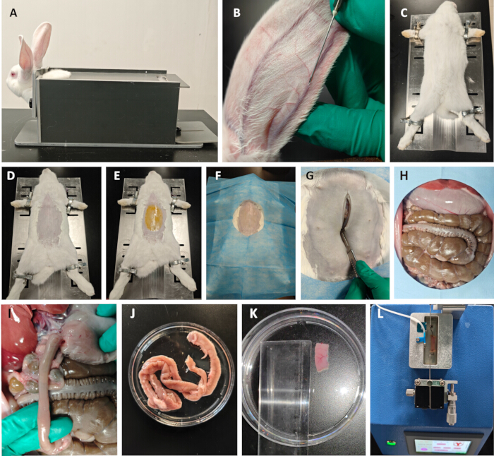
Figure 3: Preparation of rabbit in vitro duodenal smooth muscle specimen. (A) Rabbit fixation. (B) Rabbit euthanasia. (C) Supine fixation. (D) Remove abdominal hair to expose the surgical site. (E) Disinfect the surgical site with iodine. (F) Place a sterile drape to expose the surgical field. (G) Abdominal incision site localization and incision. (H) Laparotomy and abdominal cavity exposure. (I) Dissection of stomach and duodenum. (J) Completely dissected duodenum. (K) Cutting of duodenal smooth muscle strip. (L) Mounting muscle strips onto perfusion apparatus. Please click here to view a larger version of this figure.
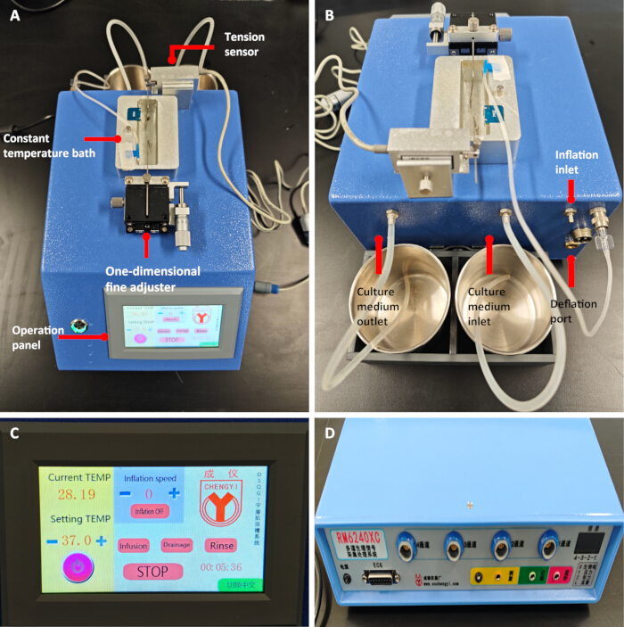
Figure 4: Key experimental instruments. (A) Front view of Horizontal Constant-Temperature Smooth Muscle Experiment System main unit. (B) Rear view of the system main unit. (C) Operation panel (D) Main unit of Multi-Channel Physiological Recording and Processing System. Please click here to view a larger version of this figure.
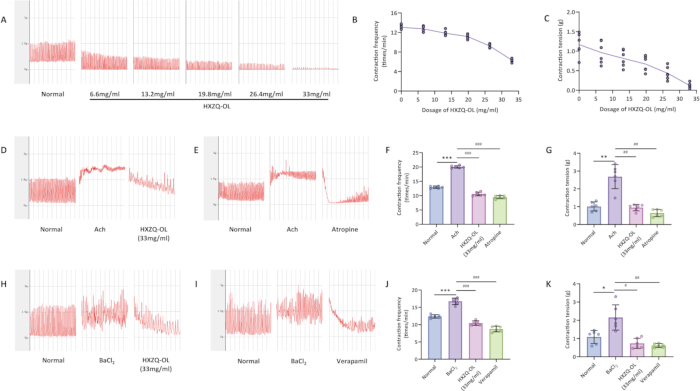
Figure 5: Effects of HXZQ-OL on contractions of isolated rabbit duodenum. (A–C) Effects of different concentrations of HXZQ-OL on spontaneous contractions of isolated duodenum and dose-response curves. (D–G) Inhibitory effects of HXZQ-OL on ACh-induced contractions in isolated duodenum. (H–K) Inhibitory effects of HXZQ-OL on BaCl₂-induced contractions in isolated duodenum. Data are presented as mean ± SD. n = 6 preparations per group. Statistical significance was determined using one-way ANOVA followed by Tukey's post hoc test. * p < 0.05, ** p < 0.01, *** p < 0.001 vs. Normal. # p < 0.05, ## p < 0.01, ### p < 0.001 vs. ACh/BaCl₂. Please click here to view a larger version of this figure.
Supplemental Figure S1: Key operational steps of multi-channel physiological recording and processing system software. (A) Experiment with channel selection and parameter configuration. (B) Signal baseline zeroing operation. (C–D) Opening the floating window for real-time data display. (E) Analyzing average tension using the region measurement tool. (F) Analyzing contraction frequency using the period measurement tool. Please click here to download this file.
Discussion
Loading...
$$\rightleftharpoonup{xx}$$
$$\longleftharp{xx}$$,
$$\longrightharp{xx}$$,
Isolated organ perfusion and tension recording techniques represent classic methods for studying smooth muscle physiology and pharmacology. Their findings hold significant importance for elucidating the pathophysiological mechanisms underlying gastrointestinal motility disorders, vascular diseases, and uterine dysfunction12,13,14,15. Using a standardized rabbit ex vivo duodenal model, this study demonstrated that HXZQ-OL induces concentration-dependent relaxation of intestinal smooth muscle. Because HXZQ-OL reduced both acetylcholine- and barium chloride-induced contractions, the findings are consistent with inhibition of cholinergic- and calcium-dependent contractile responses16,17,18. However, this organ-bath protocol does not directly identify molecular targets such as M3 receptors or L-type calcium channels; receptor- or channel-specific assays would be required to confirm these mechanisms.
The standardized operating procedures employed in this experiment—encompassing precise dissection, specimen preparation, standardized suspension, and real-time signal acquisition—support data reproducibility. This technical platform also has practical versatility. With minor adjustments to the core methodology, it can be directly applied to: (1) Functional studies of isolated vascular loops (e.g., aorta, mesenteric artery) in rats and mice to evaluate vasoactive drugs and endothelial function19; (2) Contractile mechanics analysis of uterine smooth muscle strips for uterine contraction regulation mechanisms and drug screening20; (3) Isolated studies of other hollow organs (e.g., trachea, bladder). This provides a shared technical foundation for cardiovascular, reproductive, and respiratory pharmacology.
The high success rate and high fidelity of the tension signals in this experiment hinge on the meticulous maintenance of specimen viability, the correct suspension of muscle strips, and the rigorous determination of baseline stability. First, when rapidly isolating intestinal segments, mechanical damage such as direct traction must be avoided, and the excised tissue must be immediately placed in continuously oxygenated Tyrode's solution at 4 °C to maximize the preservation of its physiological activity (Steps 2.4.2 and 2.4.3); Second, when suspending the muscle strips, it is essential to ensure that the hook precisely penetrates the smooth muscle layer, and that the tissue maintains a natural, untwisted, vertical cylindrical shape within the bath to prevent specimen tearing and distortion of the tension measurement vector (Steps 5.1–5.8); finally, troubleshooting is typically required during the equilibration phase (Step 5.9). The specimen must undergo at least 60 min of isothermal perfusion equilibration at 37 °C, and the baseline drift, amplitude, and frequency fluctuations of its spontaneous contractions must strictly meet the preset quantitative steady-state criteria. If the smooth muscle strip exhibits an unstable baseline or excessive noise in the tension recording, the researcher should first check whether the gas flow rate is too high, causing turbulence in the solution, and adjust it to a lower level (Step 3.7). If the tissue still does not exhibit a stable spontaneous contraction rhythm after 60 min, it is necessary to verify the pH and osmolarity of the Tyrode's solution, ensure that the temperature has accurately reached 37 °C, or gently rinse the tissue with fresh, preheated Tyrode's solution to remove potential endogenous inhibitory metabolites.
The Horizontal Constant-Temperature Smooth Muscle Experiment System used in this study offers significant advantages over traditional vertical smooth muscle baths, representing a methodological innovation. Traditional methods rely on surgical sutures to secure the tissue, which is cumbersome and prone to causing mechanical damage or localized ischemia at the specimen ends. In contrast, the protocol in this study employs adjustable tissue hooks to achieve direct suspension fixation (Steps 5.1–5.8). Furthermore, while traditional baths typically have large volumes (50–100 mL), this system utilizes a small-volume sample chamber (20 mL) equipped with automated, one-button perfusion and drainage interfaces (Steps 3.3–3.6). This not only reduces the consumption of perfusion solutions and test drugs but also shortens equilibration and elution times. Moreover, the equilibration and repeated perfusion steps are expected to minimize potential residual effects of the anesthetic before data acquisition, thereby enhancing experimental efficiency and reproducibility. This improvement may be suitable for pharmacodynamic screening and mechanism-oriented studies of multi-component samples, such as traditional Chinese medicine formulas. Of course, this study has limitations: in vitro conditions cannot fully simulate the complete neuro-humoral regulatory network; the specific active components and molecular targets of HXZQ-OL remain to be elucidated through subsequent chemical fractionation and cellular and molecular experiments21. In summary, this study demonstrates the relaxing effect of HXZQ-OL on isolated intestinal muscle and provides a standardized experimental methodology for pharmacological screening and studies of drug effects on gastrointestinal smooth muscle.
Disclosures
Loading...
$$\rightleftharpoonup{xx}$$
$$\longleftharp{xx}$$,
$$\longrightharp{xx}$$,
The authors have no conflicts of interest to disclose.
Acknowledgements
Loading...
$$\rightleftharpoonup{xx}$$
$$\longleftharp{xx}$$,
$$\longrightharp{xx}$$,
This work was supported by the Xinglin Scholar Research Promotion Project of Chengdu University of TCM (QJRC2022031), Xinglin Scholar Nursery Talent Project of Chengdu University of TCM (MPRC2023027), and Chengdu University of TCM—Affiliated Hospital Joint Innovation Fund (LH202402016).
Materials
| Name | Company | Catalog Number | Comments |
|---|---|---|---|
| 0.5-10 µL Pipette | Dalong Laboratory Instruments Co., Ltd. | 7010101004 | |
| 10 µL Pipette tips | LABSELECT | T-001-10 | |
| 10 mL Volumetric flask | Sichuan Shuniu Glass Instrument Co., Ltd. | B-043804 | |
| 100 mL Volumetric flask | Sichuan Shuniu Glass Instrument Co., Ltd. | B-043807 | |
| 100-1000 µL Pipette | Dalong Laboratory Instruments Co., Ltd. | 7010101014 | |
| 1250 µL Pipette tips | LABSELECT | T-001-1250 | |
| 20 mL Volumetric flask | Sichuan Shuniu Glass Instrument Co., Ltd. | B-010105 | |
| 200 µL Pipette tips | LABSELECT | T-001-200 | |
| 50 mL Conical centrifuge tube | LABSELECT | CT-002-50A | |
| 5-50 µL Pipette | Dalong Laboratory Instruments Co., Ltd. | 7010101006 | |
| Acetylcholine chloride | Supelco | PHR1546 | |
| All-in-One Lab Computer | Lenovo | R5-3500U | |
| Atropine sulfate | Macklin | A822893 | |
| Barium chloride | Sigma-Aldrich | 342920 | |
| Calcium chloride | Sigma-Aldrich | 499609 | |
| D-Glucose | Sigma-Aldrich | G5767 | |
| Freezing point osmometer | Shanghai Precision Instruments Co., Ltd. | BS100 | |
| Horizontal Constant-Temperature Smooth Muscle Experiment System | Chengdu Instrument Factory | DSQG-1 | |
| Huoxiang Zhengqi Oral Liquid | Taiji Group Co., Ltd. | Z50020409 | |
| Magnesium chloride | Sigma-Aldrich | M8266 | |
| Multi-Channel Physiological Recording and Processing System | Chengdu Instrument Factory | RM6240XC | |
| New Zealand White rabbits | Pizhou Dongfang Breeding Co., Ltd. (License No.: SCXK[Su]2022-0004) | either sex (weighing 2.0–2.5 kg) | |
| pH meter | Shanghai Leici | PHS-25 | |
| Potassium chloride | Sigma-Aldrich | P5405 | |
| Povidone-iodine | H&H Medical Co., Ltd. | 3180132 | |
| Precision balance (0.1 mg) | Sartorius | BCE64i | |
| Saline solution | H&H Medical Co., Ltd. | 100014438029 | |
| Sodium bicarbonate | Sigma-Aldrich | S6014 | |
| Sodium chloride | Sigma-Aldrich | S9888 | |
| Sodium dihydrogen phosphate | Supelco | 1063700250 | |
| Sodium Pentobarbital | Sigma-Aldrich | P3761 | |
| Verapamil Hydrochloride | Macklin | V820460 | |
| Vortex mixer | Kylin-Bell | XW-80A |
Reprints and Permissions
Request permission to reuse the text or figures of this JoVE article
Request Permission