A step-by-step guideline to generate primordial germ cell-like cells from human induced pluripotent stem cells in a robust manner using a 2D monolayer-based method with basement membrane extract overlay.
Method Article
Protocol For Differentiating Pluripotent Stem Cells Into Primordial Germ Cell-like Cells
In This Article
Summary
Abstract
Using human pluripotent stem cell differentiation in vitro has significantly broadened the understanding of lineage specification events that occur during early development in vivo, a period that is extremely challenging to investigate. One of the first embryonic lineages to be specified in the human embryo is the germ cell lineage, marked by the induction of the primordial germ cells (PGCs). The PGCs are the sole founder population of the gametes later in life, and their specification is a critical first step towards the organism's fertility. Here, a robust protocol for generating PGC-like cells (PGCLCs) from human induced pluripotent stem cells (hiPSCs) is described, starting with the initial evaluation of the pluripotent state and cellular characteristics of the hiPSCs to ensure effective PGCLC differentiation. For this, the effects of different cell densities, plate coatings, and hiPSCs used for PGCLC differentiation are discussed. Finally, a workflow for characterization and quantification of hPGCLCs, using immunofluorescence and flow cytometry, is provided. The differentiation protocol focuses on conditions that are easy to establish and test in most laboratories and can be adjusted as necessary. Moreover, this protocol is fast, allowing determination of whether hiPSCs are suitable for PGCLC differentiation. The protocol aims to promote consistency and compatibility in hPGCLC outcomes across different hiPSC lines and culture platforms, ensuring robust yields that support reliable characterization and further optimization of downstream differentiation steps toward successful in vitro gametogenesis in humans.
Introduction
Human germline development is a highly coordinated process that initiates early in embryonic development and begins with the specification of primordial germ cells (PGCs), the population that ultimately gives rise to oocytes and sperm cells1. In vivo, PGC specification occurs shortly after implantation2,3 in response to inductive signals from neighboring cells4,5. The development of PGCs has been extensively investigated in rodents, particularly through genetic studies that have demonstrated the involvement of signaling pathways, such as BMP, WNT, and TGFβ6,7,8,9. Following specification, the PGCs undergo epigenetic reprogramming, characterized by genome-wide DNA demethylation and chromatin remodeling, to later establish (sex-specific) germline identity10,11,12,13,14.
In mammals, PGC specification is driven by coordinated crosstalk between signaling pathways, and some aspects are evolutionarily conserved across humans, non-human primates, pigs, and mice, including the involvement of pluripotency factors such as POU5F1 and NANOG9,15,16,17,18. Despite shared features, species-specific differences in PGC differentiation mechanisms and gene-regulatory networks limit the direct translation from animal models to humans9,19,20,21.
To understand gametogenesis in humans, studies using human material are necessary, but they remain challenging due to limited access to early human embryos. Continuous advances in protocols enabling the in vitro differentiation of human primordial germ cell-like cells (hPGCLCs) from human pluripotent stem cells, such as induced pluripotent stem cells (hiPSCs), have reduced reliance on human gonadal tissue while enabling upscaling and genetic studies18,22. Most established strategies for hPGCLC differentiation rely on three-dimensional (3D) aggregates or embryoid bodies18. Using these platforms, multiple studies have shown that hPGCLCs recapitulate key epigenetic features of early human PGCs, including germline transcriptional programs23,24 and genome-wide DNA methylation reprogramming25. Together, these in vitro platforms have substantially advanced knowledge of human PGC biology, including lineage commitment and epigenetic resetting. However, aggregation-based hPGCLC differentiation protocols remain challenging as they are time-consuming, and the total yield of hPGCLCs for robust epigenetic characterization, further differentiation assays, or use in high-throughput experimental applications remains low.
Recently, a 2D monolayer-based feeder-free method using diluted basement membrane extract (BMEx) in combination with low levels of BMP4 to differentiate hiPSCs into hPGCLCs proved relatively easy to scale up in tissue culture plates and achieved high efficiencies (~55%) within a short differentiation window (5 days)26. Compared to other 2D systems that required an intermediate Activin A priming step before induction and used higher BMP4 concentrations (40–50 ng/mL)27,28, this BMEx approach did not require a pre-induction step and used lower BMP4 levels (10 ng/mL), while maintaining comparable efficiency and improved scalability.
Here, a detailed, step-by-step guide to the 2D hPGCLC BMEx-differentiation protocol is provided, starting with the initial evaluation of hiPSC maintenance, including pluripotency and cellular characteristics, followed by testing the effects of different hiPSC plate coatings and cell densities on hPGCLC differentiation. Also, the downstream characterization by flow cytometry analysis and immunofluorescence is described. Critical handling points and troubleshooting guidance to reduce laboratory deviations are provided, enabling researchers to consistently reproduce hPGCLC differentiation in this 2D format.
Protocol
This protocol follows guidelines and regulations of the Leiden University Medical Center (LUMC), the LUMC Biobank & Biomaterials (TCBio), and the LUMC Leiden hiPSC Center (under BB24.037). All materials and control male (XY) hiPSC lines (M54 and M199) used in this protocol are listed in the Table of Materials. All tissue culture procedures were performed in a dedicated Biosafety Level 2 tissue culture room, equipped with a humidified normoxic incubator (37 °C, 5% CO2), a multipurpose benchtop centrifuge (swing-bucket rotor), and a Class II biosafety cabinet with a stereomicroscope.
CAUTION: Not all hiPSC lines can differentiate into hPGCLCs26,29. However, if other hiPSCs were used for PGCLC differentiation and PGCLCs were observed, the method described here (Figure 1A) will allow scaling up hPGCLC production.
1. Coating tissue-culture plates for hiPSC culture
This protocol describes coating tissue culture plates with both diluted BMEx and vitronectin.
- Basal membrane extract (BMEx) coating
- Thaw an aliquot of BMEx on ice. This can be Geltrex, Cultrex, or Matrigel.
- Dilute the aliquot of basal membrane extract in cold DMEM/F12 to a final concentration of 1% (v/v) and resuspend gently.
- Coat the desired number of wells of a 6-well plate with 1 mL of the diluted BMEx per well.
- Incubate the coated plate for at least 30 min at 37 °C.
NOTE: Coated plates can be stored at 2–8 °C for up to 1 week. When using coated plates stored at 2–8 °C, incubate the plate for at least 10 min at 37 °C before further use.
- Vitronectin coating
- Thaw an aliquot of vitronectin at room temperature (RT) for 10–15 min.
- Dilute vitronectin to a final concentration of 5 µg/mL in DPBS without calcium and magnesium (DPBS -/-) and resuspend gently.
- Coat the number of desired wells of a 6-well plate with 1 mL of the diluted vitronectin per well.
- Incubate the coated 6-well plate for 1 h at RT.
2. Passage for maintenance of hiPSCs
- Prepare culture medium: serum-free cell culture medium mTeSR Plus supplemented with mycoplasma contamination-prevention solution (MycoZap (1:1000)).
- Aspirate the culture medium from the cells that are ready to be passaged.
- Add 0.5 mL human PSC selection and passaging agent (ReLeSR) to each well and aspirate this within 1 min. Make sure a very thin layer is left on the cells.
- Incubate the cells for 6 min at 37 °C.
- Tap the sides of the 6-well plate a few times.
- Carefully detach the cells with 1 mL culture medium by pipetting up and down with a p1000 pipette, and make sure to check the size of colony fragments regularly to keep their size between 50–200 µm.
- Take the pre-coated 6-well plates and remove the coating solution (Step 1).
- Add 2 mL culture medium to each well.
- Transfer the culture medium containing the detached hiPSCs colony fragments to the new well in the desired ratio.
NOTE: The ideal ratio for splitting hiPSC colony fragments is 1:6-1:50, allowing passaging every 4–7 days. - Place the cell culture plate with the passaged hiPSCs colony fragments in a humidified normoxic incubator (37 °C, 5% CO2).
- Refresh the culture medium every other day with 2 mL culture medium until the hiPSCs are ready to be passaged again.
NOTE: Add 4 mL culture medium instead of 2 mL to each 6-well to prevent the need to refresh over the weekend.
CAUTION: Do not passage hiPSCs when confluence is too low, and discard the culture when confluence is too high to maintain a healthy hiPSC culture and to avoid spontaneous differentiation.
3. hPGCLC differentiation
- Preparation prior to hPGCLC differentiation (Day 0).
- Refresh the culture medium of the hiPSCs 1 h before starting hPGCLC differentiation.
- Prepare the coating solution: 1% BMEx solution diluted v/v in cold DMEM/F12.
- Coat the desired number of wells of a 12-well plate with 0.5 mL coating solution.
- Incubate the coated plate for at least 30 min at 37 °C before starting differentiation.
- Prepare the plating medium: 2% (v/v) BMEx solution in cold culture media and supplement this with a post-thaw recovery medium (RevitaCell) (1:100).
CAUTION: Keep the plating medium on ice during the preparation and plating of hiPSCs.
- hiPSCs plating for hPGCLC differentiation (Day 0).
- Remove the culture medium from the 6-well containing the hiPSCs to use for hPGCLC differentiation. Start with high-quality hiPSCs at 50–80% confluency, with no signs of spontaneous differentiation.
- Add 1 mL trypsin solution (TrypLE) per well.
- Incubate hiPSCs for 5 min at 37 °C.
- Make a single-cell suspension by carefully resuspending the hiPSCs colonies 2–3 times with a p1000 pipette.
- Check under an inverted microscope whether a single-cell suspension is obtained. If not, resuspend the hiPSCs colonies another 1–2 times with a p1000 pipette.
- Transfer the hiPSC cell suspension to a 15 mL tube.
- Add 4 mL DMEM-F12 medium to the 15 mL tube.
- Centrifuge the hiPSC cell suspension at 200 x g for 5 min.
- Aspirate the supernatant and carefully resuspend the hiPSC cells in 0.5–1.0 mL plating medium.
- Count the number of hiPSCs using a cell counter or hemocytometer and calculate the number of hiPSCs needed per well for the hPGCLC differentiation.
NOTE: As a starting point, a plating density of 40–80K cells/cm2 can be used. However, for each cell line and at each passage number, plating density optimization is recommended to achieve optimal yield. Here, hiPSCs were plated at a plating density of 10K to 100K cells/cm2. - Carefully resuspend the required number of hiPSCs in the appropriate volume of plating medium (1 mL per well of a 12-well plate), and ensure a homogeneous cell suspension is obtained using a p1000 pipette.
- Take the coated 12-well plate from the incubator and remove the coating solution.
- Add 1 mL of the cell suspension to each well of a 12-well plate.
- Carefully shake the plate to ensure the cells are evenly distributed in the well.
- Place the 12-well plate in a humidified, normoxic incubator (37 °C, 5% CO2) for 16–24 h.
- hPGCLC differentiation (Day 1–2)
- Prepare RB27 basal medium: advanced RPMI160 with B-27 (1:100), 1X aminoacid substitute (Glutamax), 1X MEM non-essential amino acids, and mycoplasma contamination-prevention solution (1:1000).
- Prepare differentiation medium: 2% (v/v) BMEx solution in cold RB27 basal medium and add BMP4 to a final concentration of 10 ng/mL. The basal membrane extract is required to induce lumenogenesis and trigger PGCLC specification in vivo.
CAUTION: Keep the differentiation medium on ice or at 4 °C. The differentiation media can be prepared for Days 1 and 2 of the differentiation protocol. - On Day 1 (within 16–24 h after plating hiPSCs), take the plate from the incubator and remove the plating medium.
- Wash the wells by adding 0.5 mL RB27 basal medium per well.
- Aspirate the RB27 basal medium and add 1 mL differentiation medium per well.
- Place the plate with the cells back in the humidified normoxic incubator (37 °C, 5% CO2).
- On Day 2 of the differentiation, refresh the cells with 1 mL differentiation medium per well.
- hPGCLC differentiation (Day 3–5)
- Prepare switch medium: RB27 basal medium supplemented with BMP4 (10 ng/mL), LIF (10 ng/mL), SCF (50 ng/mL), and EGF (50 ng/mL). Switch medium mirrors the in vivo shift from hPGC specification to survival and proliferation. BMEx is omitted because lumenogenesis is no longer necessary.
- On Day 3 and Day 4, refresh each well with 1 mL switch medium.
- On Day 5 of differentiation, the hPGCLCs can be characterized using immunofluorescence or Flow Cytometry.
4. Characterization of hPGCLCs by immunofluorescence and flow cytometry
- Characterization by immunofluorescence.
- Prepare permeabilization solution: 0.3% Triton X-100 diluted in PBS.
- Prepare washing solution: 0.05% Tween 20 in PBS (PBST)
- Prepare blocking solution: 1% bovine serum albumin (BSA) in PBST.
- Remove the switch medium from the wells used for hPGCLC differentiation.
- Wash the cells with 1 mL per well DPBS (-/-).
- Fix cells with 1 mL per well 4% paraformaldehyde (PFA) in PBS for 15 min at RT.
- Wash the cells 3 times for 5 min with PBS, using 1 mL per well.
NOTE: At this point, plates can be stored at 4 °C prior to starting immunofluorescence. - Permeabilize cells with permeabilization solution for 15 min at RT, using 1 mL per well.
- Wash cells 3 times for 5 min with washing solution, using 1 mL per well.
- Block cells with blocking solution for 1 h at RT, using 1 mL per well.
- Prepare primary antibody solution: dilute primary antibodies of interest in blocking solution.
NOTE: Here, the following two antibody combinations for PGC markers were used: goat anti-SOX17 and rabbit anti-TFAP2C, and goat anti-SOX17 and mouse anti-POU5F1 (Table of Materials). - Remove the blocking solution, add 0.4 mL of primary antibody solution per well, and incubate overnight at 4 °C.
NOTE: It is important to include negative controls to assess nonspecific antibody binding. For this, keep one well overnight in blocking solution, omitting the primary antibodies, but subsequently stain that well with the secondary antibody solution. - The next day, remove the primary antibody solution and wash the cells 3 times for 5 min with PBST.
- Prepare secondary antibody solution: dilute secondary antibodies of interest (Table of Materials) and DAPI (1:500) in blocking solution.
NOTE: The secondary antibody solution is light sensitive and should be kept in the dark. - Remove PBST and add 0.4 mL of secondary antibody solution per well for 1 h at RT in the dark.
- Wash the cells 3 times for 5 min with PBS, using 1 mL per well.
- Do imaging of the cells (20x objective lens).
NOTE: Immunofluorescence images were acquired using a 20x objective on the Airyscan microscope, while bright-field images were captured using a 4x objective as well as a 10x objective on the EVOS microscope.
- Characterization by flow cytometry
- Prepare FACS buffer: 0.5% BSA in DPBS (-/-)
- Remove the switch medium from the wells with the hPGCLCs.
- Wash cells with 1 mL DPBS (-/-).
- Add 0.5 mL pre-warmed Accutase per well and incubate for 15 min at 37 °C. For dissociation, Accutase is preferred to trypsin solution because the hPGCLCs are highly sensitive to dissociation.
- Make a single-cell suspension by carefully resuspending the cells 2–3 times with a p1000.
- Check under an inverted microscope whether a single-cell suspension is obtained. If not, resuspend the cells another 1–2 times with a p1000 pipette.
- Filter the cell suspension through a 40 µm strainer into a 15 mL tube.
- Wash the 40 µm strainer with 5 mL FACS buffer.
- Centrifuge the cell suspension at 300 x g for 5 min.
- Remove the supernatant.
- Resuspend the cell pellet in 0.3 mL FACS buffer in a 1.5 mL tube.
NOTE: It is optional to count cells using a cell counter or a hemocytometer. If the cells are counted, expect around 1 million cells per well of a 12-well plate. - Prepare adequate control samples for flow cytometry (unstained and single-stain controls). For this, transfer 15–30 µL of the cell suspension into 1.5 mL tubes and fill to 100 µL with FACS buffer.
NOTE: For this study, 3 single-stains were prepared: EPCAM-PEcy7 (1:200), ITGA6-BV421 (1:200), and 7AAD (1:200). The solutions containing the conjugated antibodies and 7AAD are light sensitive and should be kept in the dark. - Add the conjugated antibodies of interest directly to the cell suspension to achieve adequate dilution. Here, the following conjugated antibodies for PGC markers were used together: EPCAM-PEcy7 (1:200) and ITGA6-BV421 (1:200) (Table of Materials).
- Incubate the cells with the conjugated antibodies for 30 min on ice in the dark.
- Add 1 mL FACS buffer to the samples, unstained control, and single-antibody controls, and centrifuge at 300 x g for 5 min.
- Remove the supernatant.
- Resuspend the samples in 0.5 mL, and the control samples in 0.2 mL, of FACS buffer containing 1 mM EDTA. This prevents cells from clumping.
- Add the samples and controls to a 5 mL FACS tube with a cell strainer cap, then pass them through the strainer.
- Add 7AAD directly to each sample several minutes before measuring in the flow cytometer.
- Measure samples using a Flow Cytometer. Here, the fluorescence-based cell analyzer was used to measure the samples, data was collected using FACSDiva software (v9.0), and FlowJo software (v10.10.0) was used to analyze the data.
- Use the control samples (unstained and single-antibody controls) to set up the gating strategy and perform compensation if necessary.
- Vortex the samples before putting them in the machine.
- Extract the cell population from all events by first setting a gate based on area [forward scatter-area (FSC-A) and side scatter-area (SSC-A)].
- From this cell population, extract single-cells based on size [FSC-width (FSC-W) and FSC-height (FSC-H)] and granularity [SSC-W and SSC-H].
- Exclude dead cells from further analysis by setting a gate for 7AAD-negative cells.
- Use the sample to check whether the gates are properly set, and adjust them if necessary.
- Analyze the markers of interest from this live-cell population. Here, the gated cells positive for both EPCAM and ITGA6 corresponded to hPGCLCs.
Results
The quality and stable state of the hiPSCs are important for further differentiation
hiPSCs were maintained in 6-well plates for at least 3 passages (in the different coatings) prior to differentiation experiments and analysis. hiPSCs were passaged when cell colonies reached a confluency of 70–80% (Figure 1B), which is also the optimal hiPSC confluency to initiate hPGCLC differentiation. Healthy hiPSC cultures displayed uniform colony morphology without dark or differentiated areas, visible in bright field images (Figure 1B). However, the quality of the hiPSCs was affected when cell colonies were not maintained at their optimal confluency or showed signs of differentiation (Figure 1B). At low confluence, hiPSC continued to proliferate, and more time was needed for colony formation until the confluency was suitable for passaging or differentiation experiments. At high confluency, hiPSCs grew on top of each other, while differentiated regions appeared. The pluripotency state of the hiPSCs was confirmed by expression of SOX2 or POU5F1, both transcription factors essential for maintaining pluripotency and self-renewal in hiPSCs30, and most importantly, SOX2 is not expressed by hPGCLCs31. Phalloidin staining was used to assess cytoskeletal organization and colony morphology, indicative of hiPSCs' quality. Note that differences in cell morphology are associated with the reduction of SOX2 or POU5F1 (Figure 1B).
Effect of the hiPSC maintenance substrate on hPGCLC differentiation
To assess whether maintaining hiPSCs on different coating substrates affected differentiation to hPGCLCs, the hiPSCs were cultured on different substrates (Geltrex, Matrigel, Cultrex, and vitronectin) (Figure 2). On all substrates, hiPSCs showed robust and uniform nuclear SOX2 or POU5F1 levels, indicative of their pluripotent state. hiPSC colonies maintained on Geltrex, Matrigel, and Cultrex had comparable morphology, including well-defined colony borders and irregular overall colony shapes, compared with the round colonies on vitronectin (Figure 2). hiPSCs that reached 70–80% confluency were plated at a density of 40K cells/cm2 and differentiated to hPGCLCs on Geltrex26. On Day 5, brightfield images showed characteristic hPGCLC colonies in all conditions (Figure 3). Moreover, triple-positive SOX17/TFAP2C/PDPN or double-positive SOX17/POUF51 hPGCLCs were identified in all conditions (Figure 3). This suggested that the differentiation protocol could be used with hiPSCs cultured under different conditions.
Effect of initial hiPSC cell density on hPGCLC differentiation
It is important to check the optimal seeding density for optimal hPGCLC differentiation (on Geltrex) as different hiPSCs have different proliferating rates. Moreover, in case the efficiency in hPGCLC differentiation drops as hiPSCs are continuously passaged, it is advised to adjust the hiPSC seeding cell density. To evaluate the impact of the seeding cell density on the efficiency of hPGCLC differentiation, hiPSCs were seeded at different densities, from 10K to 100K cells/cm2 (Figure 4A). At Day 5, the percentage of hPGCLCs was assessed by flow cytometry using two different conjugated antibodies for human PGC-markers, EPCAM and integrin α6 (ITGA6)32. Flow-cytometry analysis followed a standard sequential gating strategy, shown for M199 hiPSCs seeded at 40K cells/cm2 (Figure 4B). Briefly, the total cell population (all events) was gated to exclude debris (particle area), then single cells were selected based on size (FSC) and granularity (SSC), and finally, dead cells were excluded based on 7AAD staining. Thereafter, the gate to quantify cells expressing EPCAM-cy7 and ITGA6-BV421 was set using stained and unstained samples (Figure 4B).
Comparing the hPGCLC differentiation efficiency using different seeding cell densities and using hiPSC cultured on different substrates revealed a density-dependent trend (Figure 4C). At the seeding density of 20–40K cells/cm2, M54 and M199 hiPSCs showed the highest proportion of EPCAM+/ITGA6+ double-positive hPGCLCs, achieved from hiPSCs maintained on Geltrex and Cultrex. Interestingly, higher densities resulted in lower differentiation efficiency. Immunofluorescence results using PGC markers SOX17/TFAP2C/PDPN or SOX17/POUF51 on the hPGCLCs validated the flow cytometry results (Figure 4D). These results suggested that adjusting the seeding density to the specific hiPSCs is important to obtain optimal differentiation efficiency. Combining immunofluorescence and flow cytometry enhances the robustness of the hPGCLC differentiation assay.
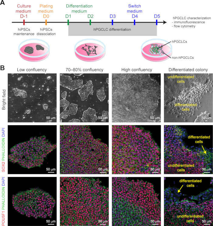
Figure 1: hPGCLC differentiation workflow and quality of hiPSCs. (A) Schematic of the workflow used in this study. hiPSCs were maintained (in culture medium) and reached 60–80% confluency on the day of differentiation (Day 0, D0). On D0, the hiPSCs were dissociated into single cells and plated in the desired seeding density (in plating medium). The next day, the cells were treated with differentiation medium (D1–D3), followed by switch medium (D3–D5). On D5, hPGCLCs were used for immunofluorescence or flow cytometry. (B) Brightfield images (top row- 4x magnification); immunofluorescence for SOX2, PHALLOIDIN-staining, and DAPI-staining (middle row); and for POU5F1, PHALLOIDIN-staining, and DAPI-staining (bottom row) showing hiPSCs with different confluency. Scale bars = 50 µm. Drawings in Figure 1A were created by the authors using basic drawing tools in Adobe Illustrator; no paid software or tools were used. Hence, no copyright license was required. Please click here to view a larger version of this figure.
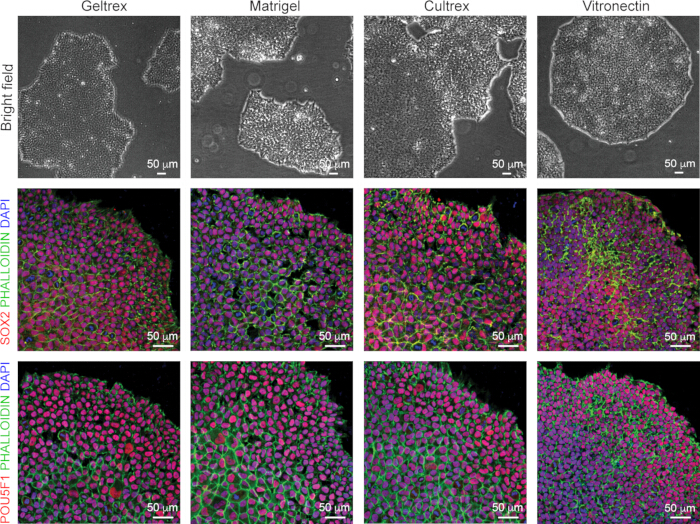
Figure 2: Effect of various coating substrates during hiPSC culture. Brightfield images (top row); immunofluorescence for SOX2, PHALLOIDIN-staining, and DAPI-staining (middle row); and for POU5F1, PHALLOIDIN-staining, and DAPI-staining (bottom row) showing hiPSCs cultured on different coating substrates. Scale bars = 50 µm. Please click here to view a larger version of this figure.
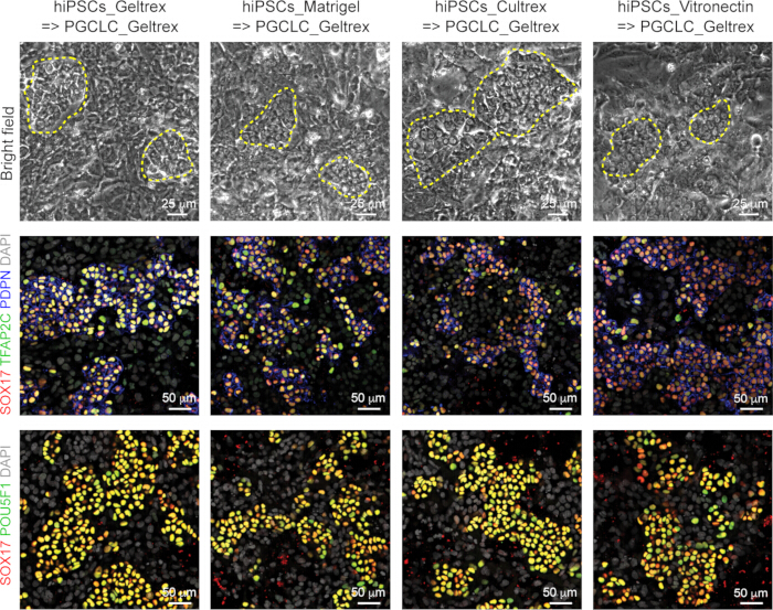
Figure 3: Effect of various coating substrates during hiPSC culture on hPGCLCs differentiation. Brightfield images (top row-10x magnification); immunofluorescence for SOX17, TFAP2C, PDPN, and DAPI-staining (middle row); and for POU5F1, SOX17, and DAPI-staining (bottom row) of hPGCLCs differentiation (using Geltrex) at Day 5, starting from hiPSCs cultured on different coating substrates. The yellow dashed line highlights regions with hPGCLCs. Scale bars = 25µm (top row) and 50 µm (middle and bottom rows). Please click here to view a larger version of this figure.
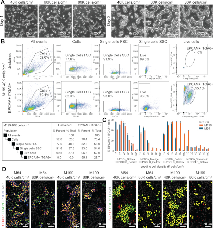
Figure 4: Effect of hiPSC seeding density on hPGCLCs differentiation. (A) Brightfield images at Day 1 (D1) and D3 of hPGCLCs differentiation using different hiPSC seeding densities (40K cells/cm2, 60K cells/cm2, 80K cells/cm2). Scale bars = 250 µm. 4x magnification. (B) Representative flow cytometry plots showing the gating strategy to quantify the percentage of EPCAM+/ITGA6+ hPGCLCs. For this, M199 hiPSCs (on Geltrex) were used at a seeding density of 40K cells/cm2. (C) Bar plot depicting the percentages of EPCAM+/ITGA6+ hiPGCLCs at D5 when using hiPSCs (M199 and M54) cultured on different coatings and seeded at different densities. (D) Immunofluorescence for SOX17, TFAP2C, PDPN, and DAPI-staining (left panels); and SOX17, POU5F1, and DAPI-staining (right panels) at D5 of hPGCLCs differentiation using hiPSCs (M199 and M54, on Geltrex) seeded at 40K and 80K cells/cm2. Scale bars = 50 µm. Please click here to view a larger version of this figure.
Discussion
In vitro gametogenesis constitutes a powerful experimental platform for investigating human germline development, reproductive toxicity, and infertility under controlled culture conditions and on a patient-specific basis. Although multiple protocols for differentiating hPGCLCs from hiPSCs have been reported18, progress in the field has been hindered by the limited availability of standardized, scalable, and reproducible approaches across laboratories. Recent studies have described simplified and efficient approaches for hPGCLC induction, including BMP4-based micropatterned differentiation27, a rapid 2D monolayer platform enabling hPGCLC generation within 3.5 days28, and a 3D culture system that models peri-implantation development and paracrine niche signaling33. Even though these protocols efficiently supported hPGCLC differentiation, they still lack the ability to serve as high-throughput platforms. For hPGCLC-based studies to be broadly applicable, particularly for reproductive toxicological screenings, protocols must be robust, technically accessible, and compatible with downstream quantitative assays. Here, a step-by-step analysis of the hPGCLCs BMEx-differentiation protocol is presented, emphasizing critical technical parameters that determine reproducibility and performance.
A major determinant for successful differentiation is the initial pluripotency state of the hiPSCs. Passage number, cellular health, line-specific characteristics, and pre-differentiation culture conditions can influence hPGCLC differentiation outcomes. Initially, during hiPSC maintenance, attention can go to the coating substrate, seeding cell density, and culture plate format, which together define colony architecture and signaling environments at the onset of hPGCLC differentiation. The present study demonstrates the effect of using hiPSCs cultured on different coatings and different seeding densities on hPGCLCs differentiation (on Geltrex). Both hiPSC lines tested here showed maximal differentiation efficiency at 20,000–40,000 cells/cm2 seeding density. This underscores the need to determine optimal seeding densities when introducing new cell lines, following extended passaging, or altering experimental layouts or culture plate formats. Even seemingly minor changes in surface area-to-volume ratios or growth kinetics may need optimization to preserve high differentiation efficiency. Accordingly, it is recommended that users perform small-scale pilot experiments to first determine optimal conditions for hPGCLC differentiation.
Despite its robustness, the method has limitations that should be acknowledged. Although compatible with multiple coatings and densities, differentiation efficiency remains sensitive to hiPSC intrinsic properties, including epigenetic state and passage history, which are not fully controllable. Several tested hiPSCs seemed unable to differentiate into hPGCLCs26. Most importantly, unlike vitronectin, which is a fully defined product, BMEx is an animal-derived product subject to batch-to-batch variation, which may affect reproducibility. Although different batches of Matrigel, Cultrex, and Geltrex were previously tested and yielded consistent results26, this remains a concern, and fully defined alternatives to BMEx should be considered to ensure future reproducibility and transferability. Here, two male (XY) hiPSC lines were used, but this protocol has been tested with both female (XX) and male (XY) lines26. Finally, while the protocol resulted in hPGCLCs, which have been shown to be transcriptionally similar to hPGCs and to have competence for further development26,34, further maturation into later germ cell stages was not addressed here. Although scalability was demonstrated for screening-style applications, automation-based adaptation and inter-laboratory validation across a broader panel of hiPSC lines will further strengthen translational utility.
Relative to existing hPGCLC differentiation strategies, this protocol offers several advantages. In contrast to highly specialized or matrix-restricted approaches, it accommodates commonly used hiPSC maintenance substrates and culture plate formats, thereby lowering technical barriers. The emphasis on density optimization and early-stage quality control further improves reproducibility, a persistent challenge in the field of in vitro gametogenesis.
The methodological framework described here is particularly relevant for reproductive toxicology, infertility modeling, and developmental biology. Its compatibility with scalable formats enables medium- to high-throughput compound screening, while its reproducibility supports cross-study comparisons and collaborative consortia efforts. More broadly, systematic standardization of hPGCLC induction protocols across laboratories, comparing characterization and functional endpoints18,35, will be essential for advancing in vitro gametogenesis toward predictive human-relevant platforms.
Disclosures
The authors have no conflicts of interest to declare and confirm that no artificial intelligence tools were used in the preparation of this manuscript.
Acknowledgements
We would like to acknowledge present and past members of the Chuva de Sousa Lopes group for inspiring discussions; the Flow Cytometry Core Facility of the LUMC, the LUMC hiPSC Hotel, and the Imaging Facility of the LUMC for their expertise. This study was supported by the Novo Nordisk Foundation (NNF21CC0073729, reNEW to EK, CLdM and SMCSL), the EC (HORIZON-MSCA-2023-DN MSCA 101169308-IMPLANTEU to SL and SMCSL); the China Scholarship Council (CSC 20230920054 to CF and CSC 202308410196 to DW), and the Dutch Organization for Health Research and Development (ZonMW PSIDER-2021-10250022120001) to MP and SMCSL.
Materials
| Name | Company | Catalog Number | Comments |
|---|---|---|---|
| 7AAD Viability Staining Solution | BioLegend | 420403 | Dilution 1:200 |
| Accutase | STEMCELL Technologies | 7920 | |
| Advanced RPMI 1640 Medium | Gibco | 12633012 | |
| Alexa Flour 488 donkey anti-mouse IgG | Invitrogen | A21202 | Dilution 1:500 |
| Alexa Flour 594 donkey anti-rabbit IgG | Invitrogen | A21207 | Dilution 1:500 |
| Alexa Flour 647 donkey anti-goat IgG | Invitrogen | A21447 | Dilution 1:500 |
| Alexa Fluor Phalloidin 594 | Invitrogen | A12381 | Dilution 1:400 |
| Alexa Fluor Phalloidin 647 | Invitrogen | A30107 | Dilution 1:400 |
| Antibody anti-CD49f (ITGA6), Brilliant Violet 421 | Biolegend | 313624 | Dilution 1:200 |
| Antibody anti-CD326 (EPCAM1), PE/Cyanine7 | Biolegend | 324222 | Dilution 1:200 |
| Antibody anti-SOX2, rabbit polyclonal | Merck | ab5603 | Dilution 1:200 |
| Antibody anti-SOX17, goat polyclonal | R&D Systems | AF1924 | Dilution 1:500 |
| Antibody anti-OCT3/4 (POU5F1), mouse monoclonal | Santa Cruz Biotechnology | SC-5279 | Dilution 1:200 |
| Antibody anti-Podoplanin (PDPN), mouse monoclonal | Abcam | ab256561 | Dilution 1:200 |
| Antibody anti-AP-2γ (TFAP2C), rabbit polyclonal | Cell Signaling Technology | 2320S | Dilution 1:300 |
| B-27 Supplement, serum free | Gibco | 17504044 | Diltuion 1:100 |
| BD FACSCanto | BD Biosciences | Equipment for flow cytometry | |
| BD FACSDiva software version 9.0 | BD Biosciences | Software | |
| Bovine Serum Albumin (BSA), fractie V | Roche | 10735086001 | Dissolve in PBST |
| CELLSTAR 12 Well Cell Culture Plate | Greiner | 665180 | |
| CELLSTAR 15 mL Tube | Greiner | 188271 | |
| CELLSTAR 6 Well Cell Culture Plate | Greiner | 657160 | |
| Corning 40 µm Cell Strainers | Corning | 431750 | |
| Cultrex Stem Cell Qualified, Reduced Growth Factor Basement Membrane Matrix | R&D systems | 3434-101-02 | Dilute to in cold DMEM-F12 |
| DAPI (4′,6-Diamidino-2-Phenylindole) | Invitrogen | D3571 | Dilution 1:500 |
| DMEM:F12, GlutaMAX supplement | Gibco | 31331028 | |
| DPBS, no calcium, no magnesium | Gibco | 14190144 | |
| EVOS M5000 | Invitrogen | Equipment for imaging | |
| EVOS M5000 software version 1.3.660.548 | Invitrogen | Software | |
| Falcon 5 mL Round Bottom Polystyrene Test Tube, with Cell Strainer Snap Cap | Corning | 352235 | |
| FlowJo software version 10.10.0 | BD Biosciences | Software | |
| Geltrex Reduced-Growth Factor Basement-Membrane Matrix, LDEV-Free, stem-cell qualified | Gibco | A1413302 | Dilute to in cold DMEM-F12 |
| GlutaMAX Supplement | Gibco | 35050061 | Dilution 1:100 |
| FlowJo software version 10.10.0 | BD Biosciences | Software | |
| Geltrex Reduced-Growth Factor Basement-Membrane Matrix, LDEV-Free, stem-cell qualified | Gibco | A1413302 | Dilute to in cold DMEM-F12 |
| GlutaMAX Supplement | Gibco | 35050061 | Dilution 1:100 |
| hiPSCs M54 | LUMC hiPSC Core Facility | LUMC0054iCTRL03 | hiPSCs can be distributed upon request with MTA |
| hiPSCs M199 | LUMC hiPSC Core Facility | LUMC0199iCTRL01 | hiPSCs can be distributed upon request with MTA |
| LUNA-II Automated Cell Counter | Logos Biosystems | Equipment for cell counting | |
| LUNA-II software version 2.3.5 | Logos Biosystems | Software | |
| Matrigel hESC-Qualified Matrix, LDEV-free | Corning | 354277 | Dilute to in cold DMEM-F12 |
| MEM Non-Essential Amino Acids Solution | Gibco | 11140050 | Dilution 1:100 |
| mTeSR Plus | STEMCELL Technologies | 100-0276 | |
| MycoZap Plus-CL | Lonza | VZA-2011 | Dilution 1:1000 |
| Paraformaldehyde (PFA) | Sigma-Aldrich | 1040051000 | Dissolve to 8% PFA in water first; and dilute to 4% (1:1) with 0.2 M phosphate buffer |
| PIPETMAN P1000, 100–1000 µL, Metal Ejector | Gilson | F144059M | |
| PIPETMAN P200, 20–200 µL, Metal Ejector | Gilson | F144058M | |
| PIPETMAN P10, 1–10 µL, Metal Ejector | Gilson | F144055M | |
| Recombinant Human BMP4, CF | R&D Systems | 314-BP-050 | Stock concentration of 100 µg/mL in 4 mM HCL containing 0.1% BSA |
| Recombinant Human EGF, CF | R&D Systems | 236-EG-200 | Stock concentration of 50 µg/mL in PBS |
| Recombinant human LIF | Gibco | 300-05 | Stock concentration of 100 µg/mL in 0.1% BSA |
| Recombinant Human SCF, CF | R&D Systems | 11010-SC-100 | Stock concentration of 100 µg/mL in PBS |
| ReLeSR | STEMCELL Technologies | 5872 | |
| RevitaCell Supplement | Gibco | A26445-01 | Dilution 1:100 |
| Triton X-100 | Sigma-Aldrich | T8787 | Dilute to 0.3% solution in PBS |
| TrypLE Express Enzyme | Gibco | 12604021 | |
| Tubes 1.5 mL | Eppendorf | 05-402-27 | |
| TWEEN 20 | Sigma-Aldrich | 822184 | Dilute to a 0.5% solution in PBS |
| UltraPure 0.5M EDTA | Invitrogen | 15575020 | Dilute 1:500 in FACS buffer |
| Vitronectin | Gibco | A14700 | Dilute to 5 µg/mL in DPBS -/- |
| Zeiss ZEN software version 3.12.107.04000 | Carl Zeiss Microscopy GmbH | Software | |
| Zeiss LSM980 AiryScan 2 | Carl Zeiss Microscopy GmbH | Equipment for imaging |