The zebrafish (Danio rerio) is a widely used laboratory model with significant genetic similarity to humans and is accessible for genetic manipulation. Standard genotyping involves anesthetizing the animal to obtain a fin clip, which may cause pain. We evaluate skin swabbing as a refinement for automated genotyping of adult zebrafish.
Method Article
Skin Swabbing as a Noninvasive Alternative to Fin Clipping for Commercial Automated Genotyping
In This Article
Summary
Abstract
Fin clipping remains the most common sampling method for genotyping zebrafish, yet it requires anesthesia and tissue removal, raising animal welfare concerns and potentially impacting behavior in the short term. To address this, we have validated a non-invasive skin swabbing technique that eliminates the need for fin clipping adults while maintaining genotyping accuracy during automated genotyping.
This method involves gently swabbing the fish’s skin to collect mucus and epidermal cells for DNA extraction. This approach avoids anesthesia, reduces handling stress, and is much less invasive than traditional fin clipping with limited tissue injury. We conducted a comparative analysis of swab-derived DNA against traditional fin clip samples sent for automated genotyping. Swab and fin clips samples taken from the same animal matched genotype results, demonstrating substantial promise as a refinement for automated genotyping under the 3Rs framework. Further testing of the swabbing technique, transport, and sample processing was optimized across nine sessions. Key aspects of the collection technique include focusing on consistent swabbing pressure and sample dry time. Proper technique is critical to ensuring robust and reproducible genotyping outcomes.
Our findings indicate that skin swabbing offers a welfare-improving alternative to fin clipping for automated genotyping. Broad adoption of this technique has the potential to enhance animal welfare standards while maintaining scientific rigor, aligning with the principles of the 3Rs and advancing non-invasive research practices in zebrafish facilities worldwide. While the current success rate indicates some limitations relative to fin clip genotyping results with automated genotyping, swabbing remains a promising refinement that can substantially improve welfare in zebrafish facilities.
Introduction
Zebrafish (Danio rerio) are the second most widely used laboratory animal model worldwide, after mice, owing to their high fecundity, externally fertilized transparent embryos, and 72% of human genes have a zebrafish ortholog1. Increasing evidence demonstrates that fish are capable of experiencing pain2, prompting continued efforts within the zebrafish research community to refine experimental procedures and minimize animal pain and distress. Zebrafish fins are capable of regeneration, and fin clipping remains a commonly performed procedure for tissue collection, primarily for genotyping purposes3. Analogous to ear punching in mice, fin clipping provides a reliable source of DNA but raises welfare concerns4, as the procedure is considered invasive and is expected to be of mild severity.
Genotyping samples obtained via fin clipping may be processed in-house or submitted to third-party service providers for automated genotyping. At the Francis Crick Institute, most fin clip samples are outsourced for automated genotyping, allowing researchers to optimize their time by continuing other experimental work while genotyping is performed externally.
Skin swabbing is an advancement in the 3Rs for zebrafish, relying on a noninvasive sampling technique rather than an invasive one5. It has been proven that mucus obtained from a swab of the skin can provide enough genetic material to run genotyping assays in-house4,6, and the collection method also works on mice and Xenopus laevis7. Skin swabbing of adult zebrafish avoids the need for analgesia or anesthesia8, sparing the animal pain and the adverse effects of MS-2229, while also saving the technician preparation time.
As we do not typically run genotyping samples in-house, we wanted to determine if sending skin swabs for automated genotyping would provide reliable genotype information. Swabbing zebrafish provides variable DNA collection due to the nature of the collection technique as compared to tissue collection via fin clipping.
During automated genotyping, skin swab samples are processed in a similar manner to fin clips, with a reduction in the concentration of NaOH in the lysing solution. Genotyping results for each sample are calculated via relative copy number (RCN) and other metrics using a software analysis package. We improved sample handling across several sessions to reduce the reported error rates during automated genotyping.
Protocol
This work was in compliance with the Francis Crick Institute’s animal welfare and ethical review board and Home Office licensing.
1. Set up
- Identify adult zebrafish (>3 months of age, at least 20 mm long) that need genotyping.
- Set up an area with a soft, wet sponge in a plastic container, a 96-well plate provided by the commercial vendor, a swab, scissors for cutting the swab, and a final isolation tank for swabbed fish.
NOTE: One must be able to individually identify each animal to match the genotyping results. - Use system water to wet the sponge, rather than purified or tap water.
2. Swabbing4
- Net fish from the home tank onto the top of a wet sponge and secure the head gently with the net using fingers4. No analgesia or anesthesia is required8.
- Try to gently cover the eyes with the net and fingers to limit external stimulation4.
- Once the fish is in place, rub the swab gently along one side of the fish from the pectoral fin to tail4.
- Repeat four more times, then roll the swab 180 degrees and repeat five more times.
- Never swab against the scales as this could cause injury.
- Rest the swab in the corresponding well on the plate while the fish is placed in its individual housing.
NOTE: Small breeding tanks may also be used to provide individual housing (1 L tank with divider). - Ensure each container is properly labeled to avoid mix-ups.
- Using scissors, cut the swab tip off so it fits in the well plate.
- Repeat for all fish that need genotyping.
- Let the plate dry for 60 min on the open bench before sealing.
- Seal the well plate with a cap mat.
3. Submitting samples
- Prior to submission, contact an automated genotyping supplier to create an account, order shipping documentation, arrange collection, and submit order requests.
4. Automated genotyping
- Follow the steps below for the automated genotyping sample processing:
- Lyse swab samples for DNA extraction via the addition of 50 µL of 0.5 M NaOH solution.
- Incubate the samples for 60 min at 90 °C.
- Transfer 15 µL of the lysed sample to another well-plate for DNA isolation.
NOTE: The isolation process is a proprietary, ferrous bead-based isolation method10 that utilizes EtOH washes to purify the DNA that has adhered to the beads. - Use 100 µL of DNase-free H2O to elute the DNA from the beads.
- Mix the DNA with qPCR assays for the targeted mutations and an internal control assay targeted to the cJun gene.
- Use the following thermocycler settings: 1 cycle at 95 °C for 10 min, followed by 35 cycles of 95 °C for 15 s, and 60 °C for 1 min. Refer to Supplementary Table 1 for the housekeeper and reporter genes.
- Add the proprietary master mix, including Taq polymerase and buffers.
- The total reaction size is less than 1 µL. Run each reaction in duplicate wells.
- Ensure the assay and Master mix are at 1× concentration.
NOTE: The results will be emailed when ready.
Results
Genotyping results from skin swabs
Over nine different sessions, we processed 470 skin swab samples of wild type and two different genetically altered lines (Table 1). Six different animal technicians obtained the swab samples across all sessions. The overall success rate of obtaining a definitive genotyping result was 86.3%.
During session 1, all the fish were swabbed and fin clipped to verify the correct genotype results we obtained. There were no errors. The total length of the fish swabbed in this session ranged from 21.53 mm to 34.28 mm.
Session 4 was performed in a small quarantine room with limited area flow. We believe this impacted the success rate based on similar error rates with fin clips collected in the same room at later dates. To rectify this issue, we added the 60-min drying time to the rest of the sessions. Removing this data point raises the average success rate of obtaining a definitive genotyping result to 90.9% (Table 2).
From session 5, a 60-min drying time was added to this procedure. The average success rate of obtaining a definitive genotyping result from sessions 5 to 9 is 90.8% (Table 3).
During sessions 8 and 9, the same technician collected all the swab samples. The average success rate across these two sessions is 92.1% (Table 4).
Genotyping results determination
The reactions were run on a thermocycler, and the data were analyzed using the thermocycler software. The cycle thresholds from that software are then used to calculate a delta Ct using both the target and control Cts:
ΔCt = Ct (gene of interest) - Ct (housekeeping gene) = RCN
The results for each target assay are then compared with RCN ranges previously established for the presence or absence of the alleles based on zebrafish tissue samples. These calculations and results review occur in a proprietary software system developed by the automated genotyping provider.
The graphs in Figure 1A–C show the curves produced by the on-board capture and analysis software of the PCR machines, depicting the visual amplification curve for the PCR assay, which measures changes in fluorescence over the PCR cycles.
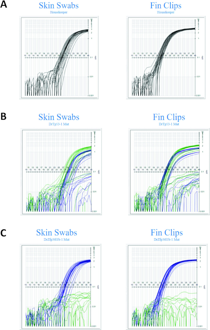
Figure 1: The visual amplification curve for the PCR assay run for comparing skin swabs versus fin clips. (A) Comparison cycle of threshold (Ct) of skin swabs to fin clips for internal control assay. Each line represents the amplification curve of a sample (each sample was run in duplicate). (B) Comparison cycle of threshold (Ct) of skin swabs to fin clips for Trp53 mutant allele assay. Each line represents the amplification curve of a sample (each sample was run in duplicate). Blue lines indicate the mutant allele and green lines the wildtype allele. (C) Comparison cycle of threshold (Ct) of skin swabs to fin clips for Zfp36l1b mutant allele assay. Each line represents the amplification curve of a sample (each sample was run in duplicate). Blue lines indicate the mutant allele and green lines the wildtype allele. Fin clips exhibit earlier Ct and a tighter range of values. Please click here to view a larger version of this figure.
| Batch # | Total Samples | Successful | UD2 | UD4 | % Success | Notes |
| 1 | 15 | 15 | 0 | 0 | 100.0 | Results compared to fin clip from same animal. Body lengths ranged from 21.53 mm to 34.28 mm. |
| 2 | 54 | 44 | 9 | 1 | 81.5 | |
| 3 | 77 | 71 | 6 | 0 | 92.2 | |
| 4 | 71 | 35 | 36 | 0 | 49.3 | Higher error rate due to location sampling took place. |
| 5 | 50 | 39 | 11 | 0 | 78.0 | |
| 6 | 71 | 65 | 6 | 0 | 91.5 | |
| 7 | 29 | 29 | 0 | 0 | 100.0 | |
| 8 | 82 | 73 | 9 | 0 | 89.0 | Technician G swabbed all fish. |
| 9 | 21 | 20 | 0 | 1 | 95.2 | Technician G swabbed all fish. |
| 470 | ||||||
| Average | 86.3 |
Table 1: Summaries of results, along with calculated averages. Summary of nine swabbing sessions held and how many errors (UD2 and UD4) occurred. Success was determined by whether a result could be reported. UD2 samples did not pass the Transnetyx internal control requirements for sample integrity. These samples will have been tested four times, but there was not sufficient DNA to determine a result. UD4 samples did not return a result, possibly due to contamination of the sample.
| Batch # | Total Samples | Successful | UD2 | UD4 | % Success | Notes |
| 1 | 15 | 15 | 0 | 0 | 100.0 | Results compared to fin clip from same animal. Body lengths ranged from 21.53 mm to 34.28 mm. |
| 2 | 54 | 44 | 9 | 1 | 81.5 | |
| 3 | 77 | 71 | 6 | 0 | 92.2 | |
| 5 | 50 | 39 | 11 | 0 | 78.0 | |
| 6 | 71 | 65 | 6 | 0 | 91.5 | |
| 7 | 29 | 29 | 0 | 0 | 100.0 | |
| 8 | 82 | 73 | 9 | 0 | 89.0 | Technician G swabbed all fish. |
| 9 | 21 | 20 | 0 | 1 | 95.2 | Technician G swabbed all fish. |
| 399 | Average | 90.9 |
Table 2: Results from 9 sessions except session 4. Results from session 4 were removed because the sample collection took place in a different room with less circulation, which is hypothesized to have impacted the error rate.
| Batch # | Total Samples | Successful | UD2 | UD4 | % Success | |
| 5 | 50 | 39 | 11 | 0 | 78.0 | |
| 6 | 71 | 65 | 6 | 0 | 91.5 | |
| 7 | 29 | 29 | 0 | 0 | 100.0 | |
| 8 | 82 | 73 | 9 | 0 | 89.0 | Technician G swabbed all fish. |
| 9 | 21 | 20 | 0 | 1 | 95.2 | Technician G swabbed all fish. |
| 253 | Average | 90.8 |
Table 3: Sessions 5–9. A 60-min drying period was added to sessions 5–9. The improved average success rate calculated here demonstrates the effectiveness of a longer drying period.
| Batch # | Total Samples | Successful | UD2 | UD4 | % Success | |
| 8 | 82 | 73 | 9 | 0 | 89.0 | Technician G swabbed all fish. |
| 9 | 21 | 20 | 0 | 1 | 95.2 | Technician G swabbed all fish. |
| 103 | Average | 92.1 |
Table 4: Sessions 8 and 9. The same technician swabbed all the fish in sessions 8 and 9, eliminating the variable of multiple technicians.
Supplementary Table 1: List of housekeeper and reporter genes used in this study. Please click here to download this file.
Discussion
It has already been shown that skin swabbing of zebrafish is an efficient DNA collection technique4,5,6,7,8. There is evidence that zebrafish that are fin-clipped experience pain11. Skin swabbing offers a refined DNA collection step for zebrafish genotyping methods, with studies showing that fish that have been fin clipped show a variety of stress markers in both behavior and the activation of stress axis marker genes, while those that have been swabbed have less changes8. Avoiding anesthesia is also a benefit of skin swabbing as opposed to fin clipping, as zebrafish find anesthetics aversive9. Reducing the amount of pain and suffering in zebrafish is an important aspect of laboratory animal technology and can lead to improved quality of collected samples and data with less variability5. In-house assays have been known to be a popular method for genotype processing. Here, we sought out outsourced genotyping services using zebrafish skin swabs to give our scientists more time to conduct primary research. We encourage all zebrafish users to learn about this refinement and implement it in their laboratories, knowing that the success rate demonstrated here is only a starting point for optimizing this outsourced service. While this protocol utilizes proprietary information for the DNA analysis, which will limit reproducibility for samples run in-house, readers should focus on sections 1 and 2 for guidance on skin swabbing. Consultation with the third-party commercial service provider may be required, depending on the gene identification needed.
The error rate observed in session 4 is hypothesized to be due to reduced circulation in the small room where the samples were collected. The quarantine room has less circulation than the large holding rooms because the door remains closed most of the time, allowing only one person to work in the room. We often observe more errors with the fin clip samples obtained in this room. In response to this, the commercial supplier recommended observing a drying time of 60 min post-skin swab collection. Dry swabs reduce the risk of bacteria growing during transport and later competing with zebrafish DNA on the beads during isolation. Prior work with oral and other epithelial mucosal swabs collected from mammals at the commercial genotyping facility has determined that DNA yield is reduced in many samples that are not fully dried (pers. comm.). Trapping a damp swab in the well plate creates a warm, moist environment that promotes the growth of mold and bacteria. These contaminants degrade and destroy the DNA before the sample reaches the lab in two days’ time. Most remote commercial labs that process buccal swab samples recommend air-drying the samples before shipment by post or courier. This commercial genotyping facility has not experienced any cross-contamination in mucosal swab samples collected into 96-well plates from mammals that have a drying time (pers. comm.)
There is a low risk of cross-contamination from other fish in the tank, as demonstrated by Breacker et al. 20174. The commercial genotyping system is very sensitive and will detect low levels of contamination from other conspecifics after DNA amplification. This is seen with an RCN for the target assay elevated above the negative/absence range, but below the presence/positive range. Such samples are not reported with a genotype and would not be counted in the reported “genotype rate.” The two UD4 results in the dataset could represent this type of eDNA contamination. It is not possible to determine the reason or source of contamination for a UD4 result. This type of contamination is observed and identified across all sample types processed through the genotyping process (including multiple tissue types and swabs from other species).
Studies have shown that different amounts of DNA are collected when comparing skin swabs to fin clips4,5,6. One way to increase the amount of DNA collected on the swab is to select larger fish, as they have more surface area from which to collect DNA. The smallest adult fish we swabbed was approximately 21 mm total length, and a genotype result was achieved. Other studies have suggested a similar minimal length4, total length is a better guide than age, as different facilities will see different growth rates of their fish. Given the lower success rate in these sessions compared to fin clip sampling, skin swabbing may be more useful for lines with a strong likelihood of achieving the desired genotype (e.g., 50% rather than 1.6%) or for lines with a large number of fish to sample. An alternative would be to collect DNA until the swab changes color12; however, the welfare impact of this technique has not been adequately tested in comparison to the current methodology4.
Going forward, we will continue to work with Transnetyx to further improve our refined success rate of 90.8% (Table 3) to over 95% by refining sample collection by trying different swab types and transport methods. After improving the success rate, the next project will be providing 3 days post fertilization (dpf) fin clips to automated genotying13.
Disclosures
Nathan Nowack is the Vice President, Scientific Affairs at Transnetyx. All other authors have nothing to disclose.
Acknowledgements
We thank the Francis Crick Institute’s Biological Resource Facility Aquatics teams, past and present. We thank the Transnetyx Research and Development team for their help and support. Transnetyx provided complimentary services for all samples submitted as part of this project.
Materials
| Name | Company | Catalog Number | Comments |
|---|---|---|---|
| 1.1 L breeding tank | Tecniplast | ZB10BTE, ZB10BTI, ZB10BTD, ZB10BTL | Four components required: outer tank, inner tank, divider and lid. This is an alternative to Gene-S stand alone |
| 96 well plate | Transnetyx | ||
| Automated Genotyping System | Transnetyx | https://www.transnetyx.com/services/genotyping | |
| Gene-S Active Blue stand alone | Tecniplast | SAAB | Stand alone system with drawers that can hold up to 288 individual adult zebrafish |
| MS-222 | Merck | 886-86-2 | Also known as Ethyl 3-aminobenzoate methanesulfonate or Tricaine |
| Plastic container to hold sponge | Can be any container that is leak proof and clean | ||
| PurFlock Ultra swab | Puritan | 25-3318-U | |
| QuantStudio real-time PCR | ThermoFisher | https://www.thermofisher.com/in/en/home/life-science/pcr/real-time-pcr/real-time-pcr-instruments/quantstudio-systems.html | |
| Scissors to cut swab | |||
| Soft car sponge | https://amzn.to/4dQwngP | ||
| Taqman genotyping assays and reagents | ThermoFisher | https://www.thermofisher.com/in/en/home/life-science/pcr/real-time-pcr/real-time-pcr-assays/snp-genotyping-taqman-assays.html | qPCR assays |
References
- NC3RS. Five reasons why zebrafish make excellent research models. 2014. At https://nc3rs.org.uk/news/five-reasons-why-zebrafish-make-excellent-research-models
- Sneddon LU, Roques JAC. Pain recognition in fish. Vet Clin North Am Exot Anim Pract. 2023;26(1):1-10.
- Xing L, et al. Rapid and efficient zebrafish genotyping using PCR with high-resolution melt analysis. J Vis Exp. 2014;(84):e51138.
- Breacker C, et al. A low-cost method of skin swabbing for the collection of DNA samples from small laboratory fish. Zebrafish. 2017;14(1):35-41.
- Tilley C, Barber I, Norton W. Skin swabbing protocol to collect DNA samples from small-bodied fish species. Version 2. F1000Research. 2021;10:1064. Updated 2024.
- Le Vin AL, et al. Validation of swabs as a nondestructive and relatively noninvasive DNA sampling method in fish. Mol Ecol Resour. 2011;11(1):107-109.
- Okada M, et al. An efficient, simple, and noninvasive procedure for genotyping aquatic and nonaquatic laboratory animals. J Am Assoc Lab Anim Sci. 2017;56(5):570-573.
- Tilley CA, et al. Skin swabbing is a refined technique to collect DNA from model fish species. Sci Rep. 2020;10(1):1-17.
- Readman GD, Owen SF, Murrell JC, Knowles TG. Do fish perceive anaesthetics as aversive? PLoS One. 2013;8(9):e73773.
- Berensmeier S. Magnetic particles for the separation and purification of nucleic acids. Appl Microbiol Biotechnol. 2006;73(3):495-504.
- Deakin AG, et al. Welfare challenges influence the complexity of movement: fractal analysis of behaviour in zebrafish. Fishes. 2019;4:8.
- Jang JY, et al. A rapid and noninvasive method that extracts polymerase chain reaction-ready genomic DNA from adult zebrafish. Zebrafish. 2022;19(4):160-164.
- Kosuta C, et al. High-throughput DNA extraction and genotyping of 3 dpf zebrafish larvae by fin clipping. J Vis Exp. 2018;(136):e58024.
Reprints and Permissions
This article has been published
Video Coming Soon