This protocol presents a Doppler-based transthoracic echocardiography approach in mice for reproducible assessment of systolic function and conventional diastolic indices.
Method Article
Doppler Echocardiography for Assessment of Diastolic Function in Mice
Opens in a new tab
In This Article
Summary
Loading...
$$\rightleftharpoonup{xx}$$
$$\longleftharp{xx}$$,
$$\longrightharp{xx}$$,
Abstract
Loading...
$$\rightleftharpoonup{xx}$$
$$\longleftharp{xx}$$,
$$\longrightharp{xx}$$,
Heart failure (HF) is a major global health burden characterized by impaired cardiac function. It may arise from reduced systolic function or abnormal diastolic relaxation, as seen in heart failure with preserved ejection fraction (HFpEF). Reliable preclinical models and quantitative tools are essential for studying disease mechanisms and therapeutic interventions. Transthoracic echocardiography is a non-invasive imaging modality widely used to measure cardiac structure and function in murine models. This protocol describes a standardized method for performing high-resolution Doppler-based echocardiography in mice using high-frequency (30–40 MHz) transducers under inhaled anesthesia. Animals are positioned on a temperature-controlled platform with continuous monitoring of heart rate, respiration, and body temperature to ensure physiological stability. The workflow includes acquisition of B-mode, M-mode, pulsed-wave Doppler, color Doppler, and tissue Doppler imaging to evaluate left ventricular structure, systolic performance, and conventional diastolic indices. Offline analysis is performed using blinded measurements averaged over multiple cardiac cycles. This method enables reproducible assessment of cardiac function and is suitable for longitudinal studies in murine models of cardiovascular disease.
Introduction
Loading...
$$\rightleftharpoonup{xx}$$
$$\longleftharp{xx}$$,
$$\longrightharp{xx}$$,
Heart failure (HF) affects more than 64 million individuals worldwide and remains a leading cause of morbidity and mortality1,2. It is clinically classified into heart failure with reduced ejection fraction (HFrEF), mildly reduced ejection fraction (HFmrEF), and preserved ejection fraction (HFpEF)3,4. The increasing prevalence of HFpEF and the lack of targeted therapies highlight the need for robust preclinical models and quantitative methods for cardiac phenotyping5. A central feature of HFpEF is impaired diastolic function, underscoring the importance of reliable tools for assessing ventricular relaxation and filling dynamics in experimental systems.
Murine models are widely used to study cardiovascular disease, including pressure overload, ischemic injury, and pharmacologic models, and are amenable to genetic modifications6,7. Accurate and reproducible assessment of cardiac function is critical in these models8,9. Transthoracic echocardiography provides a non-invasive, rapid, and reproducible method amenable to repeat measurements to document the course of disease and its response to potentially therapeutic interventions, with the ability to evaluate cardiac structure and function in vivo10. It has the additional advantage of eliminating radiation exposure for investigators or study animals, which could confound experimental results.
The primary goal of this manuscript is to provide a standardized and reproducible approach for assessing left ventricular diastolic function in mice using Doppler echocardiography. This protocol integrates conventional transmitral inflow measurements and tissue Doppler imaging to quantify parameters such as E/A and E/e′ ratios, which serve as markers of ventricular relaxation and filling pressures. By enabling longitudinal assessment in the same animal, this technique facilitates the study of disease progression and therapeutic responses in preclinical heart failure models.
The rationale for the development and widespread use of Doppler echocardiography in murine studies lies in its ability to bridge translational gaps between preclinical models and clinical cardiology. While invasive hemodynamic measurements remain the gold standard, their terminal nature precludes use in serial studies. Doppler echocardiography, adapted from clinical practice, allows investigators to obtain physiologically relevant indices of diastolic function in a minimally invasive manner. Advances in high-frequency ultrasound systems have further improved spatial and temporal resolution, enabling accurate capture of rapid murine cardiac cycles for quantitative analysis11.
Alternative techniques, such as cardiac magnetic resonance imaging, are expensive, time-consuming, and have limited availability, rendering repeat studies impractical. Pressure-volume loop analysis, although considered the gold standard for assessing diastolic function, is invasive, technically demanding, and typically a terminal procedure. In contrast, echocardiography offers a practical balance between accuracy and feasibility, making it the most accessible modality for longitudinal phenotyping in small animals. Compared to emerging approaches such as speckle-tracking strain imaging or left atrial functional analysis, conventional Doppler-based methods require less specialized expertise and are more readily implemented across laboratories, enhancing reproducibility and comparability of findings. Therefore, this method provides a robust, accessible, and translatable approach for assessing diastolic function in murine models, particularly in HFpEF or early-stage cardiac dysfunction12,13.
Protocol
Loading...
$$\rightleftharpoonup{xx}$$
$$\longleftharp{xx}$$,
$$\longrightharp{xx}$$,
All animal procedures were approved by the Institutional Animal Care and Use Committee (IACUC: Approval No. A00007272-23-R26) and conducted in accordance with institutional and national guidelines. A total of 8 C57BL/6NJ control mice were used in this study. Isoflurane anesthesia was administered using a calibrated vaporizer with proper scavenging to minimize personnel exposure. Animals were continuously monitored for heart rate and respiration to maintain physiological stability and prevent anesthetic overdose. A warming platform was used to prevent hypothermia. Investigators may choose to monitor deep body temperature using an anal thermometer. An ophthalmic lubricant was applied to prevent corneal drying. Animals were not left unattended under anesthesia and were monitored until fully awake, as indicated by recovery of upright posture and normal movement in the cage (Table of Materials).
1. Animal preparation
- Remove chest hair 24–48 h prior to imaging using depilatory cream and clean thoroughly with damp gauze.
- Ensure complete removal from the thoracic region and allow skin irritation/redness to resolve fully before imaging.
- Prepare the imaging system, anesthesia setup, and monitoring equipment before induction.
- Select the cardiac imaging preset and set imaging depth to 2–2.5 cm, frame rate ≥200 frames/s, and adjust gain to optimize myocardial visualization.
2. Anesthesia and physiological monitoring
- Induce anesthesia using 2–3% isoflurane in oxygen and maintain at 1–1.5% via nose cone.
- Place the mouse supine on a temperature-controlled platform and secure the limbs to ECG electrodes.
- Apply ophthalmic lubricant to prevent corneal drying.
- Monitor heart rate and respiratory rate continuously.
- Maintain heart rate at approximately 450–500 beats/min to ensure separation of Doppler E and A waves.
NOTE: Heart rates that are too low (<400 beats/min) may reflect deeper anesthesia and altered loading conditions, whereas higher heart rates (>500 beats/min) may result in E/A fusion. Maintain physiological parameters within ranges that preserve both normal physiology and optimal Doppler signal interpretation. A heart rate of approximately 400–500 bpm is recommended to allow clear separation of transmitral E and A waves and improve the accuracy of diastolic measurements. This target range is primarily driven by measurement considerations rather than representing a strict physiological requirement. Heart rates below 400 bpm may reflect deeper levels of anesthesia and altered loading conditions, potentially affecting diastolic indices. Conversely, heart rates above 550 bpm may result in fusion of E and A waves, limiting interpretability. Respiratory rate should be maintained at 80–120 breaths per minute, and body temperature at 37 ± 0.5 °C. Isoflurane concentration should be adjusted incrementally (±0.1–0.2%) to maintain stability.
3. Imaging workflow
- B-mode imaging
- Position the transducer along the sternum to obtain a parasternal long-axis view.
- Acquire cine loops (continuous video recordings) showing cardiac chambers and wall motion over several cardiac cycles.
- M-mode imaging
- Position the transducer to obtain a parasternal short-axis view at the level of the papillary muscles, where the left ventricle appears like a donut with the base of two papillary muscles visible at 4 o'clock and 8 o'clock positions.
- Align the M-mode cursor perpendicular to the interventricular septum and posterior wall.
- Record 3–5 cardiac cycles and save for analysis.
- LVOT Doppler
- Activate color Doppler to visualize flow at the aortic level.
- Align the Doppler beam parallel to flow in the left ventricular outflow tract.
- Record pulsed-wave Doppler signals over 3–5 cardiac cycles.
- Mitral inflow Doppler
- Obtain an apical four-chamber view.
- Place the pulse Doppler sample volume at the tips of the mitral valve leaflets.
- Record E and A waves at a speed of 200–300 mm/s.
- Tissue Doppler imaging
- Position the sample volume at the septal mitral annulus.
- Record early (e′) and late (a′) tissue diastolic velocities over multiple cardiac cycles.
4. Recovery
- Discontinue anesthesia and remove ultrasound gel.
- Place the mouse in a warm recovery cage.
- Monitor until fully ambulatory before returning to housing.
5. Image analysis
- Open analysis software and load the cine loop.
- Measure ventricular dimensions at end-diastole and end-systole.
- Measure E and A wave velocities and calculate the E/A ratio.
- Measure e′ velocity and calculate E/e′ ratio.
- Average measurements over 3–5 cardiac cycles.
Results
Loading...
$$\rightleftharpoonup{xx}$$
$$\longleftharp{xx}$$,
$$\longrightharp{xx}$$,
This protocol enables the acquisition of high-quality echocardiographic images for the reliable assessment of cardiac structure and function in mice. Proper anesthesia, positioning, and physiological monitoring are essential for consistent imaging (Figure 1). B-mode imaging provides clear visualization of cardiac anatomy. The apical four-chamber view allows assessment of all cardiac chambers (Figure 2A–B), while the parasternal long-axis view defines left ventricular structure and serves as a reference for measurements (Figure 3A–B). M-mode imaging at the papillary muscle level enables accurate measurement of left ventricular dimensions and wall thickness, including LVID, LVAW, and LVPW (Figure 4A–B), supporting evaluation of cardiac size and systolic function. Doppler imaging demonstrates distinct mitral inflow E and A waves for calculation of the E/A ratio (Figure 5), while tissue Doppler provides measurable E′ and A′ velocities for E/e′ assessment (Figure 6). Optimal heart rate and probe alignment are critical to avoid waveform fusion and ensure accurate measurements.
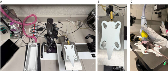
Figure 1: Mouse anesthesia and positioning for transthoracic echocardiography. (A) Imaging setup showing the temperature-controlled platform, anesthesia delivery system, ECG electrode connections, and ultrasound probe positioning apparatus. The system is arranged to allow stable physiological monitoring and precise probe manipulation during imaging. (B) Close-up of the imaging platform with integrated ECG electrodes and nose cone for isoflurane delivery, designed to secure the mouse in a standardized supine position while maintaining anesthesia. (C) Anesthetized mouse positioned supine on the imaging platform with all four paws secured to ECG electrodes using surgical tape. The nose cone delivers inhaled isoflurane (1.0–1.5% maintenance), and the chest is aligned flat to ensure optimal image acquisition and consistent cardiac monitoring. Please click here to view a larger version of this figure.
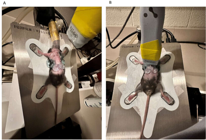
Figure 2: Apical four-chamber B-mode echocardiographic view. (A) Representative grayscale B-mode image showing all four cardiac chambers: left atrium, left ventricle, right atrium, and right ventricle in a single imaging plane. (B) Annotated image highlighting key anatomical structures. The ultrasound probe is positioned on the left lateral chest wall and angled cranially toward the cardiac apex to obtain optimal visualization. Please click here to view a larger version of this figure.
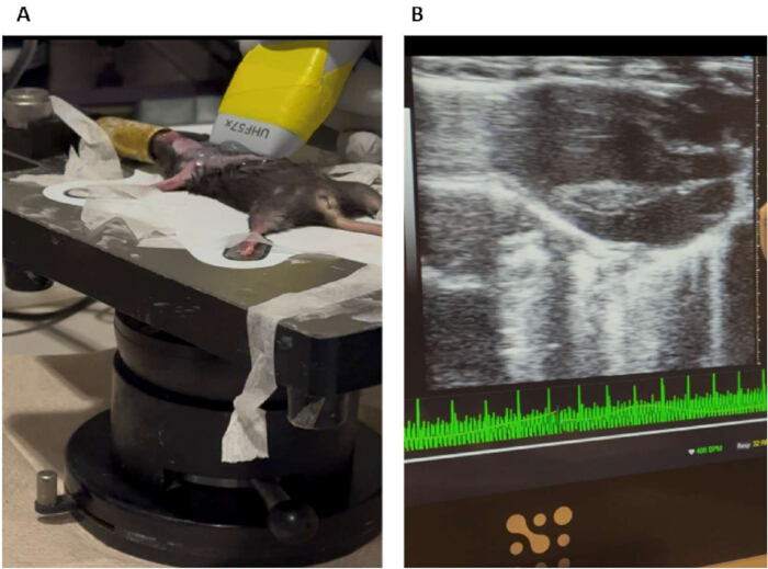
Figure 3: Parasternal long-axis B-mode imaging. (A) Representative parasternal long-axis B-mode image demonstrating cardiac anatomy, including the left ventricular chamber, the interventricular septum, and the posterior wall. (B) High-resolution image exported for quantitative analysis of cardiac dimensions. This view is used to assess global cardiac structure and guide subsequent imaging modes. Please click here to view a larger version of this figure.
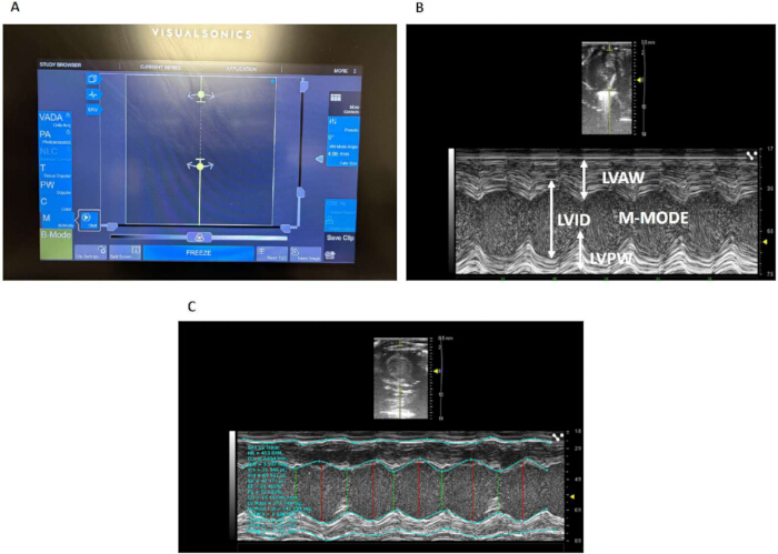
Figure 4: M-mode imaging at the papillary muscle level. (A) Parasternal short-axis view at the mid-ventricular level with papillary muscles visible. (B) Corresponding M-mode trace with the cursor aligned perpendicular to the interventricular septum and posterior wall. (C) The M-mode image with software-assisted analysis, where the system automatically detects and draws lines along the heart walls. It then calculates values like LVID, LVPW, and LVAW across cardiac cycles. Abbreviations; LVID = left ventricular internal diameter (distance between the interventricular septum and the posterior wall); LVPW = left ventricular posterior wall thickness; LVAW = left ventricular anterior wall thickness; ECG = electrocardiogram. Please click here to view a larger version of this figure.
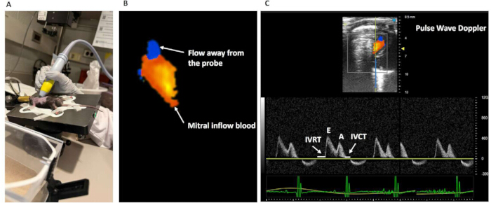
Figure 5: Experimental setup and Doppler signal overview. (A) Experimental preparation showing a small animal model positioned on a platform with an ultrasound probe placed over the thoracic region for cardiac imaging. (B) Representative color Doppler map illustrating localized blood flow signal intensity and direction. (C) Pulsed-wave Doppler tracing demonstrating sample volume placement, alongside a representative pulsed-wave Doppler waveform demonstrating early (E) and atrial (A) filling velocities. IVRT and IVCT are indicated, with simultaneous ECG tracing for cardiac cycle correlation. Abbreviations; IVRT = Isovolumetric relaxation time; IVCT = isovolumetric contraction time Please click here to view a larger version of this figure.
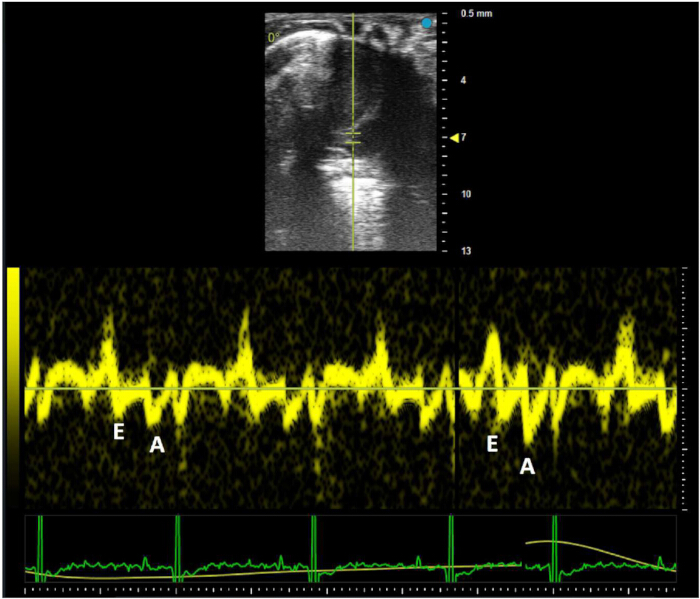
Figure 6: Tissue Doppler imaging of myocardial motion. Tissue Doppler recording illustrating myocardial velocity patterns with identifiable early (E′) and atrial (A′) components, acquired from the myocardial wall. The waveform is displayed with a synchronized ECG trace to correlate electrical and mechanical activity. Please click here to view a larger version of this figure.
Discussion
Loading...
$$\rightleftharpoonup{xx}$$
$$\longleftharp{xx}$$,
$$\longrightharp{xx}$$,
This protocol provides a standardized and reproducible approach for Doppler-based transthoracic echocardiography in mice, enabling comprehensive assessment of cardiac structure, systolic function, and conventional diastolic indices. The combined use of B-mode, M-mode, and Doppler imaging allows reliable quantification of ventricular dimensions, contractile function, and filling dynamics in preclinical models of cardiovascular disease14.
A critical consideration in murine echocardiography is the influence of inhaled anesthesia on cardiovascular physiology. Isoflurane is known to reduce heart rate, alter preload and afterload, and modulate myocardial contractility, all of which can affect Doppler-derived indices of diastolic function15. Accordingly, careful use of anesthesia and maintenance of stable physiological conditions are essential to ensure reproducible and interpretable measurements. Standardization of heart rate, body temperature, and respiratory rate minimizes variability and improves comparability across experimental groups16.
Conventional Doppler-derived indices, including transmitral inflow (E/A) and tissue Doppler-derived E/e′, are widely used for the assessment of diastolic function and are generally reproducible when standardized acquisition protocols are followed17. Reproducibility can be further enhanced by averaging measurements over multiple cardiac cycles and performing analyses in a blinded manner to reduce inter- and intra-observer variability. Consistent probe positioning and imaging planes are also essential to improve test-retest reliability across longitudinal studies18.
Maintaining an appropriate heart rate during imaging is particularly important in mice due to their high intrinsic heart rates. At lower heart rates, often associated with deeper anesthesia, altered loading conditions may influence ventricular filling patterns. Conversely, excessively high heart rates may lead to fusion of E and A waves, limiting accurate interpretation of diastolic indices. Therefore, maintaining heart rate within a range that preserves both physiological relevance and Doppler signal separation is essential for accurate assessment19.
While this protocol focuses on conventional Doppler-based indices of diastolic function, it does not incorporate more advanced echocardiographic techniques that have gained prominence in recent literature. Myocardial strain and strain rate imaging left atrial functional assessment (including reservoir, conduit, and booster pump phases), and evaluation of left ventricular untwisting provide additional mechanistic insight into myocardial relaxation and compliance19. However, these techniques require specialized acquisition protocols, higher temporal resolution, and dedicated post-processing tools, and are therefore beyond the scope of the present workflow. These modalities should be considered complementary approaches when more detailed diastolic phenotyping is required16.
In addition to echocardiography, alternative techniques such as cardiac magnetic resonance imaging (MRI) and invasive pressure-volume loop analysis provide complementary assessments of cardiac function. MRI offers high spatial resolution and accurate volumetric measurements, whereas pressure-volume analysis provides gold-standard hemodynamic data. However, these methods are limited by cost, invasiveness, and reduced suitability for longitudinal studies. In contrast, transthoracic echocardiography remains a rapid, non-invasive, and widely accessible technique for serial assessment of cardiac function in murine models20.
Overall, this protocol enables the acquisition of high-quality, reproducible echocardiographic data in mice when careful attention is paid to physiological monitoring and standardized imaging techniques. The integration of structural, systolic, and conventional diastolic measurements makes this approach well-suited for longitudinal studies investigating cardiac physiology, disease progression, and therapeutic interventions in preclinical cardiovascular research.
Disclosures
Loading...
$$\rightleftharpoonup{xx}$$
$$\longleftharp{xx}$$,
$$\longrightharp{xx}$$,
The authors declare no competing financial interests.
Acknowledgements
Loading...
$$\rightleftharpoonup{xx}$$
$$\longleftharp{xx}$$,
$$\longrightharp{xx}$$,
This work was supported by NIH/NHLBI grant HL153407.
Materials
| Name | Company | Catalog Number | Comments |
|---|---|---|---|
| Depilatory cream | Nair® | N/A | Hair removal prior to imaging |
| Heating pad | VetEquip (or equivalent) | N/A | Maintains body temperature (~37 °C) |
| High-frequency ultrasound system | FUJIFILM VisualSonics | Vevo F2 (or equivalent) | Small-animal imaging system with cardiac presets |
| Imaging platform | VisualSonics | Integrated platform | ECG and respiration monitoring |
| Induction chamber & nose cone | VetEquip (or equivalent) | N/A | Gas anesthesia administration |
| Isoflurane vaporizer | VetEquip (or equivalent) | Classic Anaesthesia System (or equivalent) | Anesthesia delivery system |
| Linear array transducer (30-40 MHz) | VisualSonics | MS550D | High-frequency probe for murine cardiac imaging |
| Ophthalmic lubricant | Generic (e.g., Refresh PM/equivalent) | N/A | Prevents corneal drying |
| Surgical tape | 3M (or equivalent) | Micropore Surgical Tape | Secures limbs for ECG |
| Ultrasound gel | Parker Labs | Aquasonic 100 (or equivalent) | Acoustic coupling |
| Vevo LAB software | VisualSonics | Version-dependent | Image acquisition and analysis |
Reprints and Permissions
Request permission to reuse the text or figures of this JoVE article
Request Permission