This protocol describes ultrasound-guided combined vacuum-assisted excision and microwave thermal ablation for symptomatic benign thyroid nodules with persistent cosmetic or compressive symptoms at least 12 months after initial thermal ablation. In this exploratory pilot study, VAE-TA was technically feasible and achieved substantial short-term nodule volume reduction without major procedure-related complications.
A subscription to JoVE is required to view this content. Sign in or start your free trial.
Research Article
Combined Vacuum-Assisted Excision and Thermal Ablation for Symptomatic Benign Thyroid Nodules: A Pilot Study
In This Article
Summary
Abstract
Thyroid nodules are a common clinical condition, while thermal ablation (TA) has emerged as a minimally invasive alternative to surgery. Single-session ablation often fails to achieve complete symptomatic resolution in large, hypervascular nodules. Post-ablation carbonization further limits volumetric resorption, necessitating optimized therapeutic strategies. This pilot study aimed to evaluate the feasibility, safety, and preliminary efficacy of combined vacuum-assisted excision and thermal ablation (VAE-TA) for symptomatic benign thyroid nodules refractory to initial ablation. From December 2023 to January 2025, five patients underwent VAE-TA at a single center. Inclusion criteria included persistent symptoms ≥12 months post-initial ablation and confirmed benignity via fine-needle aspiration. The procedure involved ultrasound-guided vacuum-assisted excision of post-ablation fibrotic tissue followed by microwave ablation. Primary outcomes included feasibility and safety, while secondary outcomes focused on volume reduction rate (VRR) and symptom improvement. Patients were followed up at 1 and 6 months. These preliminary findings suggest that VAE-TA may be a feasible, minimally invasive salvage approach for selected symptomatic benign thyroid nodules with incomplete response to initial ablation, although larger, controlled studies with longer follow-up are required.
Introduction
Thyroid nodules (TNs) represent a prevalent clinical entity, with the majority being benign and asymptomatic, warranting active surveillance alone1. However, approximately 20% of benign thyroid nodules (BTNs) exhibit progressive growth, leading to compressive symptoms or cosmetic concerns that necessitate intervention2. For such nodules, active intervention is warranted, and partial thyroidectomy remains the conventional therapeutic standard3. The 2021 guidelines for thermal ablation of thyroid nodules, jointly published by the European thyroid association (ETA) and the Korean society of thyroid radiology (KSThR), explicitly recommend thermal ablation (TA) as a therapeutic option for symptomatic benign nodules4.
Four percutaneous ultrasound-guided thermal ablation (TA) techniques have emerged as minimally invasive alternatives to surgery for patients with symptomatic benign thyroid nodules (BTNs), particularly for high-risk surgical patients, or those who decline surgery: radiofrequency ablation (RFA), laser ablation therapy (LAT), microwave ablation (MWA), and high-intensity focused ultrasound (HIFU)5,6. Although thermal ablation has been widely accepted as a promising alternative therapy, single-session ablation often fails to achieve complete symptomatic resolution in some nodules. These sizable nodules typically exhibit hypervascularity, wherein convective heat dissipation from intranodular blood flow reduces the effective thermal dose during TA, thereby compromising treatment efficacy7. post-ablation, the central portion of the thyroid nodule undergoes carbonization due to extreme hyperthermia, and this carbonized tissue impedes subsequent volumetric resorption, ultimately limiting clinical symptom relief. A study reported that nodules exceeding 20 mL required an average of 3.8 treatment sessions to attain a final volume reduction rate (VRR) of 88.2%8.
For nodules with persistent compressive symptoms after a single session of TA, additional ablation sessions may serve as a potential solution. However, repeated procedures increase the risk of complications, particularly thermal injury-related sequelae such as permanent hoarseness due to recurrent laryngeal nerve damage9. Consequently, there is an urgent need for optimized TA strategies that achieve durable clinical outcomes while mitigating procedural risks. Recently, vacuum-assisted excision (VAE)—a technique widely utilized for benign breast lesion resection or tissue biopsy—has demonstrated efficacy in removing pathological breast tissue via suction-based extraction10. In the context of thyroid nodule management, post-ablation carbonized tissue within the nodule becomes avascular and resistant to resorption, potentially perpetuating compressive symptoms. For nodules with persistent symptoms following initial TA, a critical question arises: Can combined VAE and secondary ablation address these limitations?
This is a protocol-oriented pilot study to combine ultrasound-guided vacuum-assisted excision of post-ablation fibrotic or necrotic tissue with immediate microwave ablation for symptomatic benign thyroid nodules that remain clinically problematic after prior thermal ablation. The authors hypothesized that mechanical debulking of the post-ablation tissue, followed by hemostatic and consolidative microwave ablation, would improve short-term volume reduction and symptom relief while maintaining an acceptable safety profile. Therefore, this study aimed to explore the thyroid‑applied VAE‑TA procedure for persistently symptomatic benign thyroid nodules after prior thermal ablation and evaluate its feasibility, safety, and preliminary clinical efficacy.
Access restricted. Please log in or start a trial to view this content.
Protocol
This prospective exploratory pilot study was conducted at Beijing Friendship Hospital, Capital Medical University. The study protocol was approved by the Ethics Committee of Beijing Friendship Hospital, Capital Medical University (approval number: 2022–P2–035–02). Written informed consent was obtained from all participants before enrollment and before the VAE-TA procedure. The signed consent forms were stored in the study file and the medical record. From December 2023 to January 2025, five patients with symptomatic benign thyroid nodules underwent ultrasound-guided combined vacuum-assisted excision and thermal ablation (VAE-TA). Because this was an exploratory technical pilot study designed to assess procedural feasibility, safety, and protocol reproducibility, no formal sample size calculation was performed. The results were considered preliminary and hypothesis-generating rather than confirmatory. All the materials used are listed in the Table of Materials.
Patient selection
Patients were eligible for inclusion if they had undergone previous ultrasound-guided thermal ablation, including microwave ablation or radiofrequency ablation, for a benign thyroid nodule at least 12 months before enrollment. Complete and traceable documentation of the initial ablation procedure was required, including the original procedural report, pre- and post-ablation ultrasound images, ablation modality, ablation parameters, complications, and follow-up records. Benign cytology had to be confirmed by ultrasound-guided fine-needle aspiration before the initial ablation. Eligible patients also had persistent symptoms or cosmetic concerns after the initial ablation, defined as a symptom score of ≥3 on a 10 cm visual analog scale and/or a cosmetic score of ≥3 on a 4-point physician-assessed cosmetic grading scale. In addition, patients were required to be willing to undergo VAE-TA and to provide signed written informed consent after a standardized explanation of the procedure, expected benefits, alternative treatments, and potential risks. Patients with a history of neck radiation or radioiodine therapy were not eligible.
Patients were excluded if they had severe cardiopulmonary disease that precluded tolerance of local anesthesia, supine positioning, or microwave ablation. Additional exclusion criteria included uncontrolled arrhythmia, unstable angina, severe heart failure, or severe respiratory insufficiency. Patients with severe coagulation disorder were also excluded, defined as an international normalized ratio >1.5, platelet count <50 × 109/L, or activated partial thromboplastin time >1.5 times the upper limit of normal. Pregnancy and suspected malignant thyroid nodules based on ultrasound, cytology, or clinical assessment were also exclusion criteria.
Sample size justification
This study was designed as an exploratory technical pilot study rather than a powered efficacy trial. Given the novelty of applying VAE-TA to previously ablated benign thyroid nodules, a small initial cohort was used to assess procedural feasibility, identify safety concerns, and refine the standardized protocol. Therefore, no formal sample size calculation was performed. The findings should be interpreted as preliminary and hypothesis-generating11,12,13.
Pre-ablation assessment
All patients underwent a standardized pre-procedural assessment within 1 week before VAE-TA. The assessment included medical history review, physical examination, complete blood count, thyroid function tests, coagulation profile, electrocardiography, and conventional thyroid ultrasound. The procedure was performed only when platelet count was ≥50 × 109/L, international normalized ratio was ≤1.5, activated partial thromboplastin time was ≤1.5 times the upper limit of normal, and thyroid function was within the institutional reference range (TSH: 0.49–4.91 ulU/ml; FT4: 0.59–1.25 ng/dl ) or clinically stable according to the treating physician.
Thyroid ultrasound was performed using an ultrasound system equipped with a high-frequency L9–3 linear transducer. The probe frequency was set at 3–9 MHz. Imaging depth was adjusted to include the entire thyroid nodule and adjacent critical structures, usually 3–5 cm. The focal zone was placed at or slightly below the target nodule. Gray-scale gain was adjusted to clearly delineate the nodule boundary without excessive background noise. For color Doppler imaging, pulse repetition frequency, wall filter, and color gain were adjusted to detect slow intranodular blood flow while avoiding color artifacts.
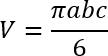
The following ultrasound features were recorded: nodule location, maximum diameter, three orthogonal diameters, echogenicity, margin, shape, calcification, vascularity, relationship to the thyroid capsule, and proximity to the trachea, esophagus, recurrent laryngeal nerve region, carotid artery, and strap muscles. Nodule volume was calculated using the ellipsoid formula:
 (1)
(1)
where V is volume, a is the largest diameter measured on the longitudinal plane, and b and c are two mutually perpendicular diameters measured on the transverse plane. Calipers were placed at the outer margin of the visible nodule boundary or residual post-ablation zone. Each measurement was performed twice, and the mean value was used for analysis.
The symptom score was self-reported by the patient using a 10 cm visual analog scale, ranging from 0 (no symptoms) to 10 (the most severe symptoms). The cosmetic score was assessed by a physician using a 4-point scale: 1, no palpable mass; 2, palpable mass without cosmetic concern; 3, cosmetic concern only during swallowing; and 4, easily visible cosmetic concern. Baseline symptom and cosmetic scores were recorded before VAE-TA and reassessed during follow-up, and ultrasound measurements were performed by sonographers blinded to the procedural details14.
Operator qualification and procedural standardization
All VAE-TA procedures were performed by one interventional radiologist with more than 10 years of experience in thyroid interventional ultrasound, including more than 500 thyroid ablation procedures and more than 500 ultrasound-guided vacuum-assisted procedures. A second interventional radiologist assisted with ultrasound monitoring, device handling, and safety assessment. To reduce operator dependency, all procedures followed a predefined checklist covering patient preparation, equipment setup, hydrodissection, vacuum-assisted excision, microwave ablation, CEUS confirmation, compression, post-procedure monitoring, and complication management. Procedural parameters, including needle approach, number of cutting passes, ablation power, ablation time, total delivered energy, CEUS findings, and complications, were recorded using a standardized case report form.
Patient preparation
Patients were instructed to fast for at least 4 h before the procedure. Routine systemic sedation was not used. Intravenous access was established before the procedure for contrast agent administration and emergency medication if required. The patient was placed in the supine position with mild neck extension. Heart rate, blood pressure, oxygen saturation, and clinical symptoms were monitored throughout the procedure. The anterior neck was sterilized and draped under aseptic conditions. Before needle insertion, the puncture route was planned under real-time ultrasound guidance. The planned route was selected to avoid the carotid artery, the internal jugular vein, the trachea, the esophagus, the recurrent laryngeal nerve region, and major cervical vessels. When feasible, a trans-isthmic or lateral cervical approach was used to maximize needle stability and continuous needle-tip visualization.
Equipment preparation
Before the procedure, the ultrasound system, vacuum-assisted excision system, and microwave ablation system were checked for normal function. The ultrasound transducer was covered with a sterile probe sheath. The vacuum-assisted biopsy device was connected to the suction system and tested before insertion. The microwave generator and applicator were assembled according to the manufacturer’s instructions, and the output power was confirmed before ablation. Vacuum-assisted excision was performed using a 12 G vacuum-assisted biopsy needle. Microwave ablation was performed using a microwave ablation system with a 14 G monopolar microwave applicator. The configuration and operational principle of the vacuum-assisted biopsy needle system are illustrated in Figure 1.
Local anesthesia and hydrodissection
Local infiltration anesthesia was administered at the skin puncture site and along the planned needle tract using 2% lidocaine. The maximum dose of lidocaine did not exceed 4.5 mg/kg without epinephrine, and the total administered volume was recorded. Hydrodissection was performed using a lidocaine-saline mixture prepared by diluting 2% lidocaine with normal saline to a final concentration of approximately 0.1%–0.2%. Under real-time ultrasound guidance, a 23 G needle was advanced into the perithyroidal space, and 20–40 mL of the mixture was injected according to nodule size, location, and proximity to critical structures. Adequate hydrodissection was confirmed when a continuous hypoechoic fluid layer was visible between the target nodule or thyroid capsule and adjacent critical structures, including the trachea, esophagus, recurrent laryngeal nerve region, carotid artery, and strap muscles. If the protective fluid layer became insufficient during the procedure, additional hydrodissection was performed before continuing VAE or MWA.
Vacuum-assisted excision
Under real-time ultrasound guidance, the vacuum-assisted needle was inserted into the post-ablation zone through the planned needle tract. The needle shaft and tip remained continuously visible throughout advancement. The insertion angle was adjusted based on the nodule's location to maintain a safe distance from the thyroid capsule and adjacent critical structures. The needle aperture was positioned within the central fibrotic, necrotic, or poorly resorbed post-ablation tissue. The aperture length was selected according to the maximum transverse diameter measured on pre-procedural ultrasound: a 2.0 cm aperture was used for nodules with a transverse diameter ≥2.0 cm, and a 1.5 cm aperture was used for nodules with a transverse diameter <2.0 cm. The allowable deviation for aperture selection was based on operator judgment when the lesion was close to critical structures.
The pre-existing ablation boundary was identified intraoperatively as the hypoechoic, heterogeneous, or fibrotic post-ablation zone on gray-scale ultrasound and was correlated with available prior ultrasound records. The cutting aperture was kept at least 2 mm within this boundary to avoid injury to the thyroid capsule and damage to adjacent structures. Tissue aspiration and rotational cutting were activated sequentially. The needle aperture was rotated in different directions within the nodule to remove central post-ablation tissue. The number of cutting passes was individualized according to nodule size, tissue resistance, residual cavity appearance, and safety considerations. The procedure was stopped when the central, poorly resorbed tissue had been sufficiently removed, the cavity had been decompressed, and the residual wall remained within the planned safety margin. Excised tissue fragments were collected from the specimen chamber, labeled with the patient study number and sampling time, and fixed in 10% neutral buffered formalin for pathological confirmation when tissue evaluation was required.
Hemostatic microwave ablation and consolidative ablation
After VAE, microwave ablation was performed to achieve hemostasis of the surgical cavity and to inactivate residual viable tissue. MWA was performed at 20 W using a 14 G monopolar microwave applicator. The applicator tip was positioned under continuous ultrasound visualization. The moving-shot technique was used. The target area was divided conceptually into multiple small ablation units. The applicator tip was first positioned in the deepest, most remote portion of the target area, then gradually withdrawn toward the superficial portion. Each unit was ablated until transient hyperechogenicity appeared, and adjacent units were overlapped to cover the entire treated zone.
Ablation was performed in repeated short applications, with duration adjusted according to nodule size, cavity morphology, echogenic change, and patient tolerance. Total ablation time, number of ablation applications, power setting, and total delivered energy were recorded for each patient. MWA was terminated when the entire planned target zone was covered by overlapping transient hyperechoic areas on gray-scale ultrasound, and no active bleeding or expanding hematoma was visible. If the patient reported intolerable pain or discomfort, ablation was paused immediately. Pain was monitored using an 11-point numerical rating scale. If the pain score was ≥4, the ablation power was reduced, the system was temporarily stopped, or supplemental lidocaine was injected around the thyroid capsule after confirming a safe needle position. A representative VAE-TA procedural workflow, including pre-procedural appearance, color Doppler imaging, vacuum-assisted excision, and thermal ablation, is shown in Figure 2.
CEUS confirmation
Contrast-enhanced ultrasound was performed immediately after VAE-TA to assess technical completeness and identify residual enhancing tissue. CEUS was performed using sulfur hexafluoride microbubbles. A bolus of 2.4 mL was injected through a peripheral vein, followed by a 5 mL saline flush. Dynamic enhancement was observed for at least 60–120 s. Residual viable tissue was defined as focal, nodular, or irregular enhancement within the treated cavity or residual nodule. If residual enhancement was detected, supplementary microwave ablation was performed until CEUS demonstrated complete absence of enhancement within the treated zone.
Compression, observation, and discharge criteria
After needle withdrawal, manual compression was applied for at least 30 min. Compression was extended in 15 min intervals if neck swelling, pain, skin tension, or sonographic evidence of hematoma was present. All patients were observed for at least 2 h after the procedure. Observation was extended to 4 h or longer if pain, hematoma, voice change, dysphagia, dyspnea, or unstable vital signs occurred. Patients were discharged only when all of the following criteria were met: stable vital signs, no progressive cervical swelling, no dyspnea, no newly developed voice change, no dysphagia, tolerable pain, and no expanding hematoma on ultrasound.
Emergency management
For suspected bleeding or hematoma, the procedure was stopped immediately, an ultrasound was used to identify the bleeding site, and manual compression was applied. If active bleeding persisted, hemostatic microwave ablation was performed under ultrasound guidance. If a hematoma expanded or airway compression was suspected, emergency airway support and surgical consultation were initiated. For voice change or suspected recurrent laryngeal nerve irritation, ablation was stopped immediately, additional hydrodissection was performed if needed, and the patient was clinically assessed. For a vasovagal reaction, the procedure was paused, the patient was kept in the supine position, and vital signs were monitored until recovery. Emergency medications and airway equipment were available in the procedure room.
Follow-up
Patients were followed up at 1 and 6 months after VAE-TA. Follow-up assessments included thyroid ultrasound, thyroid function tests, symptom score, cosmetic score, and complication assessment. Ultrasound examinations were performed by sonographers with more than 10 years of thyroid ultrasound experience and formal training in thyroid interventional follow-up assessment. Follow-up sonographers were not involved in the VAE-TA procedure and did not have access to intraoperative findings or procedural parameters. They performed standardized measurements based on follow-up ultrasound images and clinical assessment forms. At each follow-up visit, nodule volume was measured using the same ellipsoid formula. The volume reduction rate was calculated as:
 (2)
(2)
Vinitial was defined as the nodule volume measured immediately before VAE-TA on the day of treatment, rather than the volume before the initial ablation. Vfinal was defined as the nodule volume measured at each follow-up time point. Recurrence or regrowth was defined as a ≥50% increase in nodule volume compared with the smallest recorded post-procedural volume, or the reappearance of vascularized enhancing tissue within the treated zone on ultrasound or CEUS. Complications were classified as minor or major. Minor complications were defined as self-limited events requiring no invasive treatment, including mild pain, small stable hematoma, transient discomfort, or mild local swelling. Major complications were defined as events requiring hospitalization, invasive treatment, prolonged observation, or resulting in persistent functional impairment, including recurrent laryngeal nerve injury, airway compromise, severe bleeding, infection, persistent dysphagia, or thyroid dysfunction requiring treatment.
Statistical analysis
Because this was an exploratory pilot study with a very small sample size and no control group, the analysis was primarily descriptive. Continuous variables, including age, nodule diameter, nodule volume, VRR, procedure time, ablation time, and delivered energy, were summarized as mean ± standard deviation or median with range, as appropriate. Categorical variables, including sex, nodule location, technical success, and complications, were summarized as counts and percentages. Symptom and cosmetic scores before and after VAE-TA were summarized descriptively. No definitive hypothesis testing was planned because the study was not powered for efficacy comparisons. Statistical analysis and graph generation were performed using SPSS software, version 25.0.
Access restricted. Please log in or start a trial to view this content.
Results
This single-arm, exploratory pilot series included 5 patients with 5 previously ablated benign thyroid nodules. No control group was included. The cohort consisted of one man and four women. The median age was 43 years, and the baseline nodule volume measured immediately before VAE-TA was 10.42 ± 13.75 mL, with a range of 1.53–37.63 mL. All patients had persistent symptom-related and/or cosmetic concerns after prior thermal ablation and met the predefined criteria for secondary intervention. Complete and traceable docume...
Access restricted. Please log in or start a trial to view this content.
Discussion
This exploratory pilot series describes an ultrasound-guided combined vacuum-assisted excision and thermal ablation protocol for selected patients with symptomatic benign thyroid nodules after prior thermal ablation. In this initial clinical experience, VAE-TA was technically completed in all 5 patients, and short-term follow-up showed reductions in nodule volume and possible improvements in symptom-related and cosmetic outcomes. However, because the study included only 5 patients, lacked a control group, and had a limit...
Access restricted. Please log in or start a trial to view this content.
Disclosures
The authors declare that they have no known competing financial interests or personal relationships that could have influenced the work reported in this paper.
Acknowledgements
We gratefully acknowledge Ms. Xinran Cao for her guidance and support during the submission and revision process.
Access restricted. Please log in or start a trial to view this content.
Materials
| Name | Company | Catalog Number | Comments |
|---|---|---|---|
| Mindray Resona 9 ultrasound system | Mindray Medical International, Shenzhen, China | Resona 9 | Ultrasound system used for pre-procedural assessment and real-time procedural guidance. |
| High-frequency linear transducer | Mindray Medical International, Shenzhen, China | L9-3 | Linear transducer; probe frequency set at 3–9 MHz. |
| Vacuum-assisted biopsy/excision needle, 12-G | Bard, USA | ECP017 | Used for ultrasound-guided vacuum-assisted excision of central post-ablation fibrotic/necrotic tissue. |
| Microwave ablation system | Dewen Medical, Nanjing, China | DW-M5T3 | Microwave generator used for hemostatic and consolidative ablation. |
| Monopolar microwave applicator, 14 G, 10-mm active tip | Dewen Medical, Nanjing, China | DW-1805 | Microwave applicator used for post-excision cavity ablation at 20 W. |
| Sulfur hexafluoride microbubble contrast agent (SonoVue) | Bracco, Milan, Italy | N/A | Contrast-enhanced ultrasound agent; 2.4 mL bolus followed by 5 mL saline flush. |
| Lidocaine hydrochloride injection, 2% | ShanDong Hualu pharmaaceutical Co.,Ltd,China | N/A | Used for local infiltration anesthesia; maximum dose did not exceed 4.5 mg/kg without epinephrine. |
| 0.9% sodium chloride injection | Shanxi Nuocheng pharmaaceutical Co.,Ltd,China | N/A | Used for saline flush after SonoVue injection and for hydrodissection mixture preparation. |
| Lidocaine–saline hydrodissection mixture | Prepared in procedure room | N/A | Prepared by diluting 2% lidocaine with normal saline to a final concentration of approximately 0.1%–0.2%; 20–40 mL used according to nodule size/location. |
| Needle for hydrodissection, 23-G | Zhejiang Kindly MEDICAL Devices Co.,Ltd.,China | 194141574 | Used to inject the hydrodissection mixture into the perithyroidal space under real-time ultrasound guidance. |
| 10% neutral buffered formalin | MAIGE, Jiangsu, China | CC002 | Used to fix excised tissue fragments for pathological confirmation when tissue evaluation was required. |
| Specimen container | N/A | N/A | Used to collect, label, and store excised tissue fragments. |
| Manual compression materials / sterile gauze dressing | N/A | N/A | Used for at least 30 min manual compression after needle withdrawal; extended in 15-min intervals when indicated. |
| Vital signs monitor | Mindray Medical International, Shenzhen, China | N/A | Used to monitor heart rate, blood pressure, and oxygen saturation during and after the procedure. |
| Emergency airway and medication set | Procedure room emergency equipment | N/A | Available in the procedure room for bleeding, airway compromise, vasovagal reaction, or other emergencies; specify institutional emergency kit if required. |
| SPSS statistical software | IBM Corp., Armonk, NY, USA | Version 25.0 | Used for descriptive statistical analysis and graph generation. |