Here, we present a detailed protocol for remote intraoperative monitoring for cochlear implants. The prerequisites, setup, and procedure are described. The time-saving benefits, as experienced in our hospital, are shown alongside the problems encountered.
Method Article
Remote Intraoperative Monitoring of Cochlear Implants
In This Article
Summary
Abstract
Intraoperative measurements are valuable tools in cochlear implant (CI) surgery, enabling the determination of implant functionality while the patient is still in the operating room. Standard measurements include impedances for stimulating and non-stimulating contacts, triggering of the stapedial reflex with or without estimating the threshold, and assessing cochlear nerve function by measuring electrically evoked compound action potentials. Additional measurements can also be performed. For example, electrocochleography can be used to observe and help preserve residual hearing in patients. Usually, measurements are performed in situ in the operating room, which is time-consuming, especially for the engineer responsible for the measurements. This protocol describes how cochlear implant measurements can be performed remotely using a telephone in the operating room and a remotely accessible laptop. With this setup, the measurements can be completed without the engineer entering the operating room, reducing the time required from around 29 min in situ. to 8 min remotely (excluding session with ECochG).
Introduction
Intraoperative monitoring during cochlear implant (CI) surgery is a routinely performed method for checking device integrity and confirming functionality. Traditionally, most hospitals evaluate electrode impedances and electrically evoked compound action potentials (eCAPs) to ensure this functionality1.
The impedances confirm the functionality of the implant circuitry, while eCAPs provide information about the electrical stimulability of the hearing nerve. Further information can be gathered by conducting additional measurements, such as electrically evoked stapedial reflex (ESR) or electrocochleography (ECochG). ESR provides information on the hearing pathway up to the brainstem, where the stapedial reflex is triggered. ECochG is a measurement of electrical potentials in the cochlea and can provide information about residual hearing and structural preservation during and after electrode insertion2,3,4. A good overview of the intraoperative measurements has been provided by Müller et al.5. Conducting these measurements usually requires a trained professional, typically a technician or audiologist, to be present during the operation. This person is hereafter described as an audiologist. Attending the operation in person leads to a significant investment of time for the trained professional, even if they are only present during the measurements themselves.
In candidates with uncertain cochlear implant candidacy, the standard procedure for intraoperative testing is extended by electrically evoked auditory brainstem response measurements using a test electrode or the actual cochlear implant for stimulation5,6,7,8. However, since these measurements require additional hardware and are not easy to perform remotely, they are not included in this protocol.
An option to reduce this time investment is to perform the measurements remotely. The initial drive toward remote CI monitoring came from Shapiro et al.9, who showed its feasibility alongside a significant reduction in time investment for the audiologist. This has been replicated by Yanov et al.10 and Kouhi et al.11, who reported varying time savings and showed that remote measurement is both practical and reliable, with only a few cases where remote measurements were insufficient. Due to the advantages that remote measurements offer, especially in time and flexibility, multiple hospitals have already implemented this procedure. However, in most hospitals, in situ. measurement is still the preferred practice. The goal of this work is to outline the requirements and a clear methodology for remote intraoperative measurements during CI surgery, such that more hospitals can apply the techniques described.
Protocol
Written informed consent was obtained from the patients to use and publish their data where applicable.
1. Prerequisites for remote intraoperative monitoring
- Prepare the measurement equipment.
- Ensure that the programming interfaces and cables required for the selected cochlear implant system are available in the operating center and accessible to the operating room (OR) staff.
- Ensure that all hardware and software required for specialized measurements, such as electrocochleography (ECochG), are available before surgery. Refer to Figure 1 and the Table of Materials for the measurement equipment required for each manufacturer.
- Place the measurement computer in the OR in a position that allows access by OR staff without interfering with the surgical workflow. Use a device that supports remote access and allows troubleshooting when needed.
NOTE: In the authors’ setup, a laptop was used as the measurement computer because it was easy to bring into the OR, required little space, and had a built-in microphone and camera, which can be useful for troubleshooting.
- Configure connectivity and communication.
- Connect the measurement computer to the local network to enable remote access and storage of measurements in the connected database.
- Enable the remote-control software with institutional IT support before the procedure. Verify login access, network permissions, and port permissions before intraoperative use.
NOTE: In the authors’ setup, remote access was achieved using Remote Desktop on Windows. Other remote-control software may also be used. - Establish a voice communication route between the OR and the audiologist’s workstation. Confirm that the surgeon or OR staff can communicate with the audiologist throughout the monitoring procedure.
NOTE: In the authors’ setup, speech communication with the OR was established through a landline telephone in the OR.
- Train and assign OR staff.
- Assign a trained OR staff member to set up the measurement hardware and assist with troubleshooting during remote monitoring.
- Confirm that the assigned OR staff member can connect the programming interface, cables, coil or processor, and measurement computer before the procedure begins.
- Instruct the OR staff not to shut down the measurement computer or close the laptop display during the measurement.
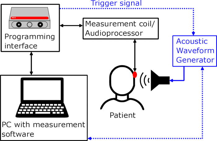
Figure 1: Schematic view of the measurement setup in the operating room. The blue components indicate the optional setup required for ECochG measurements. Please click here to view a larger version of this figure.
2. Procedure
- Additional preparation for ECochG (manufacturer-independent workflow)
- As audiologist: if ECochG will be performed, ensure that the surgeon inserts the tip of the in-ear headphone into the ear canal before the first cut. Verify correct placement before starting the measurement setup (Figure 2).
- As audiologist: inform the OR staff that the ECochG measurement must be started before insertion of the electrode array into the cochlea.
- As OR staff: confirm that the audiologist is ready to start the ECochG recording before electrode insertion begins.
- Standard preparation
- As OR staff: obtain the implant manufacturer, implant type, and serial number from the surgeon as soon as the implant has been selected. Inform the audiologist about the impending measurement and give the gathered details.
- As OR staff: turn on the measurement computer and allow the audiologist to log in remotely (Figure 3).
- As audiologist: open the measurement software and enter the patient details and implant information.
- As audiologist: confirm the connection to the implant when the surgeon and audiologist are ready to begin the measurement.
- Intraoperative monitoring (this section is from the perspective of the audiologist)
- ECochG (MED-EL workflow, adaptable to other manufacturers)
- If ECochG is indicated, perform this measurement first. Confirm with the surgeon that the electrode array has not yet been inserted into the cochlea.
- Select the appropriate ECochG settings listed in Table 1 and check the connection to the implant before starting the measurement.
- Start the recording when the first contact is inserted into the cochlea. Provide feedback to the surgeon on the ECochG responses during insertion.
- If the ECochG response decreases, advise the surgeon to pause the insertion briefly.
- After insertion is complete, stop the measurement if no ECochG responses were recorded and continue to the next measurement. If ECochG responses were recorded and the clinical workflow permits, perform an additional ECochG sweep measurement across the electrode array to assess remaining responses.
NOTE: ECochG can be used to investigate residual hearing. In general, a response amplitude of 5 µV is considered a positive response, indicating the presence of intact structures and residual hearing. Figure 4 shows an example of a clearly positive ECochG response with amplitudes exceeding 5 µV. The sweep measurement can also provide information on the location of the electrode array, with response maxima at electrodes corresponding to the frequency location.
- Impedances (this section is from the perspective of the audiologist)
- Perform the impedance measurements after ECochG, if ECochG was performed, or as the first measurement if ECochG was not performed.
- Report the results to the surgeon, including any high impedances, open circuits, or short circuits.
- Review the stimulation current-induced non-stimulating electrode voltages (SCINSEV) for anomalies related to electrode placement, including possible tip fold-overs.
- Report any placement-related anomalies to the surgeon, especially when pre-curved electrode arrays are used.
NOTE: Impedance measurements provide information about the state of the electrode array, including short circuits, open circuits, and faulty electrodes. Placement anomalies such as tip fold-overs can also be detected by evaluating SCINSEV. Figure 5A shows a normal SCINSEV result, while Figure 5B shows the characteristic off-diagonal pattern of a tip fold-over.
- Electrically evoked stapedial reflex and threshold (this section is from the perspective of the audiologist)
- After the impedance measurements, ask the surgeon whether the electrically evoked stapedial reflex (ESR) measurement is indicated based on anatomical circumstances and previous operations.
- If ESR is indicated, measure the electrically evoked stapedial reflex and, if time allows, the electrically evoked stapedial reflex threshold.
- Measure at least one apical, one medial, and one basal electrode, including electrodes near the beginning and end of the electrode array.
- Ask the surgeon to visually inspect whether the stapedial reflex is triggered and to provide oral feedback to the audiologist.
- If reflexes are absent for selected electrodes, especially the most basal electrodes, confirm with the surgeon whether the electrode array is fully inserted.
NOTE: ESR confirms the integrity of the auditory pathway up to the brainstem. Triggerable reflexes strongly support auditory pathway integrity, while completely absent reflexes do not necessarily indicate that the implant will not work for the patient.
- Electrically evoked compound action potentials (this section is from the perspective of the audiologist)
- Record the electrically evoked compound action potentials (ECAPs) after the ESR measurement. Use the recommended parameters listed in Table 1.
- Use automatic measurements for routine cases.
- If responses are difficult to evaluate or further assessment is needed, repeat the measurement using manual measurements.
NOTE: ECAPs primarily record responses of the hearing nerve, corresponding to the first waveform in acoustically evoked potentials. A positive ECAP confirms a hearing nerve response to electrical stimulation but does not provide information about the remaining auditory pathway.
- ECochG (MED-EL workflow, adaptable to other manufacturers)
- Completion of monitoring (this section is from the perspective of the audiologist)
- Communicate the final monitoring results to the surgeon.
- Repeat selected measurements if necessary.
- Save the measurements to the connected database after the surgeon confirms that no additional measurements are needed.
- Verify that the measurements have been saved before shutting down the measurement computer.
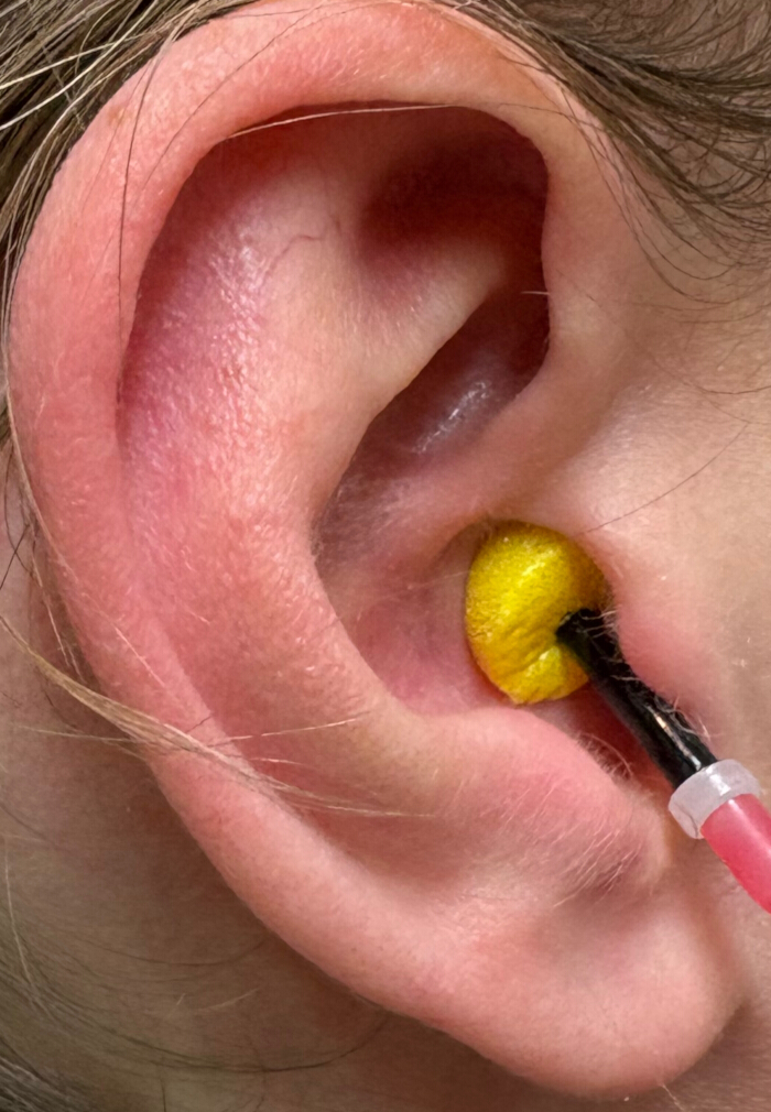
Figure 2: Example of correct tip placement for the earphones used in the ECochG measurement. Please click here to view a larger version of this figure.
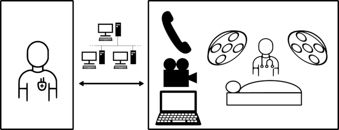
Figure 3: Schematic view of the remote monitoring setup. Please click here to view a larger version of this figure.
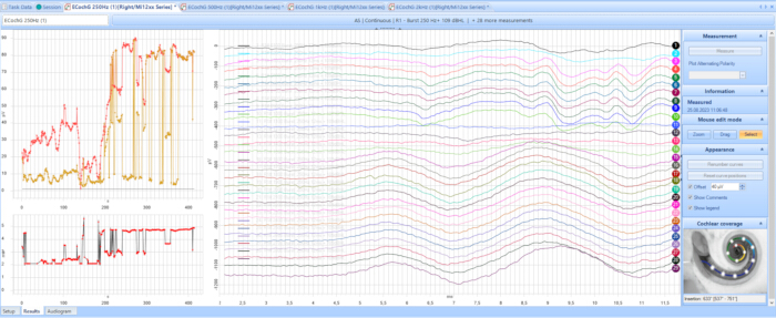
Figure 4: Example of an ECochG recording using the MED-EL system. Courtesy of MED-EL. Please click here to view a larger version of this figure.
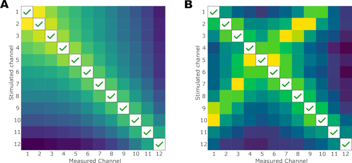
Figure 5: Example voltage matrices from MED-EL SCINSEV. (A) Voltage matrix of a normal case, from MED-EL12. (B) Voltage matrix of a conspicuous case with tip fold-over, adapted from Franke-Trieger et al.13. Please click here to view a larger version of this figure.
Results
To facilitate a comparison, the time spent by the audiologist on the operation was measured for 87 operations. Of these, 80 were performed remotely and 7 in sit.u. This stems from the fact that remote measurements are already standard practice at our hospital. To reduce waiting times for patients and minimize disruptions in the clinical routine, most intraoperative measurements are performed remotely. In Figure 6, the frequency of each measurement is shown. Impedances were the most common measurement, being conducted every time, shortly followed by ECAPs. In two operations where no manipulation of the implant occurred, only the impedances were measured. In ten cases, ESR was not performed due to surgeon feedback (e.g., no stapes). The least frequent measurement was ECochG, because it is not appropriate for every patient. Only when residual hearing is sufficient is ECochG a sensible measurement, and those patients represent only a small part.
ECochG can be used to investigate the presence of residual hearing. Generally, a response amplitude of 5 µV is considered a positive response, indicating the presence of intact structures and residual hearing. Figure 4 shows an example of a clearly positive ECochG response, with amplitudes exceeding 5 µV. The ECochG sweep can also provide information on the location of the electrode array, with response maxima at electrodes corresponding to the frequency location. Impedance measurements provide information about the state of the electrode array, including short circuits, open circuits, and faulty electrodes. Placement anomalies, such as tip fold-overs, can also be detected by evaluating the stimulation current-induced non-stimulating electrode voltages (SCINSEV). This is illustrated in Figure 5A,B; Figure 5A shows a normal SCINSEV result, while Figure 5B shows the characteristic off-diagonal pattern of a tip fold-over.
The methodology of remotely performing the intraoperative measurements is highly time-efficient, especially for the audiologist involved. If performed in situ, the audiologist has to move to the operating center, change clothes, disinfect, and go to the operating room. There, they usually have to wait until the measurements can be performed. The monitoring itself is usually the least time-consuming part, and the additional time spent walking and waiting takes longer. The measured time spent on the operation is shown in Figure 7. Figure 7 shows that measuring remotely gives a clear benefit in time spent by the audiologist. The median time spent on remote measurements was 8 min, while for in situ measurements, it increased to 29.03 min. When ECochG was measured as well, the median time spent on remote measurements was 27.75 min, and for in situ. measurements, it increased to 48.63 min. The time saved through measuring remotely, therefore, is around 20 min, which can quickly add up if the hospital performs many surgeries in a year.
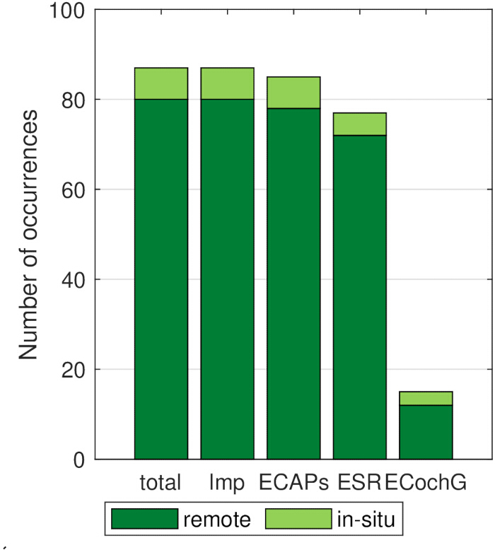
Figure 6: Number of measurements performed. Measurements are split into remote (dark green) and in situ. (light green). Please click here to view a larger version of this figure.
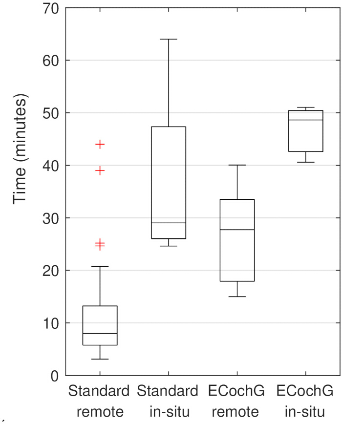
Figure 7: Time spent by the audiologist on the intraoperative measurement. Please click here to view a larger version of this figure.
| Manufacturer | Measurement | Settings name | Settings value |
| MED-EL | Impedances - IFT | No settings | |
| ESRT | Starting charge | 30 qu | |
| ECAP - AutoART | Minimum charge | 0 qu | |
| Maximum charge | 50 qu | ||
| Charge increase rate | Fast | ||
| Measurement mode | Always to maximum charge | ||
| ECAP – ART all channels | Maximum amplitude | 1000 cu | |
| Minimum amplitude | 0 cu | ||
| Phase duration | 40 µs | ||
| Iterations | 15 | ||
| Measurement gap | 0 ms | ||
| Levels | 6 | ||
| ECochG (EAEP) continuous | Stimulus Type | SPL Chirp 2 | |
| Stimulating polarity | Alternating | ||
| Stimulus Level | Audiogram threshold at relevant frequency +40 dB | ||
| Stimulus duration | 8 ms | ||
| Measurement parameters | Default | ||
| ECochG (EAEP) sequential | Protocol | All electrode sweep | |
| Stimulus Type | SPL Chirp 2 | ||
| Stimulating polarity | Condensation | ||
| Stimulus Level | Audiogram threshold at relevant frequency +40 dB | ||
| Stimulus duration | 12 ms | ||
| Measurement parameters | Default | ||
| Cochlear | Impedances | Modes | Check all boxes |
| TIM | Extracochlear electrode lead placed | Check if placed | |
| Condition Array | Check box | ||
| Settings | Default | ||
| ESRT | Number of electrodes | 5 | |
| Settings | Default | ||
| Level | 220 cl | ||
| Up | 10 cl | ||
| Down | 5 cl | ||
| ECAP - AutoNRT | Number of electrodes | 9 | |
| Starting | 150 cl | ||
| Step size | 6 cl | ||
| Settings | Default except “Perform electrode conditioning”, uncheck if TIM was measured | ||
| Advanced Bionics | Impedances | No settings | |
| ESRT | Only possible by creating a map and stimulating single channels | Starting amplitude 150 cu | |
| ECAP - NRI | Settings | Default | |
Table 1: Recommended parameters for each intraoperative measurement.
Discussion
Remote intraoperative measurements for cochlear implants are feasible and efficient. This was first shown by Shapiro et al.9 and confirmed by Yanov et al.10 and Kouhi et al.11. The largest benefit is the time saved by the audiologist, rather than necessarily a reduction in the total duration of the surgical procedure. The amount of time saved, however, is very dependent on hospital circumstances. Shapiro et al.9 reported time savings of around 80 min, while Yanov et al.10 showed improvements of 10 min. The biggest factor influencing the time saved is the physical distance between the operating center and the workplace of the audiologist. The larger the distance, the more time can be saved by performing the measurements remotely. If the operating center is in another nearby building, as for Shapiro et al.9, or even involves commuting, as reported by Kouhi et al.11, the time saved is quite large. In our case, the distance involved is on the lower side, so the time saved, while meaningful, is not too large, and most of the time saved comes from minimizing waiting times. While those waiting times occur for both remote and in situ. measurements, they can be used more productively when measuring remotely, since the audiologist has access to their office and can perform additional tasks while waiting. A further benefit of remote measurements is the availability of colleagues if a second opinion on the measurements is required. A colleague can easily examine the measurements in person without having to come to the operating room or provide an explanation via telephone.
Most important for the success of remote measurements is ensuring a stable connection to the OR, which entails the connection to the measurement computer as well as the voice connection, if it is separate. Also very important is the training of the OR staff. They need to set up the measurement hardware themselves and help with troubleshooting. They can also compromise the measurement by prematurely shutting down the measurement computer, which leads to loss of unsaved measurements. This should be avoided by instructing the OR staff properly. Modification of the described protocol can easily be done to fit the circumstances of the hospital where it will be implemented. The use of the built-in remote desktop functionality in Windows is recommended, but any tool that allows remote connectivity and control can be used. The measurements to be performed can also be adapted to include only those usually performed by the hospital.
ESR and ECAP results should also be interpreted within their limitations. Triggerable ESRs support the integrity of the auditory pathway up to the brainstem, while completely absent reflexes do not necessarily indicate that the implant will not work for the patient. ECAPs primarily record responses of the hearing nerve, corresponding to the first waveform in acoustically evoked potentials. A positive ECAP confirms a hearing nerve response to electrical stimulation but does not provide information about the remaining auditory pathway.
There are some limitations and complications that arise from measuring remotely. The biggest downside is that troubleshooting cannot be done in person. Most cable-connection problems can be identified in the measurement software. If the measurement computer is a laptop, the built-in webcam is also helpful in solving problems. Connection issues are usually less easily solved and were reported by Shapiro et al.9. The frequency of these issues depends on the remote-connection software. If the connection is set up properly, connection issues are very rare with Windows built-in remote desktop functionality. Communication with the OR can also be difficult when measuring remotely. When using a landline telephone connection to talk to the OR, the surgeon is usually not right next to the microphone, making detailed discussions with the surgeon difficult, especially if the results are atypical or difficult to interpret. This is also mentioned by Nuwer et al.14, where it is described as one of the biggest limitations for remote monitoring. Furthermore, there is the risk of losing the measurement data when measuring remotely. If the measurement computer is shut off prematurely, any unsaved data will be lost, which should be avoided.
Even though there are some limitations, the number of benefits, especially in time saved for the involved audiologist, makes remote measurements an attractive paradigm. In our hospital, remote measurements have mostly replaced in situ. measurements because they minimize the disruption to the clinical routine.
Disclosures
D.P. received financial support for business travels from MED-EL GmbH and Cochlear Ltd., and research projects from MED-EL GmbH, and is an advisory board member for MED-EL GmbH. P.N. received financial support for business travels from MED-EL GmbH and Cochlear Ltd. J.M. received funding for research projects from MED-EL GmbH and is an advisory board member for MED-EL GmbH.
Acknowledgements
We thank Giacomo Mandruzzato (MED-EL) for lending the ECochG hardware.
Materials
| Name | Company | Catalog Number | Comments |
|---|---|---|---|
| AIM measurement system | Advanced Bionics | Not provided | Used for Advanced Bionics ECochG measurements. |
| Cochlear Objective Measurement (COM) software or Custom Sound EP software | Cochlear | Not provided | Software used for Cochlear impedance, ESR, and ECAP measurements. |
| Cochlear Programming Pod | Cochlear | Not provided | Programming interface used for Cochlear impedance, ESR, and ECAP measurements. |
| CP910 processor or CP1110 processor | Cochlear | Not provided | Processor used for Cochlear impedance, ESR, and ECAP measurements. |
| Dataman waveform generator | Not provided | Not provided | Waveform generator used for MED-EL ECochG measurements. |
| In-ear headphone/insert phone | Not provided | Not provided | Used for acoustic stimulation during ECochG measurements. |
| Landline telephone | Not provided | Not provided | Used for voice communication between the OR and the audiologist's workstation in the authors' setup. |
| MAESTRO AS RUO software | MED-EL | Not provided | Software used for MED-EL ECochG measurements. |
| MAESTRO software | MED-EL | Not provided | Software used for MED-EL impedance, ESR, and ECAP measurements. |
| MAX coil | MED-EL | Not provided | Coil used for MED-EL impedance, ESR, and ECAP measurements. |
| MAX programming interface | MED-EL | Not provided | Programming interface used for MED-EL impedance, ESR, and ECAP measurements. |
| Measurement computer/laptop | Not provided | Not provided | Computer used to run the measurement software and permit remote access. |
| Naida M90 processor | Advanced Bionics | Not provided | Processor used for Advanced Bionics impedance, ESR, and ECAP measurements. |
| Noahlink Wireless | Advanced Bionics | Not provided | Wireless interface used for Advanced Bionics impedance, ESR, and ECAP measurements. |
| Remote Desktop software | Microsoft | Not provided | Remote-control software used in the authors' Windows-based setup; other remote-control software may also be used. |
| Target CI software | Advanced Bionics | Not provided | Software used for Advanced Bionics impedance, ESR, and ECAP measurements. |
References
- Wesarg, T., et al. Intraoperative audiologisch-technische Diagnostik bei der Cochleaimplantatversorgung. HNO. 62 (10), 725-734 (2014).
- Campbell, L., et al. Intraoperative real-time cochlear response telemetry predicts hearing preservation in cochlear implantation. Otol Neurotol. 37 (4), 332-338 (2016).
- Gibson, W. P. The clinical uses of electrocochleography. Front Neurosci. 11, 274 (2017).
- Harris, M. S., et al. Real-time intracochlear electrocochleography obtained directly through a cochlear implant. Otol Neurotol. 38 (6), e107-e113 (2017).
- Müller, A., et al. Empfehlungen zur Auswahl von Zielparametern und Prozessempfehlungen bei audiologisch-technischen Funktionsprüfungen des Cochlea-Implantats: erarbeitet von der Arbeitsgruppe ERA (AG-ERA) der ADANO in Kooperation mit dem Fachausschuss „Cochlea-Implantate und implantierbare Hörsysteme“ der DGA; bestätigt vom Vorstand der ADANO am 31.01.2025; englische Version. HNO. 73 (Suppl 3), 348-356 (2025).
- Cinar, B. C., et al. The role of eABR with intracochlear test electrode in decision making between cochlear and brainstem implants: preliminary results. Eur Arch Otorhinolaryngol. 274 (9), 3315-3326 (2017).
- Lassaletta, L., et al. Usefulness of electrical auditory brainstem responses to assess the functionality of the cochlear nerve using an intracochlear test electrode. Otol Neurotol. 38 (10), e413-e420 (2017).
- Polterauer, D., et al. Applications of pre- and intraoperative eAEP like eABR in cochlear implant candidacy. Curr Dir Biomed Eng. 10 (4), 502-505 (2024).
- Shapiro, W. H., et al. Remote intraoperative monitoring during cochlear implant surgery is feasible and efficient. Otol Neurotol. 29 (4), 495-498 (2008).
- Yanov, Y., et al. Successful application and timing of a remote network for intraoperative objective measurements during cochlear implantation surgery. Int J Audiol. 57 (9), 688-694 (2018).
- Kouhi, A., Swanson, A., Fitzgerald, M. B., Blevins, N. H. Remote intraoperative neural response telemetry: technique and results in cochlear implant surgery. Otol Neurotol. 43 (6), 638-642 (2022).
- . Neu in MAESTRO: IFT-Resultate auf einen Blick Available from: https://blog.medel.pro/de/audiology/neu-in-maestro-ift-resultate/ (2024)
- Franke-Trieger, A., et al. Voltage matrix algorithm for intraoperative detection of cochlear implant electrode misplacement. Audiol Neurotol. 30 (3), 252-262 (2025).
- Nuwer, M. R., Cohen, B. H., Shepard, K. M. Practice patterns for intraoperative neurophysiologic monitoring. Neurology. 80 (12), 1156-1160 (2013).
Reprints and Permissions
This article has been published
Video Coming Soon