Monogenic type I interferonopathies establish a direct mechanistic link between dysregulated innate immunity and vasculitis. Recognizing characteristic clinical features, integrating interferon gene signature analysis with genetic testing, and implementing targeted therapies such as Janus kinase inhibitors are essential for accurate diagnosis and precision management of these disorders.
Review Article
Type I Interferonopathies in the Differential Diagnosis of Vasculitis: A Comprehensive Review
In This Article
Summary
Abstract
Type I interferonopathies are a heterogeneous group of monogenic autoinflammatory disorders characterized by dysregulated type I interferon (IFN-I) signaling due to pathogenic variants that affect nucleic acid sensing, processing, or downstream signaling pathways. Mutations in genes including TREX1, RNASEH2A/B/C, SAMHD1, ADAR1, STING1 (TMEM173), PSMB8, COPA, and DNASE1L3 lead to persistent activation of innate immune pathways, particularly the cGAS–STING, MDA5, and Toll-like receptor pathways, with subsequent JAK–STAT signaling and sustained overexpression of interferon-stimulated genes. Chronic IFN-I activation promotes endothelial dysfunction, vascular inflammation, and tissue injury, providing a mechanistic link between interferonopathies and vasculitic disorders. Clinically, these conditions present with diverse manifestations, including chilblains, livedo reticularis, necrotizing cutaneous vasculopathy, panniculitis, interstitial lung disease, cerebral vasculopathy, and glomerulonephritis, often resembling autoimmune diseases such as primary central nervous system vasculitis, systemic lupus erythematosus (SLE), poliarteritis nodosa (PAN), immune complex vasculitis, and ANCA-associated vasculitis (AAV). A persistently elevated interferon gene signature represents a valuable diagnostic biomarker that distinguishes these disorders from most classical autoimmune vasculitides and facilitates early recognition. Timely genetic testing is essential to establish an accurate diagnosis, guide patient management, and avoid treatment delays. The present review summarizes the molecular mechanisms linking IFN-I dysregulation to endothelial injury and vasculitis, discusses the clinical spectrum and diagnostic challenges of monogenic interferonopathies, and highlights emerging targeted therapies, particularly Janus kinase inhibitors, that support precision medicine approaches for interferon-driven inflammatory diseases.
Introduction
Type I interferonopathies, a concept introduced in 2011, are a heterogeneous group of rare monogenic autoinflammatory disorders characterized by dysregulated type I interferon (IFN-I) signaling resulting from pathogenic variants affecting nucleic acid sensing, processing, or immune regulatory pathways. Mechanisms including nucleic acid accumulation (e.g., TREX1 deficiency), persistence of ribonucleotide hybrids (RNASEH2 defects), receptor hyperactivation (IFIH1 and STING1), proteasome dysfunction (PSMB8), and impaired negative regulation (ISG15 and USP18) lead to sustained IFN-I production1,2. A common pathogenic mechanism underlying these disorders is the aberrant recognition of self-derived cytosolic nucleic acids as viral ligands, resulting in constitutive activation of the cGAS–STING–TBK1–IRF3 signaling pathway (Figure 1)3.
The clinical spectrum of interferonopathies reflects this persistent activation of innate immunity and varies according to the underlying genetic defect. In Aicardi–Goutières syndrome (AGS), pathogenic variants in TREX1, RNASEH2A/B/C, SAMHD1, ADAR1, LSM11, and RNU7-1 result in defective nucleic acid metabolism or replication-dependent histone pre-mRNA processing, promoting nucleic acid accumulation and cerebral vasculopathy1. Genotype–phenotype correlations further illustrate this heterogeneity: STING1, COPA, and PSMB8 mutations predominantly cause small-vessel vasculopathy; TREX1, ADAR1, and RNASEH2 mutations are strongly associated with cerebral vasculopathy; DNASE1L3, DNASE1, and DNASE2 variants predispose to urticarial or systemic lupus erythematosus (SLE)-like vasculitis; whereas ISG15 and USP18 deficiencies are associated with necrotizing skin lesions and neurological manifestations4,5,6,7. In addition, STAT4 and SAMHD1 variants have been linked to interferon-γ-mediated endothelial inflammation and cerebral vasculopathy, while STAT1 and STAT2 gain-of-function mutations exhibit overlapping IFN-I activation and are therefore considered within the broader spectrum of type I interferonopathies1,5,8,9,10,11,12,13,14.
From a rheumatologic perspective, interferonopathies commonly manifest with dermal necrotizing vasculopathy, chilblains, livedo reticularis, panniculitis, interstitial lung disease, and cerebral vasculopathy. These manifestations frequently resemble autoimmune diseases, particularly ANCA-associated vasculitis (AAV) and SLE. Early-onset necrotizing vasculitis has therefore been proposed as an "interferon alarm signal"5. Such clinical overlap may result in delayed diagnosis and prolonged exposure to ineffective immunosuppressive therapies, particularly in pediatric patients. Accordingly, the interferon score derived from interferon-stimulated gene (ISG) expression has emerged as a valuable biomarker for identifying interferonopathies and supporting differential diagnosis6.
Despite these clinical similarities, important pathogenic differences distinguish interferonopathies from classical autoimmune vasculitis. Comparative transcriptomic studies have demonstrated sustained overexpression of interferon-regulated genes, including IFI27, IFI44L, ISG15, MX1, and OAS1, in interferonopathies, whereas AAV is characterized predominantly by neutrophil extracellular trap (NET) formation and granulocyte-mediated inflammatory pathways6,15,16,17. Although systemic IFN-I activation is not considered a primary driver of classical AAV, recent studies suggest that tissue-specific interferon signatures may contribute to disease heterogeneity, particularly in microscopic polyangiitis, where increased IFN-I activity has been associated with renal fibrosis and adverse outcomes15,16. These observations highlight the importance of distinguishing monogenic interferonopathies from autoimmune vasculitis to facilitate accurate diagnosis and appropriate patient management.
This review summarizes the molecular mechanisms linking IFN-I dysregulation to vascular inflammation, discusses the genotype–phenotype spectrum and rheumatologic manifestations of monogenic interferonopathies, and highlights current diagnostic approaches and emerging targeted therapies. By emphasizing the distinctions between interferon-mediated vasculopathy and classical autoimmune vasculitis, this review aims to facilitate earlier recognition of these disorders and promote precision-medicine strategies in rheumatology.
Review and Perspective
For this narrative review, a comprehensive literature search was conducted in PubMed/MEDLINE, Web of Science, Scopus, Embase, and Google Scholar from 2014 to 2026. Additionally, the Directory of Open Access Journals (DOAJ) was screened to identify relevant open-access publications.
The search strategy combined Medical Subject Headings (MeSH) terms and free-text keywords, including but not limited to: “type I interferonopathy,” “interferon signature,” “cGAS–STING pathway,” “STING1,” “TREX1,” “RNASEH2,” “Aicardi–Goutières syndrome,” “SAVI,” “PRAAS,” “CANDLE,” “COPA syndrome,” “DNASE1L3,” “ANCA-associated vasculitis,” “microscopic polyangiitis,” “systemic lupus erythematosus,” “vasculopathy,” “endotheliopathy,” and “JAK inhibitors.” Boolean operators (AND/OR) were applied to refine the search combinations.
The literature search, screening, and study selection were performed independently by the three authors. Potential disagreements regarding study eligibility or relevance were resolved through discussion and consensus among the authors. The final selection of references and the overall scientific content of the review were subsequently evaluated by three senior supervisors to ensure scientific accuracy, completeness, and consistency.
IFN-related monogenic disease studies were included if they were published in English, involved human participants, and addressed the conditions defined in the research question. The publication date range was from 2014 to 2026. Articles were excluded if they did not fit within the conceptual framework of the study. Non-scientific reviews and opinion articles were also excluded.
Clinical and pathophysiological distinctions between type I interferonopathies and autoimmune vasculopathic disorders
Type I interferonopathies represent a genetically defined and mechanistically distinct group of autoinflammatory disorders that frequently present with vasculitic phenotypes that mimic classical autoimmune diseases, including primary central nervous system vasculitis, SLE, PAN, immune complex vasculitis, and AAV. However, unlike conventional autoimmune vasculitides, these conditions are driven by monogenic defects in nucleic acid sensing, proteasome function, endoplasmic reticulum stress pathways, or interferon regulatory mechanisms, resulting in chronic and constitutive activation of IFN-I signaling18. Rheumatologic vasculitic manifestations of interferonopathies are clearly distinct from classical autoimmune diseases such as AAV and SLE, as they arise from monogenic mechanisms including dysregulation of nucleic acid sensing, proteasome dysfunction, endoplasmic reticulum (ER) stress, and direct endothelial injury. In STING-associated vasculopathy with infancy onset (SAVI), STING1 mutations lead to necrotizing dermal vasculopathy and pulmonary fibrosis; in proteasome-associated autoinflammatory syndrome (PRAAS), PSMB8 mutations result in panniculitis and lipodystrophy-associated dermal inflammation; in COPA syndrome, ANCA-positive or negative small-vessel vasculitis, interstitial lung disease, and a broad spectrum of autoantibodies (including rheumatoid factor) are predominant features that frequently lead to misdiagnosis as juvenile idiopathic arthritis (JIA); and in AGS, mutations cause cerebral vasculopathy and progressive neurological damage. The downstream consequences, persistent ISG expression, endothelial activation, procoagulant remodeling, and microvascular injury, define a unique interferon-mediated endotheliopathy that underlies their vascular manifestations. Accumulating transcriptomic and molecular evidence demonstrates that interferonopathies and AAV progress along distinct immunological axes. Interferonopathies are characterized by persistent type I interferon (IFN-I) signaling, sustained interferon-stimulated gene (ISG) expression, and monocyte–dendritic cell–driven inflammation, whereas AAV is predominantly associated with granulocyte-mediated pathology and neutrophil extracellular trap (NET) formation (NETosis)6. Accordingly, systemic IFN-I activation is not a universal driver of classical autoimmune vasculitis, and the lack of correlation between systemic IFN responses and disease activity represents a key distinguishing feature of AAV. Nevertheless, in MPA, an enhanced IFN-I signature has been associated with renal fibrosis and poorer prognosis15,16. Similar considerations apply to the differential diagnosis with SLE, in which interferon signatures are also prominent but occur in the absence of the monogenic defects that define type I interferonopathies. These molecular and genetic differences underscore the importance of recognizing monogenic type I interferonopathies as a distinct category within the vasculitis spectrum, increasingly conceptualized under the framework of inborn errors of immunity–related vasculitis (Table 1)1,5,8,9,11. This phenotypic divergence is rooted in genetically defined upstream activation mechanisms that lead to chronic, constitutive IFN-I production. In particular, ligand-independent STING dimerization and sustained TBK1–IRF3 activation in STING1 GOF mutations initiate a persistent inflammatory transcriptional program in endothelial cells14,18,19. Clinically, interferonopathies may exhibit features such as leukocytoclastic vasculitis, and SAVI is increasingly recognized as a prototypical autoinflammatory vasculitis11. At the molecular level, IFN-I upregulates endothelial expression of ICAM-1, VCAM-1, and E-selectin, enhances the procoagulant phenotype, and promotes a microthrombotic tendency20,21. These mechanisms are pathophysiologically consistent with the necrotizing vasculopathy and pulmonary capillary obliteration observed in interferonopathies. Moreover, clinical “IFN alarm” signals, such as chilblain-like vasculitis, provide important diagnostic clues5.
In PRAAS/CANDLE, mutations beyond PSMB8, including PSMA3, PSMB4, PSMB9, POMP, and PSMG2, may produce digenic or autosomal dominant loss-of-function defects in proteasome or immunoproteasome subunits, triggering ER stress and endothelial vasculopathy1. Accumulation of ubiquitinated proteins secondary to proteasome dysfunction amplifies the cellular stress response and IFN-I signaling, leading to chronic inflammatory gene expression through the JAK–STAT pathway2,9. This represents a unique autoinflammatory signature not observed in classical autoimmune vasculitis. Because genetically driven IFN-I dysregulation promotes vasculitis through endothelial injury, early genetic screening and a multidisciplinary approach are critical in rheumatologic management. Particularly in early-onset, ANCA-negative, or treatment-refractory vasculitis, whole-exome sequencing (WES) or targeted interferonopathy gene panels may substantially reduce diagnostic delay1,4. In addition, the “IFN score,” based on ISG expression, serves as a valuable quantitative biomarker for differential diagnosis22. Recent advances suggest that standardized interferon-signature assays are likely to become integral components in the diagnosis of interferon-mediated diseases. Quantitative assessment of ISG expression may facilitate patient stratification, predict therapeutic response, and enable longitudinal monitoring of disease activity. Babadei et al. demonstrated that the transcriptional regulation of ISGs extends beyond the canonical JAK–STAT signaling pathway and is orchestrated by the coordinated interplay among chromatin organization, epigenetic regulation, and cell-type-specific transcription factors. This multilayered regulatory network enables context-dependent, dynamic, and tightly controlled interferon responses, thereby optimizing antiviral defense while limiting excessive immune activation23,24,25. However, further multicenter studies are required to establish standardized methodologies, clinically validated cut-off values, and assay harmonization across laboratories23,24.
Therapeutic approaches and emerging treatment opportunities
Clinical improvements observed with JAK inhibitors, the first targeted therapies addressing IFN-I signaling, support a pathogenic role for IFN-I in these disorders. However, currently available JAK inhibitors (tofacitinib, baricitinib, ruxolitinib, peficitinib) inhibit multiple JAK isoforms, and JAK1 is not exclusive to the IFN-I receptor pathway; therefore, interpretation of therapeutic responses remains complex. At present, no clinical evidence supports the preferential use of one JAK inhibitor over another. While first-generation JAK inhibitors have demonstrated efficacy in suppressing IFN-I signaling, newer agents targeting specific kinases, particularly JAK1 and TYK2, may offer comparable efficacy with improved safety profiles by minimizing off-target immunosuppression. Second-generation agents with greater selectivity, including JAK1-selective inhibitors such as filgotinib, are under investigation and may allow more targeted intervention with potentially improved safety profiles. Although JAK inhibitors suppress IFN-I signaling and have demonstrated promising improvements in dermal lesions and systemic manifestations, their long-term efficacy in treating interstitial lung disease remains uncertain, and the infectious risk requires careful monitoring2,9,26,27. The cGAS–STING pathway has emerged as a central regulator of type I interferon production and represents an attractive therapeutic target for interferon-mediated diseases28. JAK1/2 inhibitors such as baricitinib and ruxolitinib have been shown to reduce the IFN signature and systemic inflammation, leading to improved outcomes, particularly in SAVI and PRAAS cohorts29,30. Upstream targeting strategies, including STING inhibitors, may offer more specific and potentially safer therapeutic approaches by directly modulating interferon activation3,31. Beyond kinase inhibition, gene-targeted therapies represent an exciting future direction. Antisense oligonucleotides, RNA interference technologies, and CRISPR/Cas-based genome editing have shown encouraging preclinical results by directly modulating genes involved in aberrant interferon signaling. Although these approaches remain largely experimental, they have the potential to provide durable, disease-specific therapeutic effects, paving the way for personalized treatment strategies25,32.
Phenotypic heterogeneity of monogenic vasculitis
An important feature of monogenic vasculitis is its marked phenotypic heterogeneity, whereby identical or related pathogenic variants may lead to distinct clinical manifestations, including systemic vasculitis, lupus-like disease, or other immune-mediated phenotypes. Disease onset also varies considerably, with most monogenic vasculitides presenting in early childhood, whereas disorders such as VEXAS syndrome typically manifest in late adulthood. This variability reflects differences in disease penetrance, modifier factors, and the underlying immunopathogenic mechanisms, including activation of myeloid cells, type I interferon-driven immune responses, and endothelial dysfunction. Consequently, genetic findings should always be interpreted in the context of the clinical phenotype, as variable penetrance may complicate diagnosis and genetic counseling. Recognition of characteristic clinical features, such as early-onset stroke or other atypical manifestations, together with increased awareness of these disorders, may facilitate earlier diagnosis and timely initiation of targeted therapies33,34.
A major limitation in the field is the rarity of these disorders, which limits the feasibility of large-cohort studies. Future therapeutic perspectives may include reverse transcriptase inhibitors, selective STING antagonists, and proteasome modulators to advance personalized treatment strategies1,35. Furthermore, single-cell RNA sequencing and spatial transcriptomic analyses may elucidate cellular heterogeneity within vascular target tissues and define tissue-specific interferon responses, thereby refining genotype–phenotype correlations4.
Ultimately, multidisciplinary collaboration, early pattern recognition of interferon-related clinical clues, and genetically informed management strategies have the potential to establish a new paradigm in vascular rheumatology.
Conclusions
From a clinical perspective, early-onset ANCA-negative atypical or treatment-refractory vasculitis, particularly when accompanied by chilblain-like lesions, basal ganglia calcifications, lipodystrophy, or interstitial lung disease, should raise suspicion for an underlying interferonopathy. The integration of genetic testing strategies, including whole-exome sequencing or targeted interferonopathy gene panels, together with quantitative assessment of the interferon (IFN) score, may facilitate earlier diagnosis and help reduce unnecessary exposure to ineffective or nonspecific immunosuppressive therapies.
Therapeutically, the clinical benefit observed with Janus kinase (JAK) inhibitors provides indirect evidence supporting the contribution of IFN-I signaling to disease pathogenesis. However, current data remain limited, and questions regarding long-term efficacy, safety, and optimal treatment strategies have yet to be resolved. Emerging approaches, including JAK1-selective inhibitors, STING antagonists, proteasome modulators, and reverse transcriptase inhibitors, have shown potential in preclinical or early clinical studies but require further investigation before their role in routine clinical practice can be established. In addition, advances in single-cell RNA sequencing and spatial transcriptomics may improve our understanding of tissue-specific interferon responses and genotype–phenotype relationships.
In summary, monogenic type I interferonopathies expand the current understanding of vasculitides by highlighting genetically driven, interferon-mediated mechanisms of vascular inflammation. Improved recognition of these disorders in rheumatology practice may enhance diagnostic accuracy, inform individualized therapeutic decisions, and support the continued development of precision medicine approaches.
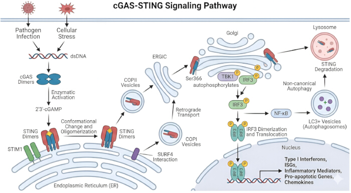
Figure 1: Overview of the cGAS–STING signaling pathway. Double-stranded DNA (dsDNA), generated during pathogen infection or cellular stress, activates cytosolic cyclic GMP–AMP synthase (cGAS) through dsDNA-dependent dimerization, resulting in the synthesis of 2′3′-cyclic GMP–AMP (2′3′-cGAMP). cGAMP binds to stimulator of interferon genes (STING) dimers located on the endoplasmic reticulum (ER) membrane, inducing conformational changes that promote STING oligomerization, dissociation from anchoring proteins such as stromal interaction molecule 1 (STIM1), interaction with trafficking factors, and packaging into coat protein complex II (COPII) vesicles. STING is subsequently transported through the ER–Golgi intermediate compartment (ERGIC) to the Golgi apparatus, where it recruits TANK-binding kinase 1 (TBK1). TBK1 undergoes autophosphorylation, phosphorylates STING at Ser366, and activates interferon regulatory factor 3 (IRF3). Phosphorylated IRF3 dimerizes and translocates to the nucleus, inducing the transcription of type I interferons (IFN-I), interferon-stimulated genes (ISGs), pro-inflammatory cytokines, chemokines, and pro-apoptotic genes. In parallel, STING signaling activates the NF-κB pathway and promotes non-canonical autophagy, resulting in the formation of LC3-positive autophagosomes. Ultimately, STING is trafficked to lysosomes for degradation. Under basal conditions, STING trafficking is regulated by coat protein complex I (COPI)-mediated retrograde transport from the Golgi to the ER, a process facilitated by the interaction between STING and SURF4, thereby maintaining signaling homeostasis3,28. The figure summarizes the key molecular events regulating cGAS–STING activation, intracellular trafficking, downstream signaling, and signal termination. Abbreviations: cGAS = cyclic GMP–AMP synthase; cGAMP = cyclic GMP–AMP; COPI = coat protein complex I; COPII = coat protein complex II; dsDNA = double-stranded DNA; ER = endoplasmic reticulum; ERGIC = endoplasmic reticulum–Golgi intermediate compartment; IFN-I = type I interferons; IRF3 = interferon regulatory factor 3; ISGs = interferon-stimulated genes; LC3 = microtubule-associated protein 1 light chain 3; NF-κB = nuclear factor kappa B; STIM1 = stromal interaction molecule 1; STING = stimulator of interferon genes; SURF4 = Surfeit locus protein 4; TBK1 = TANK-binding kinase 1. Please click here to view a larger version of this figure.
| Disease | Gene(s) | Inheritance | Most Typical Clinical Features |
| ADAR1 deficiency | ADAR1 | AR / AD | Chilblain lupus, cerebral vasculopathy |
| Aicardi–Goutières syndrome (AGS) | TREX1, RNASEH2A/B/C, SAMHD1, ADAR1, IFIH1, LSM11, RNU7-1 | AR / AD | Early-onset encephalopathy, basal ganglia calcifications, cerebral vasculopathy, chilblains |
| COPA syndrome | COPA | AD | ANCA-negative small-vessel vasculitis, ILD, arthritis, alveolar hemorrhage |
| DNASE1L3 deficiency | DNASE1L3 | AR | Urticarial vasculitis, hypocomplementemia, SLE-like phenotype |
| DNASE2 deficiency | DNASE2 | AR | Autoinflammation, cytopenias, vasculopathy, liver involvement |
| IFIH1 (GOF) syndrome | IFIH1 (MDA5) GOF | AD | AGS-like neuroinflammation, chilblains, lupus-like autoimmunity |
| IRF7 deficiency (GOF) | IRF7 | AD | IFN-driven inflammation, lupus-like manifestations |
| ISG15 deficiency | ISG15 | AR | Intracranial calcifications, mycobacterial susceptibility, IFN signature elevation |
| OAS1 (GOF) | OAS1 GOF | AD | Autoinflammation, pulmonary involvement, hypogammaglobulinemia |
| PRAAS / CANDLE | PSMB8, PSMA3, PSMB4, PSMB9, POMP, PSMG2 | AR / AD | Panniculitis, lipodystrophy, recurrent fevers, dermal inflammation |
| RNASEH2-related interferonopathy | RNASEH2A/B/C | AR | Neuroinflammation, cerebral vasculopathy |
| RNU7-1 / LSM11-associated disease | RNU7-1, LSM11 | AR | Replication stress, cerebral microangiopathy, calcifications |
| SAMHD1 deficiency | SAMHD1 | AR | Cerebral vasculopathy, strokes, chilblains |
| SAVI (STING-associated vasculopathy with onset in infancy) | STING1 (TMEM173) GOF | AD | Necrotizing dermal vasculopathy, chilblains, livedo, pulmonary fibrosis, ILD |
| STAT1 (GOF) | STAT1 GOF | AD | Autoimmunity, vasculitic features, chronic mucocutaneous candidiasis |
| STAT2 (GOF) | STAT2 GOF | AD | Early-onset systemic inflammation, severe IFN signature, vasculopathy |
| TBK1 (GOF) | TBK1 | AD | Neuroinflammation, systemic IFN activation |
| TREX1-associated familial chilblain lupus | TREX1 | AD | Chilblains, cutaneous vasculopathy, SLE-like features |
| USP18 deficiency | USP18 | AR | Severe neonatal interferonopathy, systemic inflammation, neurologic damage |
Table 1: Currently recognized monogenic type I interferonopathies and their characteristic clinical features. Monogenic type I interferonopathies share several overlapping clinical manifestations despite their distinct genetic etiologies. Cutaneous features commonly include chilblains, livedo reticularis, and necrotizing vasculopathy. Neurological involvement frequently manifests as basal ganglia calcifications, cerebral microangiopathy, developmental delay, and early-onset ischemic or hemorrhagic stroke. Pulmonary complications, particularly interstitial lung disease (ILD) and pulmonary capillary obliteration, are characteristic of disorders such as STING-associated vasculopathy with onset in infancy (SAVI) and COPA syndrome. Systemic manifestations are characterized by persistent activation of type I interferon (IFN-I) signaling, leading to an elevated interferon-stimulated gene (ISG) signature and chronic inflammation. Importantly, many monogenic type I interferonopathies closely mimic multifactorial autoimmune diseases, particularly ANCA-associated vasculitis (AAV) and systemic lupus erythematosus (SLE), which may delay diagnosis and lead to inappropriate immunosuppressive treatment if the underlying genetic defect is not recognized.
Disclosures
The authors declare no conflicts of interest.
Acknowledgements
The authors thank all investigators whose published studies have contributed to the current understanding of type I interferonopathies. No specific funding was received for the preparation of this review. Figure 1 was created by the authors using BioRender.com and Gemini (Google) as visualization tools. The figure is an original schematic and is not adapted from a previously published figure.
References
- Mendonça LO, Frémond ML. Interferonopathies: From concept to clinical practice. Best Pract Res Clin Rheumatol. 2024;38(3):101975.
- Volpi S, et al. Type I interferonopathies in pediatric rheumatology. Pediatr Rheumatol Online J. 2016;14:35.
- Decout A, Katz JD, Venkatraman S, Ablasser A. The cGAS-STING pathway as a therapeutic target in inflammatory diseases. Nat Rev Immunol. 2021;21(9):548-569.
- Uggenti C, Lepelley A, Crow YJ. Self-awareness: nucleic acid-driven inflammation and the type I interferonopathies. Annu Rev Immunol. 2019;37:247-267.
- d'Angelo DM, Di Filippo P, Breda L, Chiarelli F. Type I interferonopathies in children: An overview. Front Pediatr. 2021;9:631329.
- Adang LA, et al. IFN-signaling gene expression as a diagnostic biomarker for monogenic interferonopathies. JCI Insight. 2024;9(14):e178456.
- Martin-Fernández M, et al. Systemic type I IFN inflammation in human ISG15 deficiency leads to necrotizing skin lesions. Cell Rep. 2020;31(6):107633.
- Eleftheriou D, Brogan PA. Vasculitis and vasculitis-like manifestations in monogenic autoinflammatory syndromes. Rheumatol Int. 2018;38(1):13-24.
- Lin B, Goldbach-Mansky R. Pathogenic insights from genetic causes of autoinflammatory inflammasomopathies and interferonopathies. J Allergy Clin Immunol. 2022;149(3):819-832.
- Nesterova IV, et al. Congenital and acquired interferonopathies: Differentiated approaches to interferon therapy. In: Saxena SK, Prakash H, editors. Innate Immunity in Health and Disease. IntechOpen; London; 2020. Available at: https://doi.org/10.5772/intechopen.91723.
- An J, Marwaha A, Laxer RM. Autoinflammatory diseases: A review. J Rheumatol. 2024;51:848-861.
- Scott O, et al. STAT1 gain-of-function heterozygous cell models reveal diverse interferon-signature gene transcriptional responses. NPJ Genom Med. 2021;6:34.
- Gruber C, et al. Homozygous STAT2 gain-of-function mutation by loss of USP18 activity in a patient with type I interferonopathy. J Exp Med. 2020;217(5):e20192319.
- Liu Y, et al. Activated STING in a vascular and pulmonary syndrome. N Engl J Med. 2014;371(6):507-518.
- Batten I, et al. Investigation of type I interferon responses in ANCA-associated vasculitis. Sci Rep. 2021;11:8272.
- Brilland B, et al. A stronger type I interferon signature distinguishes ANCA-associated vasculitis phenotypes and predicts kidney prognosis. Kidney Int. 2026;109(1):139-159.
- Söderberg D, Segelmark M. Neutrophil extracellular traps in ANCA-associated vasculitis. Front Immunol. 2016;7:256.
- Triaille C, et al. The emerging concept of ANCA-associated vasculitis related to inborn errors of immunity. Autoimmun Rev. 2025;24(7):103824.
- Ng CT, Fong LY, Tan JJ, Abdullah MNH. Endothelial barrier disruptive effect of IFN-γ and TNF-α: Synergism of pro-inflammatory cytokines. Cytokine. 2025;190:156922.
- Pham PT, et al. Innate immune system regulated by stimulator of interferon genes, a cytosolic DNA sensor, regulates endothelial function. J Am Heart Assoc. 2023;12(22):e030084.
- Gairhe S, et al. Type I interferon activation and endothelial dysfunction in caveolin-1 insufficiency-associated pulmonary arterial hypertension. Proc Natl Acad Sci U S A. 2021;118(11):e2010206118.
- Cooles FAH, Isaacs JD. The interferon gene signature as a clinically relevant biomarker in autoimmune rheumatic disease. Lancet Rheumatol. 2022;4(1):e61-e72.
- Barrat FJ, Crow MK, Ivashkiv LB. Interferon target-gene expression and epigenomic signatures in health and disease. Nat Immunol. 2019;20:1574-1583.
- Nocturne G, Mariette X. Interferon signature in systemic autoimmune diseases: What does it mean? RMD Open. 2022;8(2):e002687.
- Gagne S, Sivaraman V, Akoghlanian S. Interferonopathies masquerading as non-Mendelian autoimmune diseases: Pattern recognition for early diagnosis. Front Pediatr. 2023;11:1169638.
- Shawky AM, et al. A comprehensive overview of globally approved JAK inhibitors. Pharmaceutics. 2022;14(5):1001.
- Crow YJ, Neven B, Frémond ML. JAK inhibition in the type I interferonopathies. J Allergy Clin Immunol. 2021;148(4):991-993.
- Ablasser A, Chen ZJ. cGAS in action: Expanding roles in immunity and inflammation. Science. 2019;363(6431):eaat8657.
- Sánchez GAM, et al. JAK1/2 inhibition with baricitinib in the treatment of autoinflammatory interferonopathies. J Clin Invest. 2018;128(7):3041-3052.
- Kim H, et al. Janus kinase (JAK) inhibition with baricitinib in refractory juvenile dermatomyositis. Ann Rheum Dis. 2021;80(3):406-408.
- Haag SM, et al. Targeting STING with covalent small-molecule inhibitors. Nature. 2018;559(7713):269-273.
- O'Shea JJ, Gadina M. Selective Janus kinase inhibitors come of age. Nat Rev Rheumatol. 2019;15(2):74-75.
- Gül A, et al. The pathogenesis, clinical presentations and treatment of monogenic systemic vasculitis. Nat Rev Rheumatol. 2025;21:414-425.
- Crow YJ, Stetson DB. The type I interferonopathies: 10 years on. Nat Rev Immunol. 2022;22(8):471-483.
- Babadei O, Strobl B, Müller M, Decker T. Transcriptional control of interferon-stimulated genes. J Biol Chem. 2024;300(10):107771.