Robot-assisted radical prostatectomy with patients with femoral crossover bypass grafts can be safely done through multidisciplinary perioperative management.
Case Report
Robot-Assisted Radical Prostatectomy in a Patient with Pre-existing Femoral Crossover Bypass Graft: Multidisciplinary Management and Outcomes
In This Article
Summary
Abstract
Robot-assisted radical prostatectomy (RARP) has become the standard surgical treatment for localized clinically significant prostate cancer, yet in patients with pre-existing pelvic vascular grafts, such as the iliofemoral graft, there is a significant risk of iatrogenic injury during abdominal access and pelvic dissection. This report describes a 69-year-old male with D’Amico intermediate-risk prostate cancer and a right iliofemoral polytetrafluoroethylene (PTFE) crossover bypass graft implanted for peripheral arterial disease. Major comorbidities included obesity (BMI 32 kg/m2), ASA III status, prior myocardial infarction with coronary stents, insulin-dependent type 2 diabetes, atrial fibrillation on apixaban, and Peripheral arterial disease under continuous antiplatelet therapy with aspirin. Preoperative magnetic resonance angiography enabled three-dimensional reconstruction of the graft course; vascular surgeons marked the graft trajectory directly on the abdominal wall to guide safe trocar placement. Aspirin continued perioperatively. Transperitoneal anterior RARP was performed with extended pelvic lymph node dissection (28 nodes, pN0). The graft was visualized intraperitoneally and avoided; cold scissors were used for dissection to minimize thermal injury. Operative time was 150 min, with an estimated blood loss of 300 mL. Final pathology revealed pT3a pN0 R0 disease with 4.8 cm3 tumor volume. Immediate continence was achieved after catheter removal. Graft patency was verified by Doppler ultrasound on day 1 and before discharge. PSA was undetectable (< 0.01 ng/mL) at 8 weeks and thereafter. This case illustrates that meticulous multidisciplinary planning enables safe RARP in high-risk patients.
Introduction
Robot-assisted radical prostatectomy (RARP) represents the standard of care in the treatment of localized prostate cancer, of localized prostate cancerproviding superior visualization, reduced blood loss, shorter recovery, and equivalent oncologic outcomes compared to open surgery1. Minimally invasive approaches resulted in less blood loss, transfusions, catheterization, hospital stay, and complications than open surgery, with similar oncological and functional outcomes like continence and potency recovery2.
Vascular injuries seldom happen in elective abdominal and pelvic surgeries. However, when they do, they can be fatal3. Patients with peripheral arterial disease and iliofemoral bypass grafts face higher risks of graft injury during trocar insertion, peritoneal incision, or pelvic lymphadenectomy because of anatomical distortion and closeness to major vessels4. The increasing coexistence of prostate cancer and vascular disease requires tailored surgical protocols. However, the literature on this topic is limited5,6.
Robotic technology offers clear advantages in this setting, including high-definition magnification for graft identification, tremor-filtered precision, and energy-restricted dissection. Continuation of low-dose aspirin during RARP has been shown to be safe without elevating the risk of bleeding complications7.
This case is appropriate for urologic oncologists, vascular surgeons, and multidisciplinary teams managing complex patients with significant cardiovascular comorbidities, and it illustrates the value of perioperative interdisciplinary management.
Case Presentation:
A 69-year-old male was referred with biopsy-confirmed prostate adenocarcinoma: PSA 18 ng/mL, prostate volume 60 mL on transrectal ultrasound, Gleason score 3+4=7, D’Amico intermediate-risk classification8, and negative metastatic staging. Medical history included coronary artery disease with prior bypass and coronary stents, atrial fibrillation on apixaban 5 mg twice daily, insulin-dependent type 2 diabetes mellitus, obesity (BMI 32 kg/m2), ASA III status, and >40 pack-year smoking history. Urologic obstructive symptoms were mild (International Prostate Symptom Score (IPSS) 7, quality of life (QoL) score 3). A right iliofemoral polytetrafluoroethylene (PTFE) crossover bypass graft for peripheral arterial disease. Long-term aspirin therapy was ongoing.
Diagnosis, Assessment, and Plan: Diagnosis was localized intermediate-risk prostate cancer (cT2, PSA 18 ng/mL, Gleason 3 + 4 = 7). The assessment confirmed there was no evidence of metastasis on conventional staging. The pre-existing crossover graft represented a high risk for iatrogenic injury during port placement and dissection; comorbidities increased perioperative bleeding, infection, and cardiovascular risks.
The case was discussed at our preoperative interdisciplinary tumor board. Although radiation therapy with androgen deprivation therapy (ADT) is an established treatment for intermediate-risk prostate cancer, ADT carries significant cardiovascular risks for this patient with a high-risk comorbidity profile9. Moreover, the patient's mild IPSS score (7) belied more significant voiding dysfunction, and radiation could have worsened his urinary symptoms10. After detailed counseling regarding all treatment options, the patient was determined to undergo surgery
Protocol
This study was conducted in accordance with the ethical standards of the institutional and national research committees. The patient provided written informed consent for all aspects of the treatment plan, including each surgical intervention. This study complies with the ethical standards outlined in the Declaration of Helsinki and is approved by the ethics committees of the Westfalen-Lippe Medical Association and the University of Muenster (2023–500-f-S) for the retrospective management of patient data processed within our department.
1. Preoperative Planning and Imaging
- Multidisciplinary evaluation
- The patient was evaluated in a multidisciplinary conference including urologists, vascular surgeons, cardiologists, and anesthesiologists due to the presence of a pre-existing femoral crossover bypass graft and multiple cardiovascular comorbidities.
- Cardiovascular risk stratification was performed because of the patient's history of coronary artery disease, atrial fibrillation, and insulin‑dependent diabetes mellitus.
- Anticoagulation and antiplatelet management were reviewed. Aspirin therapy continued perioperatively while apixaban was managed according to institutional perioperative protocols.
- Preoperative vascular mapping
- Magnetic resonance angiography (MRA) of the abdomen and pelvis was present to evaluate the exact anatomical course of the iliofemoral crossover PTFE graft (Figure 1).
- Three-dimensional reconstruction of the vascular structures was generated to visualize the graft trajectory relative to the abdominal wall and pelvic vasculature.
- The vascular surgery team marked the graft pathway directly on the abdominal wall preoperatively using sterile skin markers to guide safe trocar placement.
2. Patient Positioning and Surgical Setup
- Patient positioning
- The patient was placed in the supine lithotomy position with legs supported in adjustable stirrups.
- A Trendelenburg position was applied after induction of pneumoperitoneum.
- The abdomen and perineum were prepared and draped in a sterile fashion.
- Pneumoperitoneum establishment
- Pneumoperitoneum was established using a Veress needle technique at the supraumbilical site.
- Intra-abdominal pressure was maintained at 8 mmHg.
- Explorative laparoscopy showed the trajectory of the graft beneath the peritoneum, cranial to the bladder and the symphysis (Figure 2).
3. Trocar Placement and Robotic Docking
- Trocar placement strategy
- A transperitoneal anterior approach was used with a robotic surgical system.
- Trocar placement was carefully planned to maintain a distance of at least 5 cm from the pre‑marked graft pathway.
- Ports included four 8 mm robotic trocars (in the horizontal supraumbilical line), one 12 mm assistant trocar in the right lower abdominal quadrant, and one 5 mm assistant trocar in the right upper abdominal quadrant.
- Robotic docking
- The robotic cart was positioned between the patient’s legs.
- Robotic arms were docked sequentially under direct vision. The third arm, which, according to institutional standards, will be placed laterally on the left abdominal side and used to grasp, was positioned relatively medially and caudally to prevent accidental injury to the graft during both the radical prostatectomy and lymphadenectomy.
- Instruments included monopolar scissors, bipolar forceps, robotic grasping forceps and needle driver.
4. Intraoperative Identification and Protection of the Vascular Graft
- The peritoneum over the bladder was incised to enter the Retzius space.
- The bladder was mobilized to expose the anterior prostate.
- The course of the crossover bypass graft was identified beneath the abdominal wall, cranial to the os pubis.
- Cold scissors were used preferentially during dissection to minimize thermal injury.
5. Robot‑Assisted Radical Prostatectomy
- The anterior prostate approach was performed with incision of the endopelvic fascia and control of the dorsal venous complex.
- Bladder neck dissection was completed with identification of the Foley catheter.
- Vas deferens and seminal vehicles were dissected and mobilized.
- Prostatic pedicles were controlled with clips and bipolar energy.
- Nerve‑sparing was attempted where oncologically appropriate.
6. Extended Pelvic Lymph Node Dissection
- Bilateral extended pelvic lymph node dissection was performed.
- Dissection boundaries included the external iliac vein, obturator nerve, internal iliac vessels, and the common iliac bifurcation.
- Lymphatic tissue was removed en bloc.
- A total of 28 lymph nodes were retrieved.
7. Vesicourethral Anastomosis
- A running vesicourethral anastomosis was performed using a double‑armed absorbable suture.
- The anastomosis was constructed over a transurethral Foley catheter.
- The graft was identified before completion of surgery to confirm its integrity (Figure 3).
8. Postoperative Catheter Management
- Cystography performed on postoperative day 3 showed no extravasation, and the urinary catheter was removed (Figure 4)
Results
Operative time: 150 min; estimated blood loss: 300 mL. The procedure was completed without any complication. The extended lymphadenectomy yielded 28 negative lymph nodes. Final pathology: prostate weight 48 g, Gleason 3 + 4 = 7, pT3a pN0 R0, tumor volume 4.8 cm3. Postoperative hemoglobin 11.2 g/dL; no transfusion required. Cystogram on day 3 confirmed intact anastomosis without leakage (Figure 4). Immediate continence achieved (no pads at discharge). Patient ambulated on day 1 and was discharged uneventfully. Graft patency confirmed by Doppler ultrasound on postoperative day 1 and before discharge. PSA undetectable (< 0.01 ng/mL) at 8-week follow-up. Key perioperative and postoperative outcomes are summarized in Table 1.
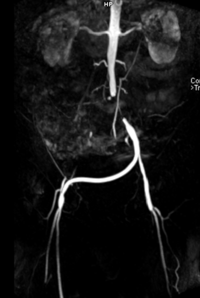
Figure 1: Coronal contrast-enhanced magnetic resonance angiography (MRA) demonstrating a patent left-to-right iliofemoral bypass graft with complete opacification of the graft and restoration of arterial inflow to the contralateral lower limb. A stent proximal to the graft from the aorta to the left iliac artery can also be seen. Please click here to view a larger version of this figure.
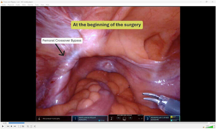
Figure 2: Intraoperative robotic view. At the beginning of the surgery, following pneumoperitoneum establishment and exploratory laparoscopy. The right iliofemoral crossover bypass graft is clearly identified coursing beneath the peritoneum, traversing cranially to the bladder dome and the pubic symphysis. The graft appears as a linear, non-pulsatile, synthetic tubular structure with a smooth surface, surrounded by mild perigraft adhesions but without evidence of kinking, compression, or previous injury. The overlying peritoneum is intact, and no anomalous vascular branches are visualized in proximity. Please click here to view a larger version of this figure.
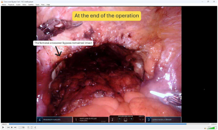
Figure 3: Intraoperative robotic view. At the end of the surgery, following completion of the robot-assisted radical prostatectomy, pelvic lymph node dissection, and vesicourethral anastomosis. The right iliofemoral crossover PTFE graft remains intact with no evidence of iatrogenic injury, thermal damage, compression, or disruption. The graft maintains its original anatomical course and structural integrity. Adjacent surgical planes are well healed without bleeding or hematoma. The pelvic dissection was successfully performed without direct manipulation or thermal energy application within 1 cm of the graft surface. Please click here to view a larger version of this figure.
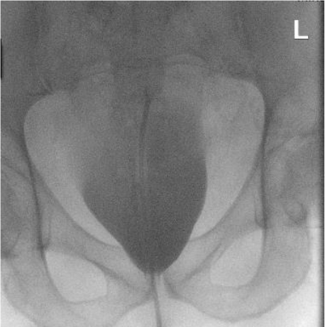
Figure 4: Postoperative cystogram. Obtained on postoperative day 3. The Foley catheter balloon is visualized within the bladder lumen. The vesicourethral anastomosis is fully intact with no evidence of contrast extravasation into the perivesical space, pelvis, or retroperitoneum. The bladder contour is smooth and symmetric, demonstrating adequate filling capacity without filling defects. This finding confirms anastomotic integrity, permitting safe removal of the transurethral catheter. Please click here to view a larger version of this figure.
| Category | Outcome Measure | Result |
| Operative Outcomes | Operative time | 150 min |
| Estimated blood loss | 300 mL | |
| Intraoperative complications | None | |
| Hemoglobin (postoperative) | 11.2 g/dL | |
| Transfusion required | No | |
| Hospital stay | Discharged uneventfully (day 5 postoperativly) | |
| Pathological Outcomes | Prostate weight | 48 g |
| Gleason score | 3 +4 = 7 | |
| Pathological stage | pT3a | |
| Surgical margins | R0 | |
| Tumor volume | 4.8 cm3 | |
| Lymph nodes retrieved | 28 (all negative) | |
| Functional Outcomes | Continence (immediate) | No pads at catheter removal / discharge |
| Cystogram result | No extravasation | |
| Catheter removal | Postoperative day 3 (after negative cystogram) | |
| Vascular Outcomes | Graft injury | None |
| Graft patency (Doppler ultrasound) | Confirmed on postoperative day 1 and before discharge | |
| Anticoagulation management | Aspirin+B1:C19 continued perioperatively; apixaban managed per protocol |
Table 1: Operative, Pathological, Functional, and Vascular Outcomes.
| Scenario | Recommended Maneuver | Key Consideration |
| Graft identified within 5 cm of planned trocar site | Consider alternative port configuration; use optical entry or open Hasson technique | Maintain minimum 5 cm distance from marked graft trajectory |
| Graft adherent to peritoneum or bladder | Use cold scissors very carefully; avoid monopolar energy within 1 cm of graft | Thermal spread can damage PTFE grafts up to 5 mm from application site |
| Graft obscuring surgical field or limiting access | Consider ipsilateral lymphadenectomy omission if graft overlies external iliac vessels | Balance oncologic necessity against graft risk |
| Inadvertent graft injury identified intraoperatively | Immediate vascular surgery consultation; apply pressure; avoid blind clamping | PTFE grafts are not self-seal; primary repair or interposition grafting typically required |
| Graft patency concern after prolonged dissection | Perform intraoperative Doppler ultrasound or graft angiography if available | Document flow before abdominal closure |
| Postoperative hemoglobin drop without visible bleeding | Emergent CT angiography to rule out graft hemorrhage or embolisation | Low threshold for vascular surgery involvement |
Table 2: Troubleshooting approach for RARP in patients with pre-existing pelvic vascular grafts.
Discussion
Robot-assisted radical prostatectomy in patients with pre‑existing vascular grafts represents a technically challenging scenario because of the risk of graft injury during trocar insertion, pelvic dissection, or lymph node removal. This case demonstrates that safe robotic prostatectomy can be achieved through meticulous preoperative planning, interdisciplinary collaboration, and careful intraoperative technique.
The robotic platform offers advantages in this complex setting. High-definition 3D visualization improves graft and structure identification, while tremor filtration and articulated instruments enable precise dissection in the pelvic space. Compared to open radical prostatectomy, robot-assisted approaches reduce blood loss, transfusion rates, hospital stays, and postoperative pain11. A recent meta-analysis of 80 studies found RARP had significantly lower positive margin rates (RR 0.893) and lower biochemical recurrence (RR 0.713 vs open, 0.672 vs laparoscopic)11. These oncologic benefits are especially relevant in reoperative or distorted anatomical fields, such as those involving vascular grafts.
Perioperative continuation of low‑dose aspirin was safe in this case and did not lead to excessive bleeding. This finding aligns with a systematic review and meta-analysis of 1,481 patients undergoing RARP, which demonstrated no significant differences between aspirin users and non-users in overall complications (10.7% vs. 15.7%, RR 0.83; p=0.45), major complications (1% vs. 3%, RR 0.98; p=0.98), estimated blood loss (278 mL vs. 307 mL), or hospital length of stay12. While a slightly higher blood transfusion rate was observed in the aspirin group (2.6% vs. 1.6%, p=0.04), this difference was small and clinically acceptable12. Of note, emerging evidence suggests aspirin may have beneficial effects on biochemical recurrence-free survival after RARP, particularly in higher-risk patients, providing additional rationale for continuation when clinically indicated13.
While open radical prostatectomy provides tactile feedback and direct vascular control, it requires a larger incision that must pass through the iliofemoral crossover graft trajectory. Laparoscopic radical prostatectomy avoids large incisions but lacks the wristed instrumentation and three-dimensional visualization of robotic systems, making delicate dissection near vascular grafts more challenging. The robotic platform's articulated instruments and magnified view are particularly advantageous when working in close proximity to grafts.
Nonetheless, preoperative three-dimensional vascular mapping using MRA angiography was essential to visualize the graft trajectory and its relationship to the abdominal wall and pelvic vasculature. Direct skin marking of the graft pathway translated imaging findings into practical intraoperative guidance for trocar placement. Based on our experience and review of the literature, we propose the following troubleshooting approach (Table 2) for RARP in patients with pre-existing pelvic vascular grafts:
Although limited to a single case, this report demonstrates the feasibility of performing complex robotic oncologic surgery in patients with significant vascular comorbidities. The described protocol may serve as a reference for surgeons managing similar cases. Future studies with larger cohorts are required to establish standardized guidelines for graft management during RARP, including optimal trocar configurations, energy dissection protocols, and postoperative graft surveillance intervals.
Disclosures
Declaration of AI use: The authors used a language model (Gemini) exclusively for language polishing and grammatical refinement. All scientific content was controlled, verified, and approved by the authors, who assume full responsibility for the final manuscript.
Materials
| Name | Company | Catalog Number | Comments |
|---|---|---|---|
| Barbed suture (4-0) | Ethicon | Absorbable barbed suture to suture the dorsal vascular Complex | |
| Double-armed barbed Suture (3-0) | Ethicon | Absorbable barbed suture to perform the vesicourethral anastomosis | |
| Monocryl (3-0) | Ethicon | Monofilament suture | |
| Indocyanine green (ICG) dye | Diagnostic Green GmbH | 25 mg vial | |
| da Vinci X robotic system | Intuitive Surgical | Robotic system | |
| Transurethral Foley catheter 18 Charriere | B. Braun Melsungen AG | 18 Fr, 2 way |