This study investigated whether acupotomy promotes cartilage repair in a rabbit model of knee osteoarthritis. Fifty rabbits were assigned to five groups. Acupotomy was administered weekly for 4 weeks. Joint swelling, histopathology, inflammatory cytokines, and circPDE4B/RIC8A/p38 MAPK axis-related markers were assessed to explore the underlying mechanism.
Research Article
Acupotomy Regulates the RIC8A/P38 MAPK Axis Based on the CircRNA Scaffold to Promote Cartilage Repair in Knee Osteoarthritis
* These authors contributed equally
In This Article
Summary
Abstract
Knee osteoarthritis (KOA) is characterized by progressive cartilage degeneration and inflammation. This study investigated the therapeutic effects of acupotomy in a rabbit model of KOA and its association with the circular RNA phosphodiesterase 4B (circPDE4B)/resistance to inhibitors of cholinesterase 8A (RIC8A)/p38 mitogen-activated protein kinase (MAPK) signaling axis. Fifty New Zealand White rabbits were randomly assigned to five groups (n = 10 per group): control, KOA model, celecoxib, acupotomy, and sham acupotomy. KOA was induced using the modified Videman method. Beginning 6 weeks after modeling, acupotomy was administered once weekly for 4 weeks, whereas celecoxib was administered daily by gavage at 24 mg/kg for 4 weeks. Knee swelling and cartilage histopathology were evaluated. Levels of interleukin-1β (IL-1β) , tumor necrosis factor-α (TNF-α) , and interleukin-6 (IL-6) in cartilage tissue homogenates were measured by enzyme-linked immunosorbent assay. Gene and protein expression associated with cartilage metabolism and the circPDE4B/RIC8A/p38 MAPK axis were assessed by quantitative reverse transcription polymerase chain reaction and Western blotting. Compared with the KOA model group, acupotomy reduced knee swelling, improved histopathological scores, decreased inflammatory cytokine levels, downregulated matrix metalloproteinase-3 (MMP-3) , matrix metalloproteinase-13 (MMP-13), RIC8A, and phosphorylated p38 MAPK, and upregulated SRY-box transcription factor 9 (SOX9), aggrecan, and relative circPDE4B. Its effects were comparable to those of celecoxib, whereas sham acupotomy produced no significant improvement. These findings indicate that acupotomy alleviates inflammation and cartilage degeneration in rabbit KOA and is associated with modulation of the circPDE4B/RIC8A/p38 MAPK axis.
Introduction
Knee osteoarthritis (KOA) is a prevalent degenerative joint disease characterized by progressive cartilage degradation, synovial inflammation, subchondral bone sclerosis, and osteophyte formation1,2,3. With global population aging, the incidence of KOA continues to increase, making it a leading cause of joint dysfunction and chronic pain among middle-aged and older adults worldwide4,5,6. Current clinical treatments primarily focus on symptomatic relief through pharmacological agents, such as nonsteroidal anti-inflammatory drugs (NSAIDs) and intra-articular corticosteroid injections. However, disease-modifying therapies capable of halting disease progression or promoting cartilage repair remain limited7,8.
Emerging evidence has highlighted the important regulatory roles of noncoding RNAs (ncRNAs) in the pathogenesis and progression of KOA9,10,11. Among these ncRNAs, circular RNAs (circRNAs) have attracted increasing attention because of their unique covalently closed-loop structure, which confers greater stability than linear RNAs, and their diverse biological functions12,13. Shen et al. reported that circPDE4B functions as a molecular scaffold that regulates resistance to cholinesterase 8A (RIC8A)/p38 mitogen-activated protein kinase (MAPK) signaling pathway inhibitors, thereby influencing chondrocyte extracellular matrix metabolism and inflammatory responses14. The RIC8A/p38 MAPK pathway plays an important role in regulating inflammation, catabolism, apoptosis, and senescence in chondrocytes, all of which contribute to osteoarthritis pathogenesis15.
Acupotomy is a minimally invasive therapeutic approach that integrates traditional Chinese medicine theory with modern anatomical principles and has shown promising efficacy in the clinical management of KOA16,17. This technique uses a specialized needle-knife device to release soft tissue adhesions, improve local circulation, and restore biomechanical balance around affected joints, thereby relieving pain and improving function in patients with KOA18. Despite these reported clinical benefits, the molecular mechanisms underlying the therapeutic effects of acupotomy, particularly its potential regulation of circRNA-mediated signaling pathways involved in cartilage repair, remain largely unexplored.
Mechanistically, acupotomy generates local mechanical stimuli through periarticular soft tissue release. Chondrocytes are mechanosensitive cells that respond to biomechanical signals, and circRNAs have been shown to participate in the mechanotransduction responses of chondrocytes19,20. Although direct evidence linking acupotomy to the regulation of articular cartilage-specific circRNAs is lacking, it is biologically plausible that mechanical signals generated by acupotomy may modulate mechanosensitive circRNAs such as circPDE4B. This possibility formed the basis of the present investigation.
Building on previous findings regarding the regulatory role of circPDE4B in the RIC8A/p38 MAPK signaling axis, the present study used a rabbit model of KOA induced by the modified Videman method to test the hypothesis that acupotomy promotes cartilage repair by modulating the circPDE4B/RIC8A/p38 MAPK signaling pathway. Acupotomy was selected for this study over other KOA interventions for several reasons. First, compared with pharmacological agents such as nonsteroidal anti-inflammatory drugs (NSAIDs) and celecoxib, which primarily provide symptomatic relief and have been associated with limited disease-modifying effects and potential long-term adverse events7,8, acupotomy offers a nonpharmacological approach that addresses both local mechanical factors and biological signaling pathways. Second, unlike intra-articular corticosteroid or hyaluronic acid injections, which require repeated intra-articular administration and carry risks of infection and joint damage, acupotomy targets periarticular soft tissues and may exert sustained biomechanical and biochemical effects21. Third, clinical evidence has demonstrated that acupotomy produces comparable or superior outcomes to conventional therapies in reducing pain and improving joint function in patients with KOA, and experimental studies have suggested that acupotomy modulates inflammatory responses and chondrocyte metabolism21,22. However, the specific molecular pathways through which acupotomy exerts its chondroprotective effects remain poorly characterized, particularly with respect to circRNA-mediated signaling. Thus, investigating the association between acupotomy and the circPDE4B/RIC8A/p38 MAPK axis may provide novel mechanistic insights and support the use of acupotomy as a disease-modifying intervention for KOA16,18. The primary endpoints were knee joint swelling, histopathological cartilage degeneration (Mankin and Moran scores), inflammatory cytokine levels (interleukin-1β, tumor necrosis factor-α, and interleukin-6), expression of cartilage metabolism-related markers (matrix metalloproteinase-3, matrix metalloproteinase-13, SRY-box transcription factor 9, and aggrecan), and expression of circPDE4B and RIC8A, as well as phosphorylation of p38 MAPK. Acupotomy was administered once weekly for 4 weeks at selected periarticular points using the four-step technique (longitudinal release, transverse separation, and incision of nodules). Histopathological evaluation, inflammatory cytokine measurement, and gene and protein expression analyses were used to characterize the effects of acupotomy and explore its potential molecular basis in KOA.
Protocol
All animal procedures were performed in accordance with the institutional guidelines for the care and use of laboratory animals and were approved by the Animal Ethics Committee of Anhui University of Chinese Medicine (approval no. AHUCM-rabbits-2023195). The reagents, chemicals, software, and tools used in this protocol are listed in the Table of Materials.
1. Experimental animals and grouping
Fifty healthy adult male New Zealand White rabbits (6 months old, weighing 2.0–2.5 kg) were obtained from Nanjing Pukou District Laifu Breeding Farm (Animal Production License No.: SCXK (Su) 2024-0007). All animals underwent a 1-week acclimatization feeding period prior to the experiment. The right knee was designated for swelling assessment and the left knee for cartilage tissue collection as a consistent procedural convention, rather than based on a specific experimental hypothesis.After acclimatization, the rabbits were randomly assigned to five groups (n = 10 per group): a control group, which received no modeling procedure or treatment; a KOA model group, in which KOA was induced using the modified Videman method; a celecoxib group, in which drug treatment was initiated 6 weeks after modeling; an acupotomy group, in which acupotomy treatment was initiated 6 weeks after modeling; and a sham acupotomy group, in which needle insertion without cutting or loosening was initiated 6 weeks after modeling.
2. Establishment of the KOA model
All rabbits except those in the control group underwent the modified Videman procedure to induce KOA. The animals were fasted for 12 h before surgery. Anesthesia was induced by intravenous administration of 3% pentobarbital sodium at 30 mg/kg through the marginal ear vein. After routine preparation of the surgical site, disinfection, and sterile draping, a longitudinal incision was made along the medial aspect of the knee joint. The medial collateral ligament and anterior cruciate ligament were sequentially transected, and the medial meniscus was completely removed. The incision was then closed in layers.
To reduce the risk of postoperative infection, penicillin was administered intramuscularly at 40,000 U/kg/day for 3 consecutive days. After surgery, the rabbits were returned to their cages and allowed unrestricted movement without joint immobilization.
3. Sham acupotomy treatment
At 6 weeks after KOA modeling, rabbits in the sham acupotomy group underwent the same palpation and point-marking procedures used for the active acupotomy treatment. A sterile disposable acupotomy needle measuring 0.35 mm × 25 mm was inserted perpendicularly through the skin at each marked point until the subcutaneous tissue was reached. No cutting, loosening, transverse manipulation, or other mechanical release maneuver was performed. The needle was withdrawn immediately after insertion without tissue manipulation. Sham treatment was administered once weekly for 4 consecutive weeks.
4. Acupotomy treatment
At 6 weeks after KOA modeling, rabbits in the acupotomy group were restrained on a treatment table. The periarticular tissues around the affected knee were palpated, particularly around the patella and the medial and lateral joint spaces, to identify cord-like structures or hard nodules. Four to six treatment points were selected according to the palpation findings and marked on the skin.
The treatment area was shaved and disinfected using routine sterile procedures. A sterile disposable acupotomy needle measuring 0.35 mm × 25 mm was inserted slowly and perpendicularly at each marked point until the bone surface was reached. The procedure was performed according to the four-step acupotomy technique. Longitudinal release was performed first, followed by transverse separation. When a hard nodule was encountered, it was incised until a distinct release sensation was perceived through the needle-knife. After the procedure, the needle was withdrawn, and each insertion site was compressed briefly with sterile gauze and covered with an adhesive dressing. Treatment was administered once weekly for 4 consecutive weeks.
5. Drug treatment
At 6 weeks after KOA modeling, rabbits in the celecoxib group received celecoxib suspension by oral gavage at 24 mg/kg once daily for 4 consecutive weeks. The suspension was freshly prepared each day in physiological saline. Supplier and catalog information are provided in the Table of Materials.
6. Assessment of knee joint swelling
At the end of the intervention period, corresponding to 10 weeks after KOA modeling, knee joint swelling was assessed. A soft silk thread was wrapped around the most swollen region of the right knee joint, typically at the level of the tibial plateau. The thread was removed and measured to the nearest millimeter with a ruler, and the recorded length was defined as the knee circumference.
The relative swelling ratio was calculated using the following equation:

7. Sample collection
At the end of the intervention period, the rabbits were anesthetized by intravenous administration of 3% pentobarbital sodium at 30 mg/kg through the marginal ear vein. The left knee joint was opened using a No. 23 scalpel blade to expose the articular surfaces of the tibial plateau and femoral condyles. Articular cartilage tissue was collected from these regions.
Each cartilage sample was divided into two portions. One portion was fixed in 4% paraformaldehyde for histopathological examination. The second portion was placed in cryogenic tubes, rapidly frozen in liquid nitrogen, and stored at −80 °C until molecular analyses were performed.Immediately after completion of all tissue collection procedures, the rabbits were euthanized by air embolism via rapid intravenous injection of air through the marginal ear vein.
Histological staining and Mankin and Moran scoring were performed using cartilage samples from all 10 rabbits per group (n = 10). Representative hematoxylin and eosin and Safranin O–Fast Green images were selected from three randomly selected rabbits per group. Immunohistochemistry and Western blotting were performed using samples from three randomly selected rabbits per group (n = 3). Quantitative reverse transcription polymerase chain reaction and enzyme-linked immunosorbent assay were performed using samples from six randomly selected rabbits per group (n = 6). All subset selections were performed randomly without prior knowledge of group assignments.
8. Hematoxylin and eosin staining
Cartilage tissue fixed in 4% paraformaldehyde was decalcified in 10% ethylenediaminetetraacetic acid (EDTA) solution at pH 7.4 for 4–6 weeks. The decalcifying solution was replaced every 3 days. After decalcification was confirmed by needle penetration, the tissue was dehydrated through a graded ethanol series of 70%, 80%, 90%, 95%, and 100%, cleared in xylene, and embedded in paraffin. The embedded tissue was sectioned at a thickness of 5 µm using a microtome, and the sections were mounted on glass slides.
For hematoxylin and eosin staining, the sections were heated at 60 °C for 60 min, deparaffinized in xylene for three changes of 5 min each, and rehydrated sequentially in 100%, 95%, 80%, and 70% ethanol, followed by distilled water. The sections were stained with hematoxylin for 5 min, rinsed under running tap water for 10 min, differentiated in 1% hydrochloric acid prepared in 70% ethanol for 2–3 s, and rinsed with distilled water. The sections were then stained with eosin for 2 min, rinsed with distilled water, dehydrated through 70%, 80%, 90%, 95%, and 100% ethanol, cleared in xylene, and mounted with neutral resin. Cartilage morphology was examined, and representative images were acquired using a light microscope.
9. Safranin O–Fast Green staining
Paraffin-embedded cartilage sections were deparaffinized and rehydrated as described in section 8. The sections were stained with Weigert iron hematoxylin for 5 min and rinsed with distilled water. The sections were then stained with 0.05% Fast Green solution for 3 min, briefly rinsed in 1% acetic acid for 10–15 s, and stained with 0.1% Safranin O solution for 5 min.
After staining, the sections were rapidly dehydrated in 95% and 100% ethanol, cleared in xylene, and mounted with neutral resin. The stained sections were examined using a light microscope. Safranin O staining intensity, which reflects proteoglycan content, was analyzed semiquantitatively using image-analysis software.
10. Histological scoring
Two pathologists who were blinded to the experimental group assignments independently evaluated the stained sections using the Mankin and Moran scoring systems to assess the degree of cartilage degeneration. Before evaluation, all sections were assigned random identification codes. The treatment allocations were not disclosed to the pathologists until all scoring was complete23.
11. Enzyme-linked immunosorbent assay
Frozen cartilage tissue was weighed, combined with phosphate-buffered saline at pH 7.4, and homogenized thoroughly on ice. The homogenate was centrifuged at 3,000 × g for 20 min at 4 °C, and the supernatant was collected for analysis. The concentrations of interleukin-1β (IL-1β), tumor necrosis factor-α (TNF-α), and interleukin-6 (IL-6) in the tissue supernatant were measured using rabbit-specific enzyme-linked immunosorbent assay kits according to the manufacturer’s instructions.
12. Quantitative reverse transcription polymerase chain reaction
Total RNA was extracted from cartilage tissue using a phenol–chloroform-based method. Briefly, the tissue was lysed in RNA extraction reagent, and chloroform was added to separate the aqueous and organic phases. The aqueous phase was collected, and RNA was precipitated with isopropanol, washed with 75% ethanol, air-dried, and dissolved in diethyl pyrocarbonate-treated water. RNA concentration and purity were measured using a spectrophotometer.
Before reverse transcription, residual genomic DNA was removed using a genomic DNA elimination reagent. Complementary DNA was then synthesized using a reverse transcription kit according to the manufacturer’s instructions. Quantitative polymerase chain reaction was performed using a SYBR Green-based detection method on a real-time polymerase chain reaction system. The cycling conditions were as follows: initial denaturation at 95 °C for 1 min, followed by 40 cycles of denaturation at 95 °C for 20 s and annealing and extension at 60 °C for 1 min. A dissociation-curve analysis was performed to assess amplification specificity.
The expression levels of matrix metalloproteinase-3 (MMP-3), matrix metalloproteinase-13 (MMP-13), SRY-box transcription factor 9 (SOX9), aggrecan, RIC8A, and circPDE4B were measured.β-actinwas used as the internal reference gene. Relative expression was calculated using the method. Primer sequences are provided in Table 1.
13. Western blot analysis
Cartilage tissue was lysed in radioimmunoprecipitation assay buffer supplemented with phenylmethylsulfonyl fluoride. The lysate was centrifuged at 12,000 × g for 15 min at 4 °C, and the supernatant was collected as the total protein fraction. Protein concentration was determined using a bicinchoninic acid assay..
Equal amounts of protein(30 µg per lane) were separated by 10% sodium dodecyl sulfate–polyacrylamide gel electrophoresis and transferred to polyvinylidene fluoride membranes that had been preactivated in methanol. Transfer was performed at 300 mA at 4 °C. Transfer times were as follows: aggrecan (250 kDa) for 90 min; MMP-3 (60 kDa), RIC8A (59 kDa), SOX9 (56 kDa), and MMP-13 (52 kDa) for 50 min; and p38 MAPK (41 kDa) and GAPDH (36 kDa) for 40 min. The membranes were blocked with 5% nonfat dry milk in Tris-buffered saline containing Tween 20 for 2 h at room temperature. The membranes were then incubated overnight at 4 °C with primary antibodies against MMP-3 (1:2,000), MMP-13 (1:1,000), SOX9 (1:1,000), aggrecan (1:1,000), RIC8A (1:1,500), phosphorylated p38 MAPK (1:1,000), and total p38 MAPK (1:2,000). GAPDH (1:2,000) was used as the loading control. After three washes with Tris-buffered saline containing Tween 20 (10 min each), the membranes were incubated with horseradish peroxidase-conjugated secondary antibodies (1:20,000) for 1.2 h at room temperature. After three additional washes, protein bands were visualized using an enhanced chemiluminescence substrate and imaged using a gel documentation system. Band intensities were quantified using ImageJ software and normalized to GAPDH.
14. Immunohistochemistry
Paraffin-embedded cartilage sections were deparaffinized and rehydrated through a graded ethanol series (three changes of xylene, 5 min each; followed by 100%, 95%, and 80% ethanol, 3 min each). Antigen retrieval was performed by heating the sections in EDTA buffer (pH 9.0) in a pressure cooker. The buffer was heated to boiling, and after pressurization, retrieval was continued for 2 min, followed by cooling to room temperature. Endogenous peroxidase activity was blocked with 3% hydrogen peroxide for 10 min at room temperature. After washing, nonspecific binding was blocked with goat serum for 20 min at room temperature in the dark.
The sections were incubated with primary antibodies against phosphorylated p38 MAPK (1:800) and phosphorylated nuclear factor kappa B (NF-κB) (1:300) for 60 min at 37 °C. After three washes with phosphate-buffered saline containing Tween 20, the sections were incubated with the appropriate horseradish peroxidase-conjugated secondary antibodies for 30 min at 37 °C. After three additional washes, diaminobenzidine was used for color development (controlled under a microscope), followed by hematoxylin counterstaining (2 min), differentiation in 1% hydrochloric acid in 70% ethanol, and bluing in lithium carbonate solution (30 s). The sections were then dehydrated, cleared, mounted, and examined using a light microscope. Staining was evaluated using ImageJ software, and integrated optical density was calculated for semiquantitative analysis.
15. Statistical analysis
Data are presented as mean ± SEM. Comparisons among multiple groups were performed using one-way analysis of variance followed by Tukey’s post hoc test. A value of P < 0.05 was considered statistically significant.
Results
Acupotomy alleviates histological alterations in articular cartilage
As shown in Figure 1A,B, knee circumference and the relative swelling ratio were significantly increased in the KOA model group compared with the control group. Both measures were significantly reduced after treatment with acupotomy or celecoxib, whereas sham acupotomy produced no significant improvement compared with the KOA model group.
As shown in Figure 1C,D, the Mankin score was significantly higher in the KOA model group than in the control group and was significantly reduced after treatment with acupotomy or celecoxib. No significant reduction was observed in the sham acupotomy group. The Moran score showed the opposite pattern.
Hematoxylin and eosin and Safranin O–Fast Green staining showed thinning and discontinuity of the articular cartilage surface, with localized erosion, in the KOA model group compared with the control group (Figure 1E,F). Acupotomy and celecoxib ameliorated these pathological changes, as indicated by clearer cartilage contours and reduced structural damage. In contrast, the sham acupotomy group showed pathological features similar to those of the KOA model group. These findings indicate that acupotomy produced effects comparable to those of celecoxib in reducing joint swelling and cartilage degeneration and improving histological scores in the KOA model.
Acupotomy reduces inflammatory cytokine levels
As shown in Figure 2A–C, the concentrations of interleukin-1β (IL-1β), tumor necrosis factor-α (TNF-α), and interleukin-6 (IL-6) in cartilage tissue homogenates were significantly higher in the KOA model group than in the control group. Treatment with acupotomy or celecoxib significantly reduced the levels of all three inflammatory cytokines. In contrast, the sham acupotomy group showed no significant reduction compared with the KOA model group.
Acupotomy modulates the RIC8A/p38 MAPK signaling pathway at the protein level
Immunohistochemical analysis showed that the expression of phosphorylated p38 mitogen-activated protein kinase (p-p38 MAPK) and phosphorylated nuclear factor kappa B (NF-κB) was significantly lower in the acupotomy and celecoxib groups than in the KOA model group (Figure 3A). No significant difference was observed between the sham acupotomy and KOA model groups.
Western blot analysis showed that the protein levels of matrix metalloproteinase-3 (MMP-3), matrix metalloproteinase-13 (MMP-13), resistance to inhibitors of cholinesterase 8A (RIC8A), and p-p38 MAPK were significantly higher in the KOA model group than in the control group, whereas the levels of the cartilage synthesis-related proteins SRY-box transcription factor 9 (SOX9) and aggrecan were significantly lower. Compared with the KOA model group, the acupotomy and celecoxib groups showed reduced expression of MMP-3, MMP-13, RIC8A, and p-p38 MAPK and increased expression of SOX9 and aggrecan. The sham acupotomy group showed protein expression patterns similar to those of the KOA model group, with no significant changes in the measured proteins (Figure 3B–I). These findings indicate that the effects of acupotomy and celecoxib on KOA were associated with modulation of the RIC8A/p38 MAPK signaling pathway.
Acupotomy modulates the RIC8A/p38 MAPK signaling pathway at the transcript level
As shown in Figure 4A–G, the expression levels of MMP-3, MMP-13, and RIC8A were significantly higher in the KOA model group than in the control group, whereas SOX9 and aggrecan expression was significantly lower. Compared with the KOA model group, acupotomy and celecoxib reduced the expression of MMP-3, MMP-13, and RIC8A and increased the expression of SOX9 and aggrecan. The sham acupotomy group showed no significant differences from the KOA model group in the measured transcript levels.
Acupotomy also increased relative circPDE4B expression, indicating that its therapeutic effects were associated with modulation of the circPDE4B/RIC8A/p38 MAPK signaling axis and with reduced inflammatory and catabolic signaling.
DATA AVAILABILITY :
The raw data supporting the findings of this study, including ELISA, H&E staining, immunohistochemistry, RT‑qPCR, Safranin O–Fast Green staining, and western blot data, are openly available in the Figshare repository at https://doi.org/10.6084/m9.figshare.33090935.
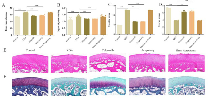
Figure 1. Acupotomy reduces knee swelling and articular cartilage degeneration in rabbits with knee osteoarthritis. (A) Knee circumference. (B) Relative knee swelling ratio. (C) Mankin histological score. (D) Moran histological score. (E) Representative hematoxylin and eosin-stained sections of articular cartilage from the control, KOA model, celecoxib, acupotomy, and sham acupotomy groups. (F) Representative Safranin O–Fast Green-stained sections from the five experimental groups. Histological scoring (Mankin and Moran) was performed using samples from all 10 rabbits per group (n = 10), whereas representative images for H&E and Safranin O–Fast Green staining were obtained from 3 randomly selected rabbits per group. Data are presented as the mean ± SEM. Statistical comparisons were performed using one-way analysis of variance followed by Tukey’s post hoc test. Horizontal bars indicate the groups compared. P < 0.05, P < 0.01, and P < 0.001. Scale bars, 100 µm. Abbreviations: KOA = knee osteoarthritis; SEM = standard error of the mean. Please click here to view a larger version of this figure.
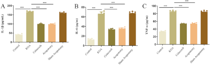
Figure 2. Acupotomy reduces inflammatory cytokine levels in rabbits with knee osteoarthritis. (A) Interleukin-1β concentration. (B) Interleukin-6 concentration. (C) Tumor necrosis factor-α concentration. Cytokine concentrations were measured by enzyme-linked immunosorbent assay in samples from six rabbits per group (n = 6). Data are presented as the mean ± SEM. Statistical comparisons were performed using one-way analysis of variance followed by Tukey’s post hoc test. Horizontal bars indicate the groups compared. P < 0.05, P < 0.01, and P < 0.001. Abbreviations: IL-1β = interleukin-1β; IL-6 = interleukin-6; KOA = knee osteoarthritis; SEM = standard error of the mean; TNF-α = tumor necrosis factor-α. Please click here to view a larger version of this figure.
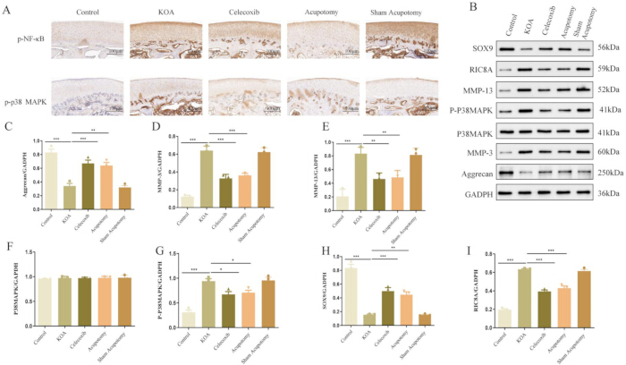
Figure 3. Acupotomy modulates inflammatory and cartilage metabolism-related proteins in rabbits with knee osteoarthritis. (A) Representative immunohistochemical images showing phosphorylated nuclear factor kappa B and phosphorylated p38 mitogen-activated protein kinase expression in articular cartilage from the control, KOA model, celecoxib, acupotomy, and sham acupotomy groups. Scale bars, 100 µm. (B) Representative Western blots showing SOX9, RIC8A, MMP-13, phosphorylated p38 MAPK, total p38 MAPK, MMP-3, aggrecan, and GAPDH expression. (C) Densitometric quantification of aggrecan normalized to GAPDH. (D) Quantification of MMP-13 normalized to GAPDH. (E) Quantification of MMP-3 normalized to GAPDH. (F) Quantification of total p38 MAPK normalized to GAPDH. (G) Quantification of phosphorylated p38 MAPK normalized to GAPDH. (H) Quantification of SOX9 normalized to GAPDH. (I) Quantification of RIC8A normalized to GAPDH. Western blot analyses were performed using samples from three rabbits per group (n = 3). Data are presented as the mean ± SEM. Statistical comparisons were performed using one-way analysis of variance followed by Tukey’s post hoc test. Horizontal bars indicate the groups compared. P < 0.05, P < 0.01, and P < 0.001. Abbreviations: GAPDH = glyceraldehyde-3-phosphate dehydrogenase; KOA = knee osteoarthritis; MAPK = mitogen-activated protein kinase; MMP-3 = matrix metalloproteinase-3; MMP-13 = matrix metalloproteinase-13; NF-κB = nuclear factor kappa B; p-NF-κB = phosphorylated nuclear factor kappa B; p-p38 MAPK = phosphorylated p38 MAPK; RIC8A = resistance to inhibitors of cholinesterase 8A; SEM = standard error of the mean; SOX9 = SRY-box transcription factor 9. Please click here to view a larger version of this figure.
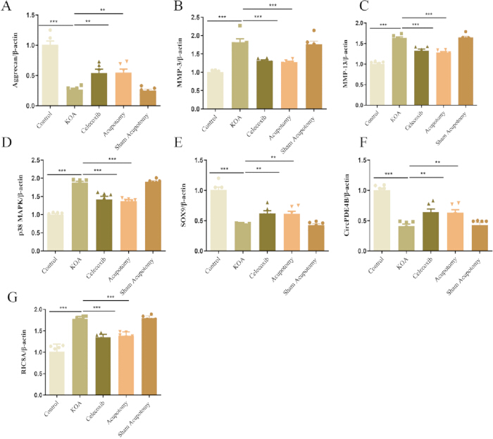
Figure 4. Acupotomy modulates cartilage metabolism-related transcripts and circPDE4B expression in rabbits with knee osteoarthritis. (A) Relative aggrecan expression. (B) Relative MMP-3 expression. (C) Relative MMP-13 expression. (D) Relative p38 MAPK expression. (E) Relative SOX9 expression. (F) Relative circPDE4B expression. (G) Relative RIC8A expression. Expression was measured by quantitative reverse transcription polymerase chain reaction and normalized to the reference gene. Analyses were performed using samples from six rabbits per group (n = 6). Data are presented as the mean ± SEM. Statistical comparisons were performed using one-way analysis of variance followed by Tukey’s post hoc test. Horizontal bars indicate the groups compared. P < 0.05, P < 0.01, and P < 0.001. Abbreviations: circPDE4B = circular RNA phosphodiesterase 4B; KOA = knee osteoarthritis; MAPK = mitogen-activated protein kinase; MMP-3 = matrix metalloproteinase-3; MMP-13 = matrix metalloproteinase-13; RIC8A = resistance to inhibitors of cholinesterase 8A; SEM = standard error of the mean; SOX9 = SRY-box transcription factor 9. Please click here to view a larger version of this figure.
| Gene | Amplicon Size | Forward primer | Reverse primer |
| (bp) | (5'→3') | (5'→3') | |
| β-actin | 113 | CAGCCCTCCTTCATCGGTAT | GACATGACGTTGTTGGCGTA |
| MMP-3 | 108 | TGTGAGTTGAAGTGGCTCAT | CCTGTTTGAACACCCGTAAC |
| MMP-13 | 119 | GGCTCCTAGGCAAGTACAAT | GTGTACGCCCATCAGGATAA |
| SOX9 | 85 | GGAGGAAGTCGGTGAAGAAT | GCCTTGAAGATGGCGTTG |
| Aggrecan | 70 | TTGATGAGTGCCTTTCAAGC | CATGTGAAAGAGTCGATGGC |
| RIC8A | 100 | ATAAACTTTCCAGAGAGGAGTTGC | TCGATGACAGAGTACTGGTTG |
| p38 MAPK | 103 | TGTTCAAGGTCTCCCACAAG | TGCAGCTCCCTTATGATCTG |
| CircPDE4B | 174 | GTCATTTTGCCGAACTCCC | CATTATCATCTGCCGTCACTG |
Table 1: Primer sequences used for quantitative reverse transcription polymerase chain reaction. The table lists the target genes, expected amplicon sizes, and forward and reverse primer sequences in the 5′ to 3′ direction. Abbreviations: bp = base pairs; circPDE4B = circular RNA phosphodiesterase 4B; MAPK = mitogen-activated protein kinase; MMP-3 = matrix metalloproteinase-3; MMP-13 = matrix metalloproteinase-13; RIC8A = resistance to inhibitors of cholinesterase 8A; SOX9 = SRY-box transcription factor 9.
Discussion
This study evaluated the therapeutic effects of acupotomy in a rabbit model of knee osteoarthritis (KOA) and identified an association between circPDE4B expression and the RIC8A/p38 mitogen-activated protein kinase (MAPK) signaling axis. Acupotomy improved joint swelling and histopathological changes and altered inflammatory and cartilage metabolism-related markers at both the transcript and protein levels.
The reduction in interleukin-1β, tumor necrosis factor-α, and interleukin-6 after acupotomy treatment was consistent with previously reported immunomodulatory effects of acupotomy21. These cytokines contribute to cartilage degeneration by promoting matrix-degrading enzymes and suppressing extracellular matrix synthesis22,24,25. The concurrent reductions in matrix metalloproteinase-3 (MMP-3) and matrix metalloproteinase-13 (MMP-13) at the transcript and protein levels further suggest that acupotomy suppresses catabolic activity associated with KOA.
A notable finding was the association between acupotomy-induced changes in circPDE4B expression and modulation of the RIC8A/p38 MAPK pathway. Abnormal activation of this pathway is associated with inflammation, oxidative stress, and extracellular matrix degradation. The reductions in RIC8A expression and p38 MAPK phosphorylation after acupotomy suggest that treatment may help restore signaling balance within this pathway. The coordinated changes in relative circPDE4B and RIC8A/p38 MAPK-related markers are consistent with the hypothesis that circPDE4B may function as a molecular scaffold, as previously proposed26,27. However, the present findings are correlative and do not establish that circPDE4B mediates the therapeutic effects of acupotomy. Loss- and gain-of-function studies are required to determine causality.
The potential association between acupotomy and circRNA regulation is an important aspect of this study. Mechanical stimulation has been shown to influence circRNA expression in several tissues28,29,30,31. However, direct evidence linking acupotomy-induced mechanical stimulation to the regulation of articular cartilage-specific circRNAs remains limited. The observed change in relative circPDE4B expression after treatment may reflect either a direct response to mechanical stimulation or an indirect response to reduced inflammation. Future studies combining controlled mechanical stimulation with chondrocyte-specific manipulation of circPDE4B would help distinguish between these possibilities.
Acupotomy and celecoxib produced similar trends across many measured outcomes despite their distinct modes of action. Several explanations may account for this convergence. First, both treatments may act on shared downstream pathways, including the RIC8A/p38 MAPK axis and nuclear factor kappa B signaling. Second, the observed similarities may reflect common features of cartilage protection rather than pathway-specific effects. Third, the selected markers may represent shared endpoints of inflammation and cartilage metabolism and may therefore be insufficient to distinguish between distinct upstream mechanisms. Similar efficacy does not imply identical mechanisms but suggests that both interventions may affect common components of the osteoarthritis pathological network26,32,33. Unbiased approaches, such as transcriptomic or proteomic profiling, may help define treatment-specific molecular signatures.
The comparable effects of acupotomy and celecoxib may also have clinical relevance. Acupotomy may offer a nonpharmacological treatment option that addresses periarticular mechanical factors while avoiding some adverse effects associated with long-term drug use. However, this interpretation should be approached with caution because the rabbit model does not fully capture the complexity of human KOA. Further translational and clinical studies are needed before these preclinical findings can support broader clinical application.
Several limitations should be acknowledged. First, the association between circPDE4B and the RIC8A/p38 MAPK axis was not tested using loss- or gain-of-function experiments. Second, the rabbit model and 4-week treatment period limit conclusions regarding human disease and long-term outcomes. Third, the molecular assays used relatively small subsets of animals, which may have limited the detection of subtle differences between groups. Fourth, the selected marker panel may not distinguish treatment-specific upstream mechanisms. Finally, the similar responses to acupotomy and celecoxib require further investigation to determine whether the two interventions act through convergent or independent pathways.
Conclusion
Acupotomy reduced joint swelling, inflammatory cytokine levels, cartilage degeneration, and catabolic marker expression in a rabbit model of KOA. These effects were associated with changes in relative circPDE4B expression and modulation of the RIC8A/p38 MAPK signaling axis. Because the present findings are correlative, further mechanistic studies are required to determine whether circPDE4B directly mediates the therapeutic effects of acupotomy.
Disclosures
The authors declare that they have no competing financial interests or personal relationships that could have influenced the work reported in this article.
Acknowledgements
This work was supported by the National Natural Science Foundation of China (grant no. 81774436), the Anhui Province Clinical Medical Research Transformation Project (grant no. 202427b10020136), the Scientific Research Project of the Anhui Provincial Department of Education (grant no. 2023AH050824), the Health Research Program of Anhui (grant no. AHWJ2023A30123), and the Anhui Association of Traditional Chinese Medicine Inheritance and Innovation Research Project (grant no. 2024ZYYXH153).
Materials
| Name | Company | Catalog Number | Comments |
|---|---|---|---|
| 3% hydrogen peroxide | Fuzhou Maixin Biotech | SP kit-A3 | Blocks endogenous peroxidase activity during immunohistochemistry. |
| 4% paraformaldehyde | Biosharp | BL539A | Fixes cartilage tissue before decalcification and histological processing. |
| Acetic acid | Merck | A6283 | Used to prepare 1% hydrochloric acid in 70% |
| Adhesive bandages | Zhejiang Renkang | RK-8802 | Used after acupotomy. |
| Aggrecan antibody | Proteintech | 13880-1-AP | Primary antibody used to detect aggrecan in cartilage protein extracts. |
| Anhydrous ethanol | Sinopharm Chemical Reagent | 10009228 | Used for tissue dehydration and section rehydration during histological processing. |
| BCA protein assay kit | Thermo Fisher Scientific | A55860 | Used to determine protein concentration. |
| Celecoxib | Biochempartner | BCP02156 | Reference pharmacological treatment administered by oral gavage. |
| Centrifuge | Anhui Jiawen Instruments | JW-3021HR | Used to clarify tissue lysates and homogenates by centrifugal separation. |
| Chloroform | Nanjing Reagent | 67-66-3 | Used for phase separation during total RNA extraction. |
| Citrate buffer, pH 6.0 | ZSGB-BIO | ZLI-9065 | Used for heat-induced antigen retrieval before immunohistochemistry. |
| Cryotubes | NEST Biotechnology | 607001 | Used for frozen sample storage |
| DAB substrate | Fuzhou Maixin Biotech | Kit-5230 | Chromogenic substrate used for visualization of HRP-labeled immunohistochemical signals. |
| DEPC-treated water | Generay Biotech | D1007 | RNase-free water used for RNA dissolution and molecular biology procedures. |
| Disposable acupotomy needle, 0.35 mm × 25 mm | Ma'anshan Bond Medical Equipment | GB3525R | Sterile needle-knife used for acupotomy and sham acupotomy procedures. |
| EDTA | biosharp | BL617A | Used to prepare the decalcification solution for cartilage-containing tissue. |
| Enhanced chemiluminescence substrate | Thermo 340958 | 340958 | Used to visualize Western blot bands. |
| Eosin solution | Zhuhai Beso | BA4022 | Counterstain used in hematoxylin and eosin staining. |
| Fast Green solution | Solarbio | G1371 | Counterstain used in Safranin O–Fast Green staining. |
| GAPDH antibody | Zsbio | TA-08 | Used as the Western blot loading control but not currently listed. |
| Gel imaging system | Shanghai Peiqing Technology | JS-1070P | Captures chemiluminescent Western blot images. |
| Glass microscope slides | CITOTEST | 188105 | Used to mount sections |
| Goat anti-mouse IgG, HRP-conjugated | ZSGB-BIO | ZB-2305 | HRP-conjugated secondary antibody for mouse primary antibodies. |
| Goat anti-rabbit IgG, HRP-conjugated | ZSGB-BIO | ZB-2301 | HRP-conjugated secondary antibody for rabbit primary antibodies. |
| Goat serum | ZSGB-BIO | ZLI-9056 | Blocks nonspecific antibody binding during immunohistochemistry. |
| GraphPad Prism, version 10 | GraphPad Software | Not applicable | Used for statistical analysis and graph preparation. |
| Hematoxylin solution | Zhuhai Beso | BA4041 | Nuclear stain used for hematoxylin and eosin staining and immunohistochemical counterstaining. |
| Homogenizer | Witeg | SGCR-5-553-722 | Homogenizes cartilage tissue for ELISA and molecular assays. |
| Hydrochloric acid | Merck | 7607 | Used to prepare 1% hydrochloric acid in 70% |
| Image Lab software | Bio-Rad | Not applicable | Used for densitometric analysis of Western blot bands. |
| Image-Pro Plus, version 6.0 | Media Cybernetics | Not applicable | Used for semiquantitative analysis of Safranin O staining intensity. |
| Isopropanol | Sinopharm Chemical Reagent | SCR40007960 | Used to precipitate RNA during total RNA extraction. |
| Light microscope | Olympus | CX41 | Used to examine and image stained cartilage sections. |
| Medical marker | edding | 497985 | Used to mark treatment points |
| Methanol | Shanghai Suyi | 20180427 | Used to activate the PVDF membrane |
| Microtome | Leica | RM2016 | Cuts paraffin-embedded tissue into 5 µm sections. |
| MMP-13 antibody | Bioss | bs-0575R | Primary antibody used to detect matrix metalloproteinase-13. |
| MMP-3 antibody | Bioss | bs-43017R | Primary antibody used to detect matrix metalloproteinase-3. |
| Neutral resin | Wuxi Jiangyuan | ZLI-9516 | Mounting medium used for permanent preparation of stained tissue sections. |
| New Zealand White rabbits (male,6 months old, weighing 2.0–2.5 kg) | Nanjing Pukou District Laifu Breeding Farm | SCXK (Su) 2024-0007 | Experimental animals used for knee osteoarthritis modeling and treatment assessment. |
| No. 23 scalpel blade | Caisson | SCX08-100PC | Used during sample collection. |
| Nonfat dry milk | Gene | 9999S | Used as the blocking reagent. |
| p38 MAPK antibody | Bioss | bs-0637R | Primary antibody used to detect total p38 MAPK. |
| Paraffin wax | Thermo | 8330 | Used for tissue embedding |
| PBS | ZSGB-BIO | ZLI-9062 | Used for tissue homogenization, washing, and reagent preparation. |
| Penicillin | Jilin Huamu Animal Health Products | Not applicable | Used postoperatively at 40,000 U/kg/day |
| Pentobarbital sodium | Beijing Chemical Reagents | 61222 | Used for anesthesia at 3%, 30 mg/kg. |
| phosphate-buffered saline | beyotime | ST448 | Used for tissue homogenization and washing during immunohistochemistry. |
| Physiological saline | servicevio | G4702 | Used to prepare the celecoxib suspension |
| PMSF | Beyotime | ST506 | Protease inhibitor added to lysis buffer during protein extraction. |
| p-NF-κB antibody | Santa | sc-136548 | Primary antibody used to detect p- NF-κB in immunohistochemical analysis. |
| p-p38 MAPK antibody | Bioss | bs-5476R | Primary antibody used to detect phosphorylated p38 MAPK. |
| PrimeScript RT Reagent Kit with gDNA Eraser | TaKaRa | RR047A | Used for genomic DNA removal and reverse transcription of RNA to cDNA. |
| PVDF membrane | Millipore | IPVH00010 | Membrane used for protein transfer during Western blotting. |
| Rabbit IL-1β ELISA kit | Jiyinmei | JYM0011Rb | Quantifies rabbit interleukin-1β in the analyzed sample. |
| Rabbit IL-6 ELISA kit | Jiyinmei | JYM0006Rb | Quantifies rabbit interleukin-6 in the analyzed sample. |
| Rabbit TNF-α ELISA kit | Jiyinmei | JYM0003Rb | Quantifies rabbit tumor necrosis factor-α in the analyzed sample. |
| Real-time PCR system | Thermo Fisher Scientific | PIKOREAL 96 | Performs fluorescence-based quantitative PCR amplification. |
| RIC8A antibody | Bioss | bs-11355R | Primary antibody used to detect RIC8A. |
| RIPA lysis buffer | Beyotime | P0013B | Extracts total protein from cartilage tissue. |
| Safranin O solution | Solarbio | G1371 | Stains cartilage proteoglycans in Safranin O–Fast Green histology. |
| SDS-PAGE reagents or precast gels | Solarbio | S8010 | Include gel percentage or product. |
| SOX9 antibody | Bioss | bs-4177R | Primary antibody used to detect SOX9. |
| Spectrophotometer | Nanjing Wuyi Technology | OD1000+ | Measures RNA concentration and purity. |
| Sterile gauze | Zhende | V505463 | Used for compression after needle withdrawal. |
| SYBR Green Master Mix | Novoprotein | E096-01B | Fluorescent reagent used for quantitative PCR. |
| Tris-buffered saline | Solarbio | T8060 | Used to prepare the washing and blocking buffer for Western blotting. |
| TRIzol reagent | Life Technologies | 15596018 | Phenol–guanidinium reagent used for total RNA extraction. |
| Tween 20 | Solarbio | T8220 | Nonionic detergent added to Tris-buffered saline and phosphate-buffered saline wash solutions. |
| Weigert iron hematoxylin solution | Merck | 1.15973 | Used in Safranin O–Fast Green staining. |
| Xylene | Sinopharm Chemical Reagent | 10023428 | Used for deparaffinization and tissue clearing. |
References
- Mobasheri A, Batt M. An update on the pathophysiology of osteoarthritis. Ann Phys Rehabil Med. 2016;59(5):333-339.
- Forestier R, et al. Prevalence of generalized osteoarthritis in a population with knee osteoarthritis. Joint Bone Spine. 2011;78(3):275-278.
- Motta F, Barone E, Sica A, Selmi C. Inflammaging and osteoarthritis. Clin Rev Allergy Immunol. 2023;64(2):222-238.
- Ghouri A, Conaghan PG. Prospects for therapies in osteoarthritis. Calcif Tissue Int. 2021;109(3):339-350.
- Roseti L, et al. Articular cartilage regeneration in osteoarthritis. Cells. 2019;8(11):1305. https://doi.org/10.3390/cells8111305
- Fujii Y, et al. Cartilage homeostasis and osteoarthritis. Int J Mol Sci. 2022;23(12):6330. https://doi.org/10.3390/ijms23126330
- Kreutzinger V, et al. Limited effects of non-steroidal anti-inflammatory drugs (NSAIDs) on imaging outcomes in osteoarthritis: observational data from the osteoarthritis initiative (OAI). BMC Musculoskelet Disord. 2025;26. https://doi.org/10.1186/s12891-025-08245-0
- Migliore A, et al. Low dose NSAIDs and sysadoas in the management of knee osteoarthritis. Aging Clin Exp Res. 2025;37(1):121. https://doi.org/10.1007/s40520-025-03015-6
- Fu M, et al. Expression profile of long noncoding RNAs in cartilage from knee osteoarthritis patients. Osteoarthritis Cartilage. 2015;23(3):423-432.
- Jiang SD, et al. Long noncoding RNAs in osteoarthritis. Joint Bone Spine. 2017;84(5):553-556.
- Xing D, et al. Identification of long noncoding RNA associated with osteoarthritis in humans. Orthop Surg. 2014;6(4):288-293.
- Wu W, Zou J, Yang Y. Studies on the role of circRNAs in osteoarthritis. BioMed Res Int. 2021;2021:6672322. https://doi.org/10.1155/2021/6672322
- Liu X, et al. Identification and comprehensive analysis of circRNA–miRNA–mRNA regulatory networks in osteoarthritis. Front Immunol. 2022;13:1018820. https://doi.org/10.3389/fimmu.2022.1018820
- Shen S, et al. circPDE4B prevents articular cartilage degeneration and promotes repair by acting as a scaffold for RIC8A and MID1. Ann Rheum Dis. 2021;80(9):1209-1219.
- Cao M, et al. A peroxiredoxin-P38 MAPK scaffold increases MAPK activity by MAP3K-independent mechanisms. Mol Cell. 2023;83(17):3140-3154.
- Lu M, et al. Promotion and mechanism of acupotomy on chondrocyte autophagy in knee osteoarthritis rabbits. Chin J Integr Med. 2024;30(9):809-817.
- Chen C, et al. Clinical effects of ultrasound-guided acupotomy in knee osteoarthritis treatment. J Vis Exp. 2024;(208):e66587. https://doi.org/10.3791/66587
- Wang Y, et al. Effect of acupotomy modulation of NLRP3/Caspase-1/GSDMD pathway on chondrocyte pyroptosis in knee osteoarthritis. J Pain Res. 2025;18:4935-4945.
- Zhang J, et al. Whole transcriptome mapping identifies an immune- and metabolism-related non-coding RNA landscape remodeled by mechanical stress in IL-1β-induced rat OA-like chondrocytes. Front Genet. 2022;13:843187. https://doi.org/10.3389/fgene.2022.843187
- Liu Q, et al. Emerging roles of circRNA related to the mechanical stress in human cartilage degradation of osteoarthritis. Mol Ther Nucleic Acids. 2017;7:223-230.
- Guo Y, et al. Acupotomy improves synovial hypoxia, synovitis and angiogenesis in KOA rabbits. J Pain Res. 2023;16:749-760.
- Ma Y, et al. Acupotomy ameliorates KOA related chondrocyte premature senescence through YAP/FOXD1 pathway. J Pain Res. 2025;18:2011-2023.
- Wang Z,et al. Electroacupuncture ameliorates cartilage damage in a rat model of knee osteoarthritis and regulates expression of miRNAs and the TLR4/NF-κB pathway, Acupuncture in Medicine. 2025, 43(2025), 164-173.
- Feng S, Cong H, Ji L. Salvianolic acid A exhibits anti-inflammatory and antiarthritic effects via inhibiting NF-κB and p38/MAPK pathways. Drug Des Devel Ther. 2020;14:1771-1778.
- Abdallah AM, et al. Pyrazole derivatives ameliorate synovial inflammation in collagen-induced arthritis mice model via targeting p38 MAPK and COX-2. Naunyn Schmiedebergs Arch Pharmacol. 2025;398(1):819-832.
- Alaaeldin E, et al. Topical nano-vesicular spanlastics of celecoxib: enhanced anti-inflammatory effect and down-regulation of TNF-α, NF-кB and COX-2 in complete Freund's adjuvant-induced arthritis model in rats. Int J Nanomedicine. 2021;16:133-145.
- Pamudurti NR, et al. Translation of CircRNAs. Mol Cell. 2017;66(1):9-21.
- Wang Z, et al. Electroacupuncture ameliorates cartilage damage in a rat model of knee osteoarthritis and regulates expression of miRNAs and the TLR4/NF-κB pathway. Acupunct Med. 2025;43(3):164-173.
- Shen J, et al. Circular RNA sequencing reveals the molecular mechanism of the effects of acupuncture and moxibustion on endometrial receptivity in patients undergoing infertility treatment. Mol Med Rep. 2019;20(2):1623-1633. https://doi.org/10.3892/mmr.2019.10386
- Wang Y, et al. Electroacupuncture alleviates perioperative hypothalamus-pituitary-adrenal axis dysfunction via circRNA-miRNA-mRNA networks. Front Mol Neurosci. 2023;16:1152861. https://doi.org/10.3389/fnmol.2023.1152861
- Xiao X, et al. Transcription profiling of a revealed the potential molecular mechanism of Governor Vessel electroacupuncture for spinal cord injury in rats. Neurospine. 2022;19(3):757-769.
- Xu C, Gu K, Yasen Y, Hou Y. Efficacy and safety of celecoxib therapy in osteoarthritis: a meta-analysis. Medicine (Baltimore). 2016;95(20):e3585. https://doi.org/10.1097/MD.0000000000003585
- 33. Jiang D, et al. Efficacy of intra-articular injection of celecoxib in a rabbit model of osteoarthritis. Int J Mol Sci. 2010;11(10):4106-4113.