This protocol provides a step-by-step guide for performing transforaminal full-endoscopic ventral facetectomy under local anesthesia for lumbar lateral recess stenosis, particularly in patients who may not tolerate general anesthesia.
Method Article
Transforaminal Full-Endoscopic Ventral Facetectomy for Lumbar Lateral Recess Spinal Stenosis under Local Anesthesia
In This Article
Summary
Abstract
This article describes a reproducible protocol for performing transforaminal full-endoscopic ventral facetectomy (TF-FEVF) under local anesthesia for lumbar lateral recess stenosis. Lateral recess spinal stenosis (LRSS) in the lumbar spine is generally treated with posterior decompression surgery under general anesthesia. The invasiveness of posterior decompression has decreased with the development of endoscopic techniques such as interlaminar full-endoscopic spine surgery and unilateral biportal endoscopy. However, these techniques still require general anesthesia. With population aging in developed countries, older patients with LRSS are becoming increasingly common. In older patients with severe comorbidities, general anesthesia may pose a substantial risk. LRSS can also be decompressed by transforaminal full-endoscopic spine surgery (TF-FESS), such as discectomy or foraminotomy, and may benefit elderly patients in poor general condition. In 2017, we developed TF-FEVF as a decompression procedure that can be performed under local anesthesia. This article outlines the surgical indications, operative workflow, and key anatomical landmarks of TF-FEVF. It also describes, step by step, the technique used to achieve effective local anesthesia. Under fluoroscopic guidance, 1% lidocaine is infiltrated in the subcutaneous tissue (3 mL), fascia/muscle (7 mL), facet joint (8 mL), and caudal endplate (2 mL) for local anesthesia. The working cannula is inserted and attached to the lateral wall of the superior articular process (SAP). The ventral and cranial portions of the SAP are extensively resected to widen Kambin’s triangle while preserving the dorsal facet complex whenever possible. Next, the ventral surface of the inferior articular process is drilled, allowing exposure and elevation of the ligamentum flavum (LF). Resection of the elevated LF enables adequate decompression of the traversing nerve root. Finally, adequate decompression of the traversing nerve root is confirmed endoscopically. At our institution, more than 200 patients have undergone TF-FEVF, including 10 patients older than 90 years without complications.
Introduction
Lumbar spinal stenosis is a common degenerative condition that significantly impairs quality of life, particularly in elderly populations. One of its subtypes is lateral recess stenosis, which results in compression of the traversing nerve root and frequently causes severe radicular pain, neurological deficits, and functional limitations1,2. Conventional surgical management typically involves posterior decompression under general anesthesia, which may require extensive muscle dissection and partial facetectomy3. Although advances in minimally invasive spine surgery have reduced surgical morbidity, many of the currently available techniques still rely on general anesthesia, which may pose substantial risks in elderly patients with multiple comorbidities.
With the rapid aging of populations in developed countries, the number of patients with lumbar spinal stenosis who are poor candidates for general anesthesia is increasing. Therefore, surgical techniques that can be safely performed under local anesthesia are becoming increasingly important. Transforaminal full-endoscopic spine surgery (TF-FESS) provides a minimally invasive corridor to the lumbar spine through Kambin’s triangle4and can be performed under local anesthesia with conscious sedation5,6,7. While transforaminal approaches have been widely used to treat disc herniation and foraminal stenosis, their application for lateral recess stenosis remains technically challenging because adequate decompression of the traversing nerve root often requires removal of ventral bony structures, including portions of the facet joint.
To address this limitation, we have developed a novel surgical technique called transforaminal full-endoscopic ventral facetectomy (TF-FEVF), which enables adequate decompression of the traversing nerve root through a transforaminal approach under local anesthesia. This technique involves strategic resection of the superior articular process (SAP) and ventral facet components to widen the surgical corridor and allow direct visualization and decompression of the affected neural structures while preserving posterior musculoligamentous integrity. Unlike conventional transforaminal endoscopic foraminoplasty, which primarily targets foraminal pathology and the exiting nerve root, TF-FEVF is designed to decompress the traversing nerve root in lateral recess stenosis through a transforaminal corridor. This technique may be particularly useful in elderly patients or patients with significant comorbidities who are poor candidates for general anesthesia, as well as in cases where the main compressive pathology can be addressed through the transforaminal route.
The objective of this protocol is to describe a reproducible transforaminal approach for performing TF-FEVF under local anesthesia in patients with lumbar lateral recess stenosis. The intended endpoint is adequate decompression of the traversing nerve root, as confirmed by intraoperative endoscopic visualization and postoperative radiographic assessment.
Protocol
The research was approved by the Institutional Review Board of Tokushima University Hospital (approval number 3642). Written informed consent was obtained from the patient for participation in this study and for publication of the clinical data and images.
1. Preoperative preparation
- Patient eligibility
- Confirm that the patient has lumbar lateral recess stenosis causing radicular symptoms that correspond to the MRI and CT findings.
- Confirm that the main compressive pathology can be addressed through the transforaminal corridor.
- Confirm that decompression under local anesthesia is desirable because of advanced age, comorbidities, or the need to avoid general anesthesia.
- Exclude patients with clear segmental instability, dynamic instability on flexion-extension radiographs, or degenerative spondylolisthesis of Meyerding grade II or greater, for whom fusion surgery is generally preferred.
- Trajectory planning and patient preparation
- Before surgery, determine the distance from the midline to the endoscope entry point and the insertion angle on lumbar computed tomography (CT) and magnetic resonance imaging (MRI).
- Identify the target point (point A) on a coronal CT image along a line connecting the medial borders of the superior and inferior pedicles (corresponding to the level of the superior endplate of the caudal vertebra on the sagittal view).
- Draw a tangential line from point A toward the SAP. The intersection of this line with the skin surface is defined as the skin entry point (point B).
- Measure the distance from the midline to the skin entry point (point B), which is the intersection of the line tangential from point A with the skin surface (Figure 1).
NOTE: Precise trajectory planning is essential to ensure safe docking on the bony surface and to avoid neural injury. On the lateral view, the target point is set along the extension line of the superior endplate of the caudal vertebra. The planned trajectory is aligned as closely as possible with the target disc plane, while the rostrocaudal and dorsoventral angles are adjusted according to the patient’s anatomy to maintain a safe transforaminal corridor. Because these angles vary with individual anatomy, including lumbar lordosis, disc inclination, iliac crest height, and target level, the planned pathway should be carefully confirmed on both axial and sagittal images before the procedure.
- Perform discography when discogenic low back pain or concomitant disc pathology is suspected, such as when the patient reports low back pain during lumbar flexion or when preoperative MRI suggests a discogenic source of pain, or when intradiscal trajectory confirmation is required.
- Before surgery, determine the distance from the midline to the endoscope entry point and the insertion angle on lumbar computed tomography (CT) and magnetic resonance imaging (MRI).
- Sedation protocol
- When the patient is positioned prone on the operating table, administer intravenous hydroxyzine hydrochloride 12.5 mg and pentazocine 7.5 mg for anxiolysis and analgesia. Continuously monitor the patient’s level of consciousness, blood pressure, heart rate, oxygen saturation by pulse oximetry, and electrocardiography throughout the procedure.
NOTE: Antihypertensive agents (e.g., nicardipine) and atropine must be readily available in the operating room for immediate management of potential hemodynamic changes, such as hypertension or vagally mediated bradycardia. - After skin disinfection and sterile draping, administer the remaining hydroxyzine hydrochloride 12.5 mg and pentazocine 7.5 mg intravenously immediately before starting the surgical procedure.
- When the patient is positioned prone on the operating table, administer intravenous hydroxyzine hydrochloride 12.5 mg and pentazocine 7.5 mg for anxiolysis and analgesia. Continuously monitor the patient’s level of consciousness, blood pressure, heart rate, oxygen saturation by pulse oximetry, and electrocardiography throughout the procedure.
- Operating room setup
- Position a C-arm fluoroscope to obtain clear anteroposterior (AP) and lateral images of the target lumbar segment throughout the procedure.
CAUTION: Use appropriate radiation protection during fluoroscopy and follow institutional radiation safety practices. - Adjust the X-ray beam so that it is parallel to the target disc space, ensuring that the vertebral endplates and the SAP are clearly visualized.
- Use pulsed fluoroscopy whenever possible and minimize fluoroscopy time by obtaining images only when confirming the needle, dilator, or cannula position.
- Collimate the fluoroscopic field to the target segment and use standard radiation protection, including lead aprons, thyroid shields, and protective eyewear.
- Prepare the endoscopic tower, including the light source, camera system, monitor, irrigation pump, and radiofrequency coagulation system. Confirm the correct functioning of all equipment before starting the procedure. Arrange a sterile surgical table with all required instruments and disposable materials.
NOTE: The fluoroscope should allow rapid switching between AP and lateral views without disturbing the sterile field.
- Position a C-arm fluoroscope to obtain clear anteroposterior (AP) and lateral images of the target lumbar segment throughout the procedure.
2. Surgical technique
- Patient positioning and initial marking
- Position the patient prone on a radiolucent operating table with appropriate thoracic and pelvic support cushions to permit free abdominal movement and reduce epidural venous pressure.
- Adjust the operating table to achieve appropriate hip and knee flexion to optimize the intervertebral space and facilitate the transforaminal approach, particularly in patients with a high iliac crest.
- Using C-arm fluoroscopy, obtain true AP and lateral views of the target lumbar segment. Ensure that the vertebral endplates are parallel on the lateral view and that the spinous processes are centered on the AP view to allow accurate localization.
NOTE: Accurate fluoroscopic alignment is essential for planning a safe needle trajectory and placement of the cannula. - Determine the skin entry site for the transforaminal approach using both preoperative imaging and intraoperative fluoroscopic confirmation, which in most cases is located several centimeters lateral to the midline, corresponding to the level of the intended surgical segment and oriented toward the SAP.
- Under fluoroscopic guidance, outline the iliac crest on the skin surface to establish an additional external landmark for orientation during the procedure (Figure 2).
NOTE: The optimal entry location may vary depending on individual anatomical factors, including the height of the iliac crest, spinal alignment, and level of pathology. In patients with a history of lumbar surgery, special attention should be paid to existing implants and postoperative scar tissue. Nevertheless, fluoroscopic identification of osseous landmarks remains the most reliable method for accurate targeting.
- Local anesthesia and skin incision
CAUTION: Handle needles and scalpel blades using standard sharps precautions and dispose of them immediately in an approved sharps container.- At the predetermined entry site, administer superficial local anesthesia using a 23-gauge needle. Infiltrate approximately 3–5 mL of 1% lidocaine into the dermal and subcutaneous layers to achieve adequate analgesia at the skin surface.
- Under intermittent fluoroscopic guidance using both AP and lateral views, advance the same 23-gauge needle along the planned trajectory toward the SAP. Gradually infiltrate approximately 7 mL of 1% lidocaine into the paraspinal musculature and surrounding soft tissues along the needle pathway to achieve adequate deep anesthesia.
- Before local anesthetic infiltration, calculate and record the maximum allowable dose and volume of 1% lidocaine based on the patient’s body weight, generally not exceeding 4.5 mg/kg.
CAUTION: Calculate the maximum allowable lidocaine dose before injection, aspirate before each injection, and monitor for signs of local anesthetic systemic toxicity. - Switch to an 18-gauge spinal needle to facilitate precise deep anesthetic infiltration. Under intermittent fluoroscopic guidance, advance the needle along the planned trajectory toward the target anatomical landmarks.
- Administer approximately 2 mL of 1% lidocaine into the facet joint capsule, followed by 2 mL at the tip of the SAP, 2 mL at the midportion of the SAP, and 2 mL at the base of the SAP.
- Infiltrate approximately 2 mL of 1% lidocaine onto the surface of the inferior vertebral endplate when the needle tip can be safely positioned on the bony surface under fluoroscopic confirmation without eliciting radicular pain.
- Advance the needle tip to the surface of the annulus fibrosus, confirm the position fluoroscopically, and inject approximately 2 mL of 1% lidocaine onto the annular surface.
- Carefully advance the needle into the intervertebral disc space and administer an additional approximately 2 mL of 1% lidocaine. To avoid local anesthetic systemic toxicity, inject slowly with repeated aspiration, avoid intravascular injections, and continuously monitor the patient’s level of consciousness, vital signs, and neurological symptoms throughout the procedure.
CAUTION: Confirm that the patient has no relevant allergy before using contrast medium or indigo carmine and avoid excessive intradiscal injection. - Inject approximately 1–2 mL of a mixture of indigo carmine and contrast medium into the intervertebral disc space. This step is performed routinely because staining of the disc material provides an intraoperative landmark if endoscopic orientation becomes difficult. Avoid excessive injections to prevent increased intradiscal pressure and patient discomfort.
- At the anesthetized entry site, make an approximately 8 mm skin incision using a No. 11 or No. 15 scalpel blade. Extend the incision through the fascia when necessary to allow uninterrupted insertion of instrumentation.
- Placement of the working cannula
- Through the approximately 8 mm skin incision, routinely insert a guidewire along the previously predetermined and anesthetized trajectory under fluoroscopic guidance. Then, introduce the first blunt-tipped dilator over the guidewire.
- Under intermittent fluoroscopic guidance using both AP and lateral views, advance the dilators sequentially with gentle rotational movements toward the lateral surface of the SAP.
NOTE: Maintain continuous contact with the bony surface while advancing the dilators to minimize the risk of neural injury. - Confirm that the tip of each dilator is positioned firmly on the bony surface of the SAP, preferably near the junction between the SAP and the pedicle. Ensure that the final dilator is securely seated on the bone to establish a safe and stable working corridor away from neural elements.
NOTE: Loss of bony contact during advancement of a dilator may increase the risk of nerve irritation or injury. - Advance a working cannula (working channel, Ø8.0 mm, length 165 mm) over the final dilator until it is firmly seated against the lateral surface of the SAP. In our practice, an elevator-type oblique cannula is generally used for L4/L5 or more cranial levels, whereas a duckbill-type cannula is used for L5/S1. The beveled tip is oriented toward the SAP to maintain stable bony docking and prevent unintended advancement toward the neural structures.
NOTE: Stable docking on the bony surface is essential before inserting the endoscope to prevent unintended movement toward neural structures. - Confirm the final position of the working cannula on both AP and lateral fluoroscopic views. On the AP view, the cannula should be aligned with the lateral margin of the SAP, with the tip projected toward the lateral portion of the foramen. On the lateral view, ensure that the cannula tip rests over the posterior boundary of the foramen at the level of the SAP.
- Avoid advancing the cannula excessively into the foramen or spinal canal at this stage.
NOTE: Excessive advancement of the cannula may cause nerve root irritation. Reconfirm fluoroscopic orientation if the patient reports radicular pain during positioning. - Carefully withdraw the final dilator while maintaining the position of the working cannula, which serves as the operative channel for subsequent insertion of the endoscope and manipulation of instruments.
- Insertion and initial orientation of the endoscope
- Insert the endoscope (25° angled optic) through the working cannula once stable docking has been confirmed.
- Initiate continuous saline irrigation with simultaneous removal using the suction channel of the endoscope. Set the irrigation pump pressure to approximately 60 mmHg and adjust the inflow and outflow to maintain a clear operative field while avoiding excessive epidural pressure.
NOTE: Excessive irrigation pressure may increase epidural pressure, leading to patient discomfort or neurological symptoms. Maintain communication with the patient throughout the procedure.
CAUTION: Avoid excessive irrigation pressure and continuously monitor the patient for discomfort or neurological symptoms during pressurized irrigation. - Perform an initial endoscopic inspection to establish orientation within the operative field. The lateral surface of the SAP, where the working cannula is positioned, serves as the primary anatomical landmark.
- Carefully clear the surface of the SAP by removing overlying soft tissues, residual muscle fibers, and fatty tissue using endoscopic forceps or a bipolar radiofrequency probe. In our practice, a bipolar radiofrequency probe is typically used at approximately 30 W to coagulate small vessels and shrink soft tissue around the facet surface.
CAUTION: Use radiofrequency devices under direct endoscopic visualization and avoid activation near neural structures unless the probe tip is clearly identified.
NOTE: Ideally, the bony surface of the facet joint should be visible immediately after insertion of the endoscope. If soft tissue obscures the bony landmark, it should be cleared promptly with endoscopic forceps and the radiofrequency probe, and SAP resection should generally be initiated within approximately 5 min after endoscope insertion. Maintain continuous visualization of the cannula tip during removal of soft tissue to avoid inadvertent injury to adjacent neural structures. - When the lateral surface of the SAP is adequately exposed, examine the surrounding bony anatomy to identify key orientation points, including the junction with the pedicle caudally and the trajectory toward the intervertebral foramen cranially and ventrally. Establishing this anatomical orientation provides a roadmap for the subsequent foraminoplasty.
NOTE: Accurate identification of bony landmarks before drilling helps prevent disorientation and reduces the risk of neural injury.
- Foraminoplasty: resection of the SAP
- When the lateral surface of the SAP is clearly visualized, initiate foraminoplasty using a high-speed endoscopic drill fitted with a 3.5-mm coarse diamond burr. The same non-articulated burr is used for SAP resection, partial IAP resection, and LF detachment; an articulated burr is not used.
- Begin bone resection at the caudal base of the SAP, near its junction with the pedicle. Starting from this confirmed bony landmark allows controlled bone removal and maintenance of a safe distance from the exiting nerve root.
NOTE: Maintain constant visualization of the drill tip and surrounding structures during bone removal to avoid injury to neural elements. - Progressively remove the ventral and cranial portions of the SAP. Drill in a caudal-to-cranial direction, gradually thinning the SAP from its base toward the tip (Figure 3). The objective is to unroof the foramen dorsally and laterally, enabling exposure of the underlying LF and facilitating decompression of the exiting nerve root.
NOTE: Bone removal should proceed gradually while maintaining orientation to adjacent neural structures. - Continue bone removal until approximately 80%–90% of the involved portion of the SAP has been resected or until adequate exposure of the foraminal ligamentum flavum (LF) and the shoulder of the exiting nerve root is achieved.
NOTE: The goal is not to completely resect the SAP. Rather, the ventral and cranial portions of the SAP are resected sufficiently to create a safe working corridor and expose the LF and traversing nerve root. The most dorsal portion of the facet complex should be preserved whenever possible to maintain segmental stability. The extent of resection should be guided by preoperative CT, intraoperative endoscopic findings, and confirmation of adequate neural decompression. - Following adequate resection of the SAP, identify whether the hypertrophied LF remains covered by the inferior articular process (IAP) of the cranial vertebra. Perform partial resection of the ventral IAP to widen the working corridor, expose the LF adequately, and allow direct visualization of the neural structures before proceeding with LF detachment.
NOTE: Bone should be removed incrementally to avoid excessive facet resection and potential instability.
- Exposure of the LF by IAP resection
- Identify the IAP of the cranial vertebra, located ventrally and cranially within the operative field.
- Using a high-speed endoscopic drill, perform partial resection of the ventral portion of the IAP with careful bone removal.
NOTE: Excessive resection of the facet joint should be avoided to preserve segmental stability. - Continue partial bone removal until the LF is clearly and completely visualized within the foramen (Figure 4).
NOTE: Adequate exposure of the LF is essential before attempting detachment to avoid unintended neural injury.
- Detachment technique: releasing the LF from the SAP
- When the LF is fully exposed, identify its caudal attachment to the remaining ventral edge of the SAP.
- Perform the detachment technique using the cranial portion of the same 3.5-mm coarse diamond burr. Apply the burr to the bony margin of the residual SAP and use controlled rotational movements with a “pulling” or “shaving” motion to undercut the attachment and gradually detach the LF from its deep bony insertion point (Figure 5).
- Continue the undercutting maneuver until the hypertrophied LF becomes completely mobile and appears to be floating, indicating full detachment from the SAP.
NOTE: Complete separation is essential for subsequent safe removal. Pulsation of the detached LF may be observed at this stage, reflecting pulsation in cerebrospinal fluid. LF detachment is not routinely extended to the midline; rather, the extent of detachment is determined by the location of lateral recess stenosis and the degree of traversing nerve root compression. The goal is to achieve adequate decompression of the traversing nerve root while avoiding unnecessary medial dissection or excessive facet resection. Therefore, preservation of part of the SAP on postoperative CT is intentional and consistent with the surgical concept. During undercutting, the burr should always remain in contact with bone to avoid unintended injury to neural structures.
- Removal of the LF and final decompression
- When the LF has been completely detached from the SAP, proceed with its removal.
- Grasp the detached, free-floating LF using a Kerrison punch (e.g., 3.5-mm) or endoscopic forceps and remove it carefully.
NOTE: Remove the LF under direct visualization to avoid unintended contact with the nerve root or dura. Following removal of the LF, the lateral margin of the thecal sac should become clearly visible (Figure 6). - Carefully inspect to ensure that there is no residual compression by bony fragments or ligamentous tissue.
- Confirm adequate decompression by observing free mobilization of the traversing nerve root, visible pulsation of the nerve root and thecal sac, and the absence of residual compression by bone, ligament, or disc material.
- Use a flexible probe or blunt dissector to gently assess the course of the nerve root and confirm that there is no residual stenosis or tethering.
NOTE: Excessive manipulation of the nerve root should be avoided to prevent postoperative dysesthesia.
- Final decompression check, hemostasis, and closure
- After removal of all compressive factors, including hypertrophied bone, LF, and any associated disc material, perform a comprehensive inspection of the exiting nerve root and the lateral margin of the thecal sac throughout the accessible foraminal region.
- Achieve meticulous hemostasis under continuous irrigation. Minor bleeding from epidural vessels or soft tissues can be controlled using a bipolar radiofrequency probe, typically at approximately 30 W. In our practice, a small amount of a flowable hemostatic agent (e.g., Floseal Hemostatic Matrix) is routinely applied to the decompressed area under endoscopic visualization after hemostasis to reduce the risk of postoperative epidural hematoma.
CAUTION: Use only a small amount of hemostatic agent and remove excess material to avoid postoperative neural irritation or compression. - Perform a final endoscopic survey of the decompressed area to confirm that no residual bone fragments, disc material, or hemostatic agent remain that could cause recurrent neural compression.
NOTE: Excessive use of hemostatic agents must be avoided because retained material may cause postoperative neural irritation. - A small surgical drain is routinely inserted through the working cannula into the decompressed area under endoscopic guidance, as the thecal sac is exposed after decompression. The drain is usually removed on the first postoperative day.
- Withdraw the endoscope and working cannula carefully while maintaining the position of the drain.
- Close the approximately 8 mm skin incision with one or two dermal sutures.
- Apply a sterile dressing over the incision site.
NOTE: The operative time varies depending on the severity of stenosis and anatomical complexity. In a typical case, the time from local anesthesia to endoscope placement is within 10 min, SAP resection requires approximately 15–20 min, IAP resection requires approximately 20–25 min, LF detachment and removal require approximately 15–20 min, and final inspection, hemostasis, drain placement, and skin closure require approximately 10 min. The total operative time is generally approximately 70–90 min.
3. Postoperative care
- After completion of the procedure and application of a sterile dressing, assist the patient in transferring from the operating table to a stretcher and return them directly to the hospital room.
NOTE: Transfer usually requires minimal assistance because the procedure is performed under local anesthesia, with the patient remaining fully conscious. - When the patient is in the ward, apply a soft lumbar corset. Encourage early standing and ambulation as tolerated, with assistance if necessary.
NOTE: Many patients experience immediate postoperative relief, with marked improvement or complete resolution of leg pain. - Postoperative pain at the incision site is usually mild and can be managed with an oral non-steroidal anti-inflammatory drug or other oral analgesics as required.
- Initiate a postoperative rehabilitation program, typically consisting of approximately 2 weeks of supervised Pilates-based exercises focusing on gentle trunk stabilization, pelvic mobility, breathing control, and progressive ambulation.
NOTE: Patients can be discharged after this initial rehabilitation period provided that they are comfortable, neurologically stable, and able to ambulate safely. - Provide discharge instructions that include guidance on wound care, continuation of prescribed exercises, and a gradual return to daily activities. Advise patients to avoid heavy lifting or strenuous activities for an appropriate period.
- Arrange outpatient follow-up visits to assess clinical outcomes, neurological status, and symptom recurrence, typically at 1 month, 6 months, and 1 year after surgery. Thereafter, provide annual follow-up based on the patient’s symptoms and preferences.
NOTE: Dispose of sharps, contaminated materials, and any unused medications or contrast media in accordance with institutional biosafety and hazardous waste regulations.
Results
A 78-year-old woman presented with pain in the right lower extremity consistent with radicular symptoms. She had no motor weakness or sensory deficits on neurological examination.
MRI demonstrated lateral recess-type lumbar spinal stenosis at the L4/L5 level with compression of the corresponding nerve root (Figure 7). The patient underwent TF-FEVF at L4/L5 under local anesthesia according to the protocol described. The procedure included stepwise foraminoplasty with resection of the hypertrophied SAP, followed by exposure and detachment of the LF using the detachment technique. Adequate decompression was achieved by removal of the compressive bony and ligamentous structures. Final endoscopic inspection demonstrated clear visualization and free mobilization of the affected nerve root with visible pulsation, indicating successful decompression. Postoperative CT confirmed adequate bony decompression at the L4/L5 level compared with the preoperative findings (Figure 8). The postoperative course was uneventful, with no new neurological deficits.
The patient's radicular pain improved markedly after surgery, and the clinical outcome was graded as excellent according to the modified Macnab criteria.
Figure 7 and Figure 8 illustrate the radiographic changes associated with successful decompression. Preoperative MRI demonstrated lateral recess stenosis with compression of the traversing nerve root, whereas postoperative CT demonstrated sufficient bony decompression of the lateral recess following ventral facetectomy.
Successful outcome after TF-FEVF should be assessed using a combination of radiographic findings, neurological examination, symptom improvement, and functional outcome measures. Conversely, persistent compression on postoperative imaging, persistent radicular or neurological symptoms, inadequate nerve root pulsation or mobility on endoscopic assessment, or postoperative segmental instability may indicate a suboptimal TF-FEVF outcome. In addition to this representative case, our institutional experience includes 205 patients who underwent TF-FEVF, with a mean follow-up period of 20.2 ± 17.0 months. No postoperative hematoma or surgical site infection was observed. Only one patient required additional surgery, which consisted of subsequent fusion for postoperative segmental instability. According to the modified Macnab criteria, 185 of the 205 patients (90.2%) achieved excellent or good outcomes at the final follow-up. Because VAS and ODI data were incomplete in some cases, modified Macnab outcomes were used as the summarized clinical outcome measure.
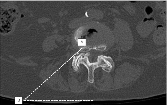
Figure 1: Preoperative planning of the skin entry point and trajectory. (A–B) Axial computed tomography image demonstrating planning for the transforaminal approach. The target point (A) is determined based on anatomical landmarks within the spinal canal, and the skin entry point (B) is defined by extending the planned trajectory line from the target point to the skin surface. The dotted line represents the planned trajectory of the endoscopic portal. Please click here to view a larger version of this figure.
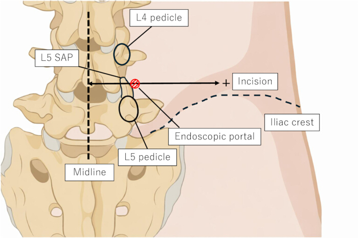
Figure 2: Schematic illustration demonstrating the anatomical landmarks and planned trajectory for the endoscopic portal. The midline, pedicles, SAP, iliac crest, and skin incision site guide safe placement of the working portal toward the facet region. SAP, superior articular process. Please click here to view a larger version of this figure.
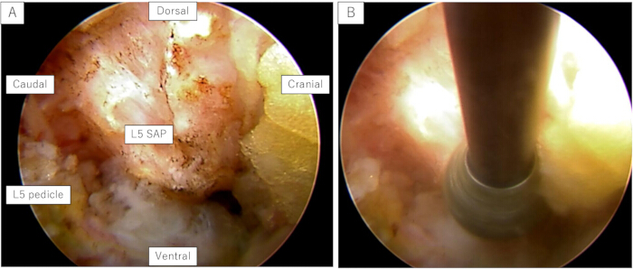
Figure 3: Initial endoscopic orientation and foraminoplasty. (A) Endoscopic view after docking on the SAP demonstrating key anatomical landmarks and orientation. (B) Resection of the SAP using a high-speed drill during foraminoplasty. SAP, superior articular process. Please click here to view a larger version of this figure.
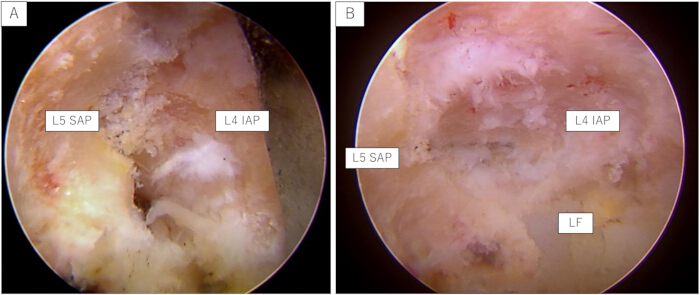
Figure 4: Progressive bone resection and exposure of the LF. (A) Endoscopic view during drilling of the SAP, demonstrating exposure of the IAP after partial resection of the SAP. (B) Further resection of the IAP revealing the underlying LF. IAP, inferior articular process; LF, ligamentum flavum; SAP, superior articular process. Please click here to view a larger version of this figure.
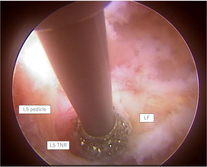
Figure 5: Detachment technique for releasing the LF. Endoscopic view demonstrating detachment of the LF from its bony attachment using a high-speed drill. The drill is applied to the bony margin to undercut the attachment and separate the LF from the underlying bone. LF, ligamentum flavum; TNR, traversing nerve root. Please click here to view a larger version of this figure.
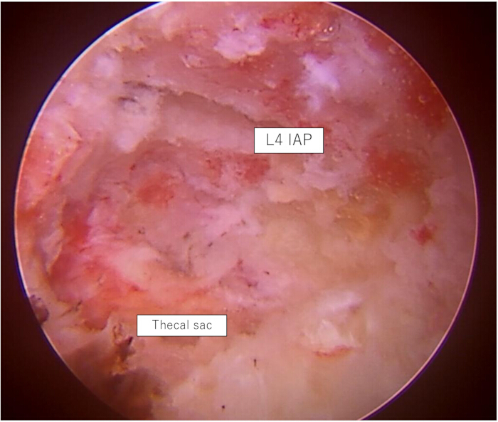
Figure 6: Final endoscopic view after decompression. Endoscopic view demonstrating the decompressed region after completion of ventral facetectomy. The neural structures, including the thecal sac, are identified following removal of the compressive elements. IAP, inferior articular process. Please click here to view a larger version of this figure.
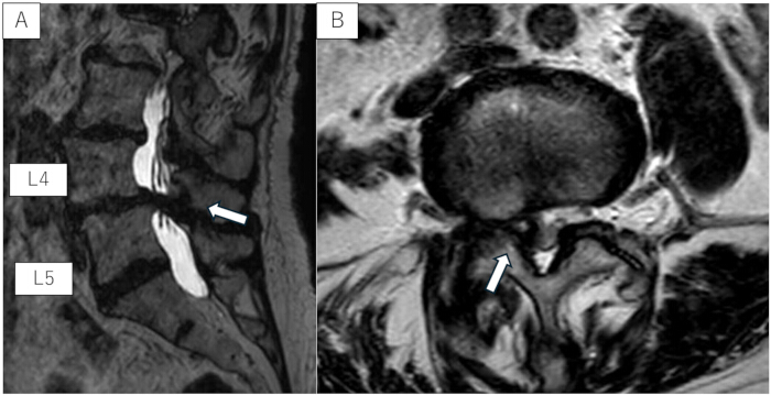
Figure 7: Preoperative magnetic resonance images. (A) Sagittal image demonstrating lumbar spinal stenosis at the L4/L5 level. (B) Axial image showing lateral recess stenosis with compression of the neural structures at the L4/L5 level. Arrows indicate the site of lateral recess stenosis and neural compression. Please click here to view a larger version of this figure.
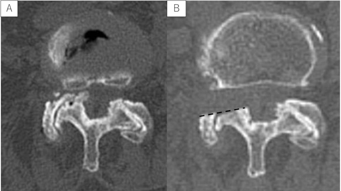
Figure 8: Preoperative and postoperative computed tomography images. (A) Preoperative axial image showing facet hypertrophy and lateral recess stenosis at the L4/L5 level. (B) Postoperative axial image demonstrating adequate bony decompression following ventral facetectomy. The dotted line indicates the extent of bone resection. Please click here to view a larger version of this figure.
| Potential complication | Prevention | Management |
| Exiting nerve root injury | Maintain continuous bony contact during needle, dilator, and cannula insertion. Avoid excessive advancement of the cannula into the foramen. Perform foraminoplasty under direct endoscopic visualization. | Stop manipulation immediately if radicular pain occurs. Reconfirm fluoroscopic and endoscopic orientation. Administer additional local anesthesia if needed and continue only after symptoms improve. |
| Traversing nerve root injury | Detach and remove the ligamentum flavum under direct visualization. Avoid blind manipulation around the lateral recess. Confirm the nerve root before using forceps or a Kerrison punch. | Stop the procedure if severe radiating pain or motor symptoms occur. Inspect the nerve root endoscopically and avoid further manipulation. Postoperative neurological evaluation and imaging should be performed if deficits are suspected. |
| Postoperative dysesthesia | Avoid excessive manipulation of the exiting or traversing nerve root. Maintain adequate irrigation pressure and avoid thermal injury from radiofrequency devices. | Most cases can be managed conservatively with observation and medication. Persistent symptoms may require careful neurological follow-up and imaging to exclude residual compression. |
| Dural tear | Perform ligamentum flavum detachment gradually and avoid blind use of sharp instruments near the thecal sac. | Small tears may be managed conservatively with bed rest and careful observation. If cerebrospinal fluid leakage is suspected postoperatively, wound assessment and additional treatment should be considered. |
| Epidural hematoma | Perform meticulous hemostasis; use of hemostatic agents, and drain placement. | Postoperative neurological deterioration requires urgent imaging. Symptomatic hematoma should be treated promptly, including surgical evacuation if necessary. |
| Inadequate decompression | Confirm decompression by direct visualization of the traversing nerve root, visible pulsation, and free mobilization with a probe. | If symptoms persist, postoperative imaging should be performed. Revision decompression may be considered when residual stenosis is confirmed. |
| Excessive facet resection | Plan the trajectory preoperatively using CT and MRI. Remove bone incrementally and preserve the remaining stabilizing structures whenever possible. | If excessive resection is suspected, postoperative CT and flexion-extension radiographs should be considered. Patients should be followed for signs of segmental instability. |
| Postoperative instability | Exclude patients with dynamic instability or Meyerding grade II or greater spondylolisthesis. Preserve dorsal facet structures and avoid unnecessary bone removal. | Flexion-extension radiographs should be obtained when instability is suspected. Fusion surgery may be required in symptomatic cases. |
| Infection | Use standard sterile technique, minimize operative time, and ensure appropriate wound care. | Treat superficial infection with antibiotics and wound care. Deep infection requires imaging, laboratory evaluation, and possible surgical debridement. |
| Conversion to other procedures | Perform careful preoperative assessment of stenosis pattern, spinal alignment, and instability. Avoid applying TF-FEVF to cases requiring fusion or wide central decompression. | If adequate decompression cannot be achieved safely, conversion to another decompression procedure or staged fusion surgery should be considered. |
Table 1: Potential complications and management strategies for TF-FEVF. This table summarizes common potential complications associated with TF-FEVF and provides corresponding prevention and management strategies to support safe procedural execution and postoperative care.
Discussion
Lumbar lateral recess stenosis is a common degenerative condition that causes radicular symptoms as a consequence of compression of the traversing nerve root1,2. Conventional posterior decompression under general anesthesia has been widely performed but may require extensive muscle dissection and partial facet joint removal, which can increase surgical invasiveness and the risk of postoperative instability3. Furthermore, some patients with multiple comorbidities may not tolerate general anesthesia well, highlighting the need for less invasive procedures that can be performed under local anesthesia8. The TF-FEVF technique described in this article was developed to address these challenges by providing direct access to the ventral facet region, enabling adequate decompression of the traversing nerve root while minimizing disruption of posterior structures.
A critical aspect of this technique is the creation of an adequate working space by stepwise ventral facetectomy. Resection of the ventral portion of the SAP, followed by partial removal of the IAP, allows direct access to the lateral recess and facilitates safe decompression of the traversing nerve root. The detachment technique for release of the LF is particularly important because it enables controlled separation of the ligament from its bony attachment while minimizing the risk of neural injury. Maintaining constant orientation using bony landmarks such as the pedicle and facet complex is also essential for procedural safety. These technical principles are consistent with the concepts of full-endoscopic spine surgery, which emphasize targeted decompression with minimal disruption of tissue while preserving stabilizing structures4,5,9. Several technical challenges may be encountered during TF-FEVF. Severe facet hypertrophy or a high iliac crest can make cannula docking difficult; in such cases, the trajectory should be reconfirmed with fluoroscopy, and stable bony contact with the SAP should be established before endoscope insertion. Limited visualization is usually caused by residual soft tissue, bleeding, bone debris, or inadequate inflow and outflow, and should be managed by careful soft-tissue clearance, meticulous hemostasis, removal of debris, and adjustment of irrigation and suction. If residual stenosis is suspected after LF removal, the decompressed area should be reassessed using endoscopic visualization and a blunt probe, and additional bone or ligament removal should be performed only under direct visualization.
An important distinction between TF-FEVF and previously reported endoscopic decompression techniques is the target and route of decompression. Conventional transforaminal endoscopic foraminoplasty primarily focuses on foraminal pathology and decompression of the exiting nerve root10. In contrast, TF-FEVF is designed to decompress the traversing nerve root in lateral recess stenosis through a transforaminal corridor. Unlike undercutting laminectomy or interlaminar endoscopic lateral recess decompression, TF-FEVF accesses the ventral facet region directly without a posterior interlaminar approach. The combination of extensive SAP resection, partial ventral IAP resection, and LF detachment creates a working corridor to the lateral recess while preserving posterior musculoligamentous structures. Therefore, TF-FEVF should be regarded as a distinct transforaminal decompression strategy for lumbar lateral recess stenosis rather than a simple extension of conventional foraminoplasty.
Despite these advantages, several limitations specific to the TF-FEVF technique should be considered. TF-FEVF is mainly indicated for patients with lumbar lateral recess stenosis causing radicular symptoms, especially when decompression under local anesthesia is desirable. Central stenosis, foraminal stenosis, scoliosis, severe facet hypertrophy, and previous lumbar surgery do not necessarily exclude patients from this procedure, provided that the main compressive pathology can be adequately addressed through the transforaminal corridor. However, TF-FEVF is not indicated for patients with clear segmental instability. Because this method involves substantial resection of the superior articular process and partial resection of the inferior articular process to expand the surgical corridor, there is a theoretical concern regarding postoperative segmental instability. Therefore, careful patient selection is essential. In our practice, TF-FEVF is not indicated for patients with dynamic instability on flexion-extension radiographs or degenerative spondylolisthesis of Meyerding grade II or greater, for whom fusion surgery is generally preferred. Regarding the extent of facet resection, a previous finite-element biomechanical study demonstrated that 50% resection of the superior articular process had the least effect on spinal stability, and that even 100% resection of the superior articular process did not result in substantial instability in severely degenerated discs11. Therefore, extensive facet resection may be acceptable in appropriately selected patients with advanced degenerative changes. In our institutional experience, TF-FEVF has been performed in 205 patients with a mean follow-up period of 20.2 ± 17.0 months. Only one patient subsequently required fusion surgery because of postoperative instability. Postoperative CT was used to confirm the extent of bony decompression, and flexion-extension radiographs were obtained when postoperative segmental instability was clinically suspected. Clinical outcomes were generally favorable. According to the modified Macnab criteria, 185 of the 205 patients (90.2%) achieved excellent or good outcomes at the final follow-up. These findings further support the safety and effectiveness of TF-FEVF when performed in appropriately selected patients. Although longer-term follow-up is necessary, these findings suggest that postoperative segmental stability can be preserved in most patients following TF-FEVF.
TF-FEVF is a technically demanding procedure and should not be considered an entry-level endoscopic technique. Surgeons should first acquire sufficient experience with transforaminal full-endoscopic discectomy and foraminoplasty before attempting TF-FEVF. Familiarity with endoscopic anatomical orientation, safe foraminoplasty techniques, and management of neural structures under endoscopic visualization is essential. Therefore, the learning curve of TF-FEVF is expected to be steeper than that of conventional transforaminal endoscopic procedures. Potential complications, along with their prevention and management strategies, are summarized in Table 1.
Future studies should focus on the biomechanical impact of ventral facet resection and determine the optimal extent of bone removal necessary to achieve adequate decompression while maintaining segmental stability. Prospective clinical studies with larger patient populations and longer follow-up are needed to clarify long-term outcomes and potential indications for this technique. In addition, objective radiological measurements of lateral recess dimensions could further validate the decompression achieved by TF-FEVF. Further refinement of surgical instruments and navigation technologies may improve the safety and reproducibility of TF-FEVF, potentially expanding its use in patients with lateral recess stenosis.
Disclosures
The authors declare no conflicts of interest.
Acknowledgements
The authors would like to thank the medical staff of Tokushima University Hospital for their support. This research received no specific grant from any funding agency in the public, commercial, or not-for-profit sectors.
Materials
| Name | Company | Catalog Number | Comments |
|---|---|---|---|
| 10K arthroscope tube set | Zimmer Biomet, Warsaw, IN | LC-10K0-100-00 | Irrigation system console |
| 10K irrigation console | Zimmer Biomet, Warsaw, IN | LC-10K0-000-00 | Irrigation system console |
| 1-Coarse Diamond Bur SPL (3.5) | Nakanishi, Kanuma, Japan | PDS-1CD330-35 | High-speed drill |
| 1% lidocaine | Various manufacturers | N/A | Local anesthetic |
| 21G PTCD needle, 200 mm | Hakko Shoji, Hyuga City, Japan | U5099-MM | Needle for local anesthesia |
| 23G Catelan needle | NIPRO, Osaka, Japan | 2022 | Needle for local anesthesia |
| 32-inch 4K LCD monitor | Sony, Tokyo, Japan | LMD-X3200MD | Monitor |
| Aquarius NET server | TeraRecon, Durham, NC | N/A | CT reconstruction and analysis software |
| Atarax-P | Pfizer, New York, NY | N/A | Hydroxyzine hydrochloride |
| Centricity Universal Viewer Zero Footprint Client | GE Healthcare, Chicago, IL | Version 6.0 SP11.2.2 | MRI visualization and analysis software |
| CLICKLINE grasping forceps | Karl Storz, Tuttlingen, Germany | 28163FSI | Intervertebral disc rongeur |
| Contrast medium | Various manufacturers | N/A | Mixed with indigo carmine for intradiscal injection |
| Curved rongeur, working length 360 mm, φ2.5 mm | RIWOspine, Knittlingen, Germany | 89240.1044 | Soft tissue rongeur |
| Dilator, Ø7.0 mm, effective length 225 mm (2 holes) | Elliquence, Baldwin, NY | 11-2311 | Dilator |
| ENDOCAM Logic 4K camera controller | Richard Wolf GmbH, Knittlingen, Germany | 55253011 86-103 | Camera |
| ENDOCAM Logic 4K camera head | Richard Wolf GmbH, Knittlingen, Germany | 85525942 86-104 | Camera |
| Fiber light cable (φ3.5 mm/3.0 mm) | Richard Wolf GmbH, Knittlingen, Germany | 806635301 86-124 | Camera |
| Flexible probe (Φ2.5 mm, WL 350 mm) | RIWOspine, Knittlingen, Germany | 892501925 | Flexible probe |
| Floseal Hemostatic Matrix | Baxter, Deerfield, IL | ADS201844 5 mL Kit | Hemostatic agent |
| Guide wire | Nihon MDM, Tokyo, Japan | 28163GWT | Dilator |
| Indigo carmine | Various manufacturers | N/A | Disc staining agent |
| Kerrison punch handle | Elliquence, Baldwin, NY | 12-1947 | Kerrison |
| Kerrison punch, angled type, Ø3.5 mm, effective length 360 mm | Elliquence, Baldwin, NY | 12-1938 | Kerrison |
| Kerrison punch, angled type, Ø4.0 mm, effective length 360 mm | Elliquence, Baldwin, NY | 12-1940 | Kerrison |
| LED light source unit for endoscopy (LED 1.2 set) | Richard Wolf GmbH, Knittlingen, Germany | 51610011 86-164 | Camera |
| Primado2 drill system | Nakanishi, Kanuma, Japan | P200-CU-100 | High-speed drill |
| Primado2 foot controller | Nakanishi, Kanuma, Japan | FC-73 | High-speed drill |
| Primado2 Slim motor handpiece | Nakanishi, Kanuma, Japan | P200-SMH-HS | High-speed drill |
| PTC needle type B, 18-gauge × 200 mm | Hakkou Shoji, Sapporo, Japan | 22411830 | Dilator |
| Rongeur, working length 360 mm, φ3.0 mm | RIWOspine, Knittlingen, Germany | 89240.1003 | Soft tissue rongeur |
| Sosegon | Maruishi Pharma, Osaka, Japan | N/A | Pentazocine |
| Step dilator set (5 pieces) | Elliquence, Baldwin, NY | CSD-5 | Dilator |
| Surgical drain | Various manufacturers | N/A | Postoperative drainage |
| Super Slim Attachment 200 | Nakanishi, Kanuma, Japan | P200-RA330-L | High-speed drill |
| Surgi-Max Air | Elliquence, Baldwin, NY | IEC4-SP | Bipolar device |
| Trigger-Flex | Elliquence, Baldwin, NY | DTF-40 | Bipolar device |
| VERTEBRIS lumbar, 25°, 6.9 mm, working length 207 mm | RIWOspine GmbH, Knittlingen, Germany | 89210.1254 | Rigid endoscope |
| Working channel, duckbill type, Ø8.0 mm, length 165 mm | Richard Wolf GmbH, Knittlingen, Germany | 11-2910 | Duckbill cannula |
| Working channel, elevator type, Ø8.0 mm, length 165 mm | Richard Wolf GmbH, Knittlingen, Germany | 11-2916 | Oblique cannula |
References
- Genevay S, Atlas SJ. Lumbar spinal stenosis. N Engl J Med. 2008;358:818-25.
- Lee CK, Rauschning W, Glenn W. Lumbar spinal stenosis: classification and pathogenesis. Spine. 1988;13:313-20.
- Weinstein JN, et al. Surgical versus nonsurgical therapy for lumbar spinal stenosis. N Engl J Med. 2008;358:794-810.
- Kambin P, Zhou L. Arthroscopic discectomy of the lumbar spine. Spine. 1992;17(1 Suppl):S1-7.
- Sairyo K, Chikawa T, Nagamachi A. State-of-the-art transforaminal percutaneous endoscopic lumbar surgery under local anesthesia: discectomy, foraminoplasty, and ventral facetectomy. J Orthop Sci. 2018;23:229-36.
- Yamashita K, et al. Percutaneous full endoscopic lumbar foraminoplasty for adjacent level foraminal stenosis following vertebral intersegmental fusion in an awake and aware patient under local anesthesia: a case report. J Med Invest. 2017;64:291-5.
- Sairyo K, et al. A novel surgical concept of transforaminal full-endoscopic lumbar undercutting laminectomy for central canal stenosis of the lumbar spine with local anesthesia. J Med Invest. 2019;66:224-9.
- Ruetten S, Komp M, Merk H, Godolias G. Full-endoscopic lumbar decompression versus conventional microsurgical technique. Spine (Phila Pa 1976). 2009;34:2247-56.
- Ahn Y. Endoscopic spine discectomy: indications and outcomes. Int Orthop. 2019;43:909-16.
- Hoogland T, Schubert M, Miklitz B, Ramirez A. Transforaminal posterolateral endoscopic discectomy. Spine J. 2006;6:226-32.
- Matsumoto K, et al. Biomechanical evaluation of a novel decompression surgery: Transforaminal full-endoscopic lateral recess decompression (TE-LRD). N Am Spine Soc J. 2020;5:100045.
Reprints and Permissions
This article has been published
Video Coming Soon