This article summarizes nurse-led strategies for managing diabetic foot ulcers, including patient education, systematic monitoring, advanced wound care, infection prevention, and emerging digital health technologies such as telemedicine and wearable sensors to improve early detection, treatment outcomes, and long-term prevention.
Review Article
Nursing Roles in Diabetic Foot Ulcer Prevention, Wound Management, and Digital Monitoring
207 views
⸱
September 11th, 2026
* These authors contributed equally
In This Article
Summary
Abstract
Diabetic foot ulcers (DFUs) remain a major cause of infection, hospitalization, impaired quality of life, and non-traumatic lower-limb amputation among people with diabetes. Effective management requires coordinated prevention, structured foot-risk assessment, pressure offloading, wound care, infection surveillance, patient education, and multidisciplinary follow-up. This narrative review examines the nurse-led and nurse-supported components of contemporary DFU care. Nurses contribute through routine foot and wound assessment, reinforcement of preventive behaviors and treatment adherence, support for offloading, monitoring after debridement and advanced wound therapies, and early recognition of infection, ischemia, osteomyelitis, or clinical deterioration. Patient and caregiver education should be practical, individualized, and reinforced over time. Current evidence most consistently supports improvements in foot-care knowledge, self-management behavior, and healthcare-provider competence, whereas effects on ulcer recurrence, hospitalization, and amputation remain less certain. Digital technologies, including telemedicine, wearable sensors, mobile health platforms, and artificial intelligence-based wound assessment, may improve access to monitoring, communication, and early risk detection. However, their evidence base and clinical readiness vary, and improved monitoring does not necessarily translate into better healing or fewer amputations. Their usefulness depends on patient training, data quality, governance, timely professional review, and integration with safe referral pathways. Digital tools should therefore support, rather than replace, direct clinical assessment and multidisciplinary decision-making. Overall, nursing practice is essential for translating evidence-based DFU management into continuous, coordinated, and patient-centered care.
Introduction
Diabetes mellitus represents one of the most significant global health challenges, with its prevalence continuing to rise rapidly worldwide1. Among its chronic complications, diabetic foot ulcers (DFUs) are particularly devastating, affecting approximately 15%–25% of individuals with diabetes during their lifetime and accounting for a substantial proportion of diabetes-related hospitalizations2. As the prevalence of diabetes increases with age, diabetic foot disease has become an important component of geriatric medicine, reflecting the complex clinical needs of older adults with chronic metabolic disorders3. DFUs are a leading cause of non-traumatic lower-limb amputation and are associated with high morbidity, mortality, and healthcare costs. The pathogenesis of DFUs is multifactorial and typically involves interactions among peripheral neuropathy, peripheral arterial disease, impaired immune function, and abnormal mechanical loading of the foot. These factors collectively compromise tissue integrity, delay wound healing, and increase susceptibility to infection, making DFUs a complex condition that requires comprehensive and coordinated management2,4 (Figure 1).
In recent years, substantial progress has been made in understanding the mechanisms underlying diabetic wound healing impairment and in developing advanced therapeutic strategies. Improvements in wound care technologies, infection control strategies, offloading techniques, and digital health tools have significantly expanded the therapeutic landscape for DFU management5,6. Despite these advances, the recurrence rate of DFUs remains high and long-term outcomes remain suboptimal in many healthcare settings. Effective management therefore requires appropriate medical and surgical interventions and also continuous monitoring, early detection of complications, and patient engagement in preventive practices5,7. Within this multidisciplinary framework, nurses play a pivotal role in the prevention, assessment, and ongoing management of diabetic foot complications. Their responsibilities extend beyond routine wound care to include patient education, risk assessment, monitoring of comorbidities, coordination of multidisciplinary care, and implementation of emerging technologies that facilitate remote monitoring and early intervention. Nursing-led strategies are particularly important in improving patient adherence, enhancing self-management behaviors, and ensuring continuity of care across different healthcare environments8,9.
This review aims to summarize recent advances in DFU care from a nursing perspective. Specifically, it highlights current strategies in patient education and monitoring, modern approaches to wound management and infection prevention, the role of comorbidity control and psychosocial support, and the growing impact of digital health technologies and multidisciplinary collaboration in optimizing DFU outcomes.
Review and Perspective
Review methodology
This narrative review was informed by a structured search of PubMed and Web of Science. Search terms included combinations of “diabetic foot ulcer”, “nursing care”, “patient education”, “foot assessment”, “offloading”, “wound management”, “infection prevention”, “pain management”, “nutrition”, “telemedicine”, “remote monitoring”, “wearable sensors”, and “artificial intelligence”. English-language clinical guidelines, systematic reviews, meta-analyses, clinical trials, observational studies, and relevant nursing studies addressing DFU prevention, treatment, or follow-up were considered. Conference abstracts, protocols, editorials, and studies that were not directly relevant to diabetic foot care were excluded. The literature was organized into education and monitoring, wound management and infection prevention, pain and nutritional support, and remote monitoring and digital technologies. As this was a narrative review, no formal PRISMA-based screening, independent dual-reviewer selection, risk-of-bias assessment, or meta-analysis was performed.
Patient education and clinical monitoring
Education and systematic monitoring are fundamental components in the prevention and early detection of DFUs. The contribution of nursing professionals in this process is particularly significant, as nurses frequently serve as primary educators, clinical monitors, and patient advocates for individuals at increased risk of diabetic foot complications. Through continuous patient engagement, nurses facilitate the translation of preventive knowledge into daily self-care practices and ensure early identification of clinical changes that may predispose patients to ulcer development10,11. Consistent with current International Working Group on the Diabetic Foot (IWGDF) and National Institute for Health and Care Excellence (NICE) guidance, structured foot-risk assessment should be performed when diabetes is diagnosed, at least annually thereafter, whenever a new foot problem develops, and during hospital admission or a clinically important change in the patient’s condition. The frequency of subsequent foot examinations should be determined by the identified risk category, with closer surveillance and timely specialist referral for patients at increased risk of ulceration12. Patient and family education represents a cornerstone of DFU prevention. Effective preventive strategies require a comprehensive educational approach delivered collaboratively by physicians and nursing staff, with the aim of improving patients' understanding of appropriate foot care practices and emphasizing the importance of vigilant foot surveillance13,14. To optimize comprehension and retention of information, multiple educational modalities can be employed. Structured lectures allow healthcare professionals to present key concepts related to diabetic foot care while providing opportunities for patient interaction and clarification of questions15. Collectively, evidence from systematic reviews, randomized trials, and quasi-experimental studies indicates that structured educational interventions can improve foot-care knowledge, preventive behaviors, and the competence of healthcare professionals involved in diabetic foot care. The systematic review and meta-analysis by Drovandi et al. provides the broadest evidence, demonstrating generally favorable effects on patient knowledge and self-care behavior, with possible reductions in ulcer and amputation risk16. Monami et al. further showed that a brief program combining theoretical instruction with supervised practical training may reduce short-term ulcer occurrence in high-risk patients17. However, the relatively small sample, limited number of ulcer events, and short follow-up restrict the certainty and generalizability of this finding. In contrast, Aalaa et al. demonstrated improved knowledge among healthcare professionals after an interactive workshop, but this provider-level outcome does not establish corresponding improvements in ulcer healing, recurrence, or amputation rates18. Overall, the findings are most consistent for improvements in knowledge, practical skills, and self-care behavior, whereas evidence for sustained effects on clinical outcomes remains less certain because the interventions differ in content, intensity, duration, target population, and follow-up. Educational programs should therefore be practical, individualized, reinforced over time, and integrated with risk assessment, appropriate footwear and offloading, wound surveillance, and multidisciplinary follow-up. Along with that, educational programs should address several essential aspects of foot health management. Patients should be instructed to perform daily foot inspection, paying particular attention to areas that are difficult to visualize, such as the plantar surface and interdigital spaces19. Proper hygiene practices, including careful washing, thorough drying, and appropriate moisturization, are important to prevent skin breakdown and fissure formation20. Education regarding safe nail care is also necessary to minimize the risk of minor trauma during trimming21. Furthermore, guidance on appropriate footwear selection is critical, as poorly fitted shoes may create pressure points that predispose patients to ulceration. Patients should also be trained to recognize early signs of tissue injury, including erythema, swelling, localized warmth, or changes in skin color21,22. In addition, education should emphasize the importance of adequate glycemic control, as persistent hyperglycemia contributes to neuropathy, vascular impairment, and delayed wound healing23. Lifestyle factors such as regular physical activity, smoking cessation, and balanced nutrition should also be addressed because of their significant influence on overall vascular and metabolic health24,25.
For patients with limited mobility, impaired vision, or other functional limitations, family involvement becomes an essential component of effective preventive care. Healthcare providers should therefore ensure that family members or caregivers receive appropriate training in foot inspection techniques and are able to recognize signs that require prompt medical evaluation. Encouraging family participation in routine clinical follow-up and reinforcing the importance of regular medical evaluations further strengthens preventive efforts26,27. Wantonoro et al. conducted a longitudinal study to evaluate the impact of 6-month interdisciplinary education on family caregivers' knowledge and self-efficacy in diabetic wound care28. The intervention involved home-based interdisciplinary education provided twice weekly by nursing, physiotherapy, nutrition, and psychology professionals to 16 caregivers of patients with diabetic ulcers. After 6 months, caregivers demonstrated a significant improvement in wound-care knowledge and self-efficacy, indicating that long-term interdisciplinary educational support can strengthen caregiver competence and enhance family involvement in diabetic ulcer management28. In addition to education, routine clinical monitoring is essential for early identification of risk factors and pre-ulcerative lesions. Nurses should perform periodic foot examinations, with the frequency determined by the patient's individual risk profile. A comprehensive foot assessment typically includes inspection for skin lesions, discoloration, deformities, and callus formation, as well as evaluation of sensory function using monofilament or tuning fork testing29,30. Vascular status should be assessed through palpation of pedal pulses and evaluation of capillary refill time. Structural abnormalities, joint mobility limitations, and gait disturbances should also be examined, as these factors may alter pressure distribution across the foot2,4. Furthermore, assessment of footwear is necessary to ensure appropriate fit and to identify potential pressure areas that may predispose patients to ulcer formation31. When additional evaluation is required, several diagnostic modalities may provide further insight into vascular and tissue status. These may include Doppler ultrasound for vascular assessment, pressure sensors for evaluating plantar pressure distribution, thermographic imaging to detect localized inflammatory changes, laser Doppler techniques to assess microcirculatory perfusion, and transcutaneous oxygen measurement (TcPO2) to evaluate tissue oxygenation32,33,34. Collaboration with specialists, including podiatrists, endocrinologists, and vascular surgeons, is often necessary to provide comprehensive management for patients at increased risk of diabetic foot complications35,36. In addition, nurses play an important role in educating patients regarding correct insulin pen administration techniques and reinforcing adherence to prescribed medication regimens and treatment plans37,38. Such support helps ensure effective glycemic control, which remains a critical factor in preventing neuropathy, vascular compromise, and impaired wound healing. Finally, through regular follow-up visits and structured monitoring programs, nurses can detect early pathological changes and initiate timely preventive interventions and further educate the patients and their families before ulceration develops39.
Offloading and mechanical pressure redistribution
Offloading is a fundamental component of DFU management because repetitive plantar loading and mechanical tissue stress can perpetuate local injury and delay wound closure, even when debridement, infection control, and appropriate dressings are provided. Current International Working Group on the Diabetic Foot guidance recommends a non-removable knee-high offloading device as the first-choice treatment for neuropathic plantar forefoot or midfoot ulcers. This may consist of either a total contact cast or a prefabricated knee-high walker rendered non-removable, with selection based on foot deformity, clinical expertise, available resources, and patient acceptability40. A contemporary systematic review and meta-analysis found that non-removable devices increased the likelihood of ulcer healing compared with removable devices, primarily because they provide continuous mechanical protection and reduce the opportunity for device removal during weight-bearing activities41. Earlier randomized evidence similarly demonstrated greater healing effectiveness with total contact casting than with less restrictive offloading modalities42. When a non-removable knee-high device is contraindicated, unavailable, or not tolerated, a removable knee-high or ankle-high offloading device may be considered. Patients should be instructed to use the prescribed device during all weight-bearing activities, including short periods of walking within the home. Conventional footwear and standard therapeutic footwear should not be used as substitutes for an appropriate offloading device during active treatment of a plantar ulcer40. However, the clinical effectiveness of removable devices is strongly influenced by adherence. Objective activity monitoring has shown that patients may wear removable cast walkers during only approximately one-third of their total weight-bearing activity43. Device weight, inconvenience, difficulty performing daily activities, occupational demands, impaired balance, and the perceived absence of pain in neuropathic ulcers may contribute to poor adherence. Therefore, selection of an offloading device should consider both its pressure-relieving capacity and the likelihood that the patient will use it consistently.
Therapeutic footwear and custom-made insoles have a more prominent role after ulcer healing, when the objective shifts from active wound treatment to recurrence prevention. Footwear should accommodate structural deformity, avoid excessive friction, and redistribute pressure away from previously ulcerated or high-risk plantar regions. Where validated in-shoe pressure-measurement systems are available, pressure-optimized footwear or insoles should achieve a clinically meaningful reduction in peak plantar pressure. The IWGDF defines a demonstrated pressure-relieving effect as either a reduction of at least 30% at high-pressure locations compared with the existing footwear or an in-shoe peak pressure below 200 kPa during walking44. Evidence suggests that pressure-optimized therapeutic footwear and insoles may reduce recurrent plantar ulceration, although their effectiveness remains dependent on regular use45. A multicenter randomized trial similarly showed that the preventive benefit of custom-made footwear was closely associated with adherence to wearing the prescribed footwear46. Nurses have an important role in supporting the safe and effective implementation of offloading. Patient education should explain that pressure redistribution is an active treatment rather than an optional adjunct and that removable devices must be worn whenever standing or walking. During follow-up, nurses should evaluate device fit, gait stability, mobility, adherence, and the condition of both the ulcerated and contralateral foot. The skin should be inspected for abrasions, blistering, maceration, edema, new pressure lesions, or device-related trauma, while the wound should be assessed for changes in size, exudate, inflammation, and infection. Patients and caregivers should be instructed to report new pain, skin discoloration, increasing swelling, fever, device damage, or difficulty walking. When offloading is poorly tolerated, repeatedly removed, associated with new lesions or falls, or fails to produce satisfactory healing progress, the multidisciplinary team should reassess device selection, plantar pressure redistribution, adherence, vascular status, infection, and the need for alternative or surgical offloading. Non-removable devices also require particular caution when frequent wound inspection is necessary because of moderate infection, ischemia, or heavy exudate40,47.
Wound management and infection prevention
Effective wound management and infection control represent central components of DFU treatment, as persistent tissue necrosis, microbial colonization, and impaired host immunity significantly delay wound healing in patients with diabetes19. Optimal care requires a structured approach that combines regular wound assessment, appropriate debridement, maintenance of a moist wound environment, and early identification and treatment of infection. Within the multidisciplinary DFU care team, nurses play a key role in performing routine wound evaluation, implementing evidence-based dressing strategies, and monitoring clinical signs of infection during follow-up care48,49. Debridement is considered a cornerstone of DFU management because the presence of necrotic tissue, biofilm, and hyperkeratotic callus increases bacterial burden and prevents the formation of healthy granulation tissue. Regular removal of devitalized tissue promotes re-epithelialization and reduces local inflammatory responses50,51. Depending on wound characteristics and patient tolerance, several debridement techniques may be applied, including sharp or surgical debridement, enzymatic debridement, mechanical methods, and autolytic approaches facilitated by moisture-retentive dressings. Among these methods, sharp debridement remains the most effective technique for rapidly removing necrotic tissue and controlling infection in chronic DFUs, particularly when extensive slough or callus is present51,52. However, less invasive approaches such as autolytic or enzymatic debridement may be preferred in patients with ischemic wounds or those who cannot tolerate aggressive procedures53. Regular reassessment is essential to determine the need for repeated debridement as the wound progresses through different stages of healing. In addition, maintaining an optimal wound environment is another critical element of DFU management. Modern wound dressings are designed to maintain moisture balance, control exudate, and protect the wound from external contamination54. Dressing selection should be individualized according to wound depth, exudate level, and presence of infection55. For example, hydrocolloid and hydrogel dressings promote autolytic debridement and support epithelial migration in relatively dry wounds, whereas foam and alginate dressings are more suitable for highly exudative ulcers due to their superior absorptive capacity56,57. Antimicrobial dressings containing agents such as silver or iodine may also be used to reduce microbial burden in wounds at risk of infection58,59. Continuous reassessment of wound characteristics is necessary to adjust dressing strategies during the healing process and prevent complications associated with excessive moisture or desiccation (Table 1). In recent years, several advanced wound therapies have been introduced to enhance the healing of complex DFUs. Negative pressure wound therapy (NPWT) has gained widespread clinical use because it removes excess exudate, decreases local edema, improves tissue perfusion, and stimulates granulation tissue formation60. Clinical studies have demonstrated that NPWT can accelerate wound closure and reduce the risk of major amputation in patients with large or deep diabetic ulcers61,62. In addition, bioengineered skin substitutes and acellular dermal matrices have shown promising results by providing structural scaffolds that support cellular migration and extracellular matrix remodeling63,64. Growth factor-based therapies, including platelet-derived growth factor formulations, may further promote fibroblast proliferation and angiogenesis in selected cases65. Selection of bioengineered skin substitutes, dermal matrices, or growth-factor therapies should be based on multidisciplinary assessment rather than nursing decision-making alone. Nurses support their safe use by preparing the wound bed, applying or maintaining the prescribed treatment according to protocol, educating patients regarding protection and adherence, and monitoring for infection, displacement, excessive exudate, allergic reactions, or failure of wound progression. Furthermore, infection remains one of the most serious complications of DFUs and is a major contributor to delayed healing, hospitalization, and lower-limb amputation66. Early recognition and prompt treatment are therefore essential (Table 2). Microorganisms are commonly present in open DFUs; however, microbial colonization does not itself indicate infection, and a positive culture without corresponding clinical inflammatory findings should not be interpreted as evidence of diabetic foot infection. Diabetes-related foot infection remains primarily a clinical diagnosis based on local or systemic manifestations of inflammation. According to the IWGDF/Infectious Diseases Society of America (IDSA) classification, an ulcer is considered uninfected when no local or systemic manifestations of infection are present67. A mild or localized infection is confined to the skin and subcutaneous tissue and is characterized by at least two local findings, including swelling or induration, erythema extending more than 0.5 cm but less than 2 cm from the wound margin, tenderness or pain, increased warmth, or purulent discharge. Other non-infectious causes of inflammation, such as trauma, gout, acute Charcot neuro-osteoarthropathy, fracture, thrombosis, or venous stasis, should be excluded67. A moderate infection is characterized by erythema extending at least 2 cm from the wound margin or involvement of structures deeper than the skin and subcutaneous tissue, including fascia, tendon, muscle, joint, or bone, without systemic manifestations. Spreading soft-tissue infection therefore generally falls within the moderate category. Severe infection is defined as a foot infection accompanied by at least two systemic inflammatory response criteria, including abnormal body temperature, tachycardia, tachypnea, or an abnormal leukocyte count67,68,69,70. Furthermore, the term limb-threatening infection is not a separate IWGDF/IDSA severity grade but describes a severe or complicated moderate infection associated with extensive gangrene, necrotizing infection, deep abscess, compartment syndrome, rapidly progressive tissue destruction, or clinically important lower-limb ischemia. These findings require urgent surgical and vascular assessment. Sepsis should not be considered synonymous with severe diabetic foot infection; it is defined as life-threatening organ dysfunction caused by a dysregulated host response to infection and requires immediate emergency management67,71. NICE similarly recommends immediate referral to acute services for limb-threatening or life-threatening diabetic foot problems, including ulceration associated with fever or sepsis, gangrene, ischemia, or suspected deep soft-tissue or bone infection12,71. In addition, when infection is clinically suspected, microbiological testing should support rather than establish the diagnosis. Following wound cleansing and debridement, an aseptically collected tissue specimen obtained by curettage or biopsy is generally more clinically informative than a superficial swab. Comparative studies have shown that concordance between superficial swabs and tissue specimens varies according to ulcer characteristics and infection severity, with superficial swabs potentially missing clinically relevant organisms in deeper or more severe infections68,72,73. Therefore, superficial swabs should not be described as universally unacceptable but should generally be avoided when a reliable deep-tissue specimen can be safely obtained. Moreover, antibiotics are indicated for clinically infected DFUs but should not be prescribed for clinically uninfected ulcers solely to prevent infection or accelerate wound healing. Antimicrobial selection should consider infection severity, likely or confirmed pathogens, previous antibiotic exposure, recent culture findings, renal function, drug interactions, adverse-effect profiles, and local antimicrobial susceptibility patterns12,67. This distinction is central to antimicrobial stewardship and reduces unnecessary antibiotic exposure without compromising appropriate treatment of confirmed infection.
Effective infection prevention also relies on strict adherence to aseptic wound care practices during dressing changes. Proper hand hygiene, use of sterile equipment, and maintenance of a clean wound environment are fundamental measures for minimizing bacterial contamination74,75. Close collaboration between nurses, infectious disease specialists, and surgeons is essential to ensure appropriate antibiotic selection, monitoring of therapeutic response, and timely surgical intervention when necessary76,77. Nurses support guideline-based infection management by monitoring local and systemic signs of infection, facilitating appropriate specimen collection, administering prescribed antimicrobial therapy, assessing treatment response and adverse effects, documenting wound progression, and promptly escalating clinical deterioration to infectious disease, vascular, surgical, or multidisciplinary diabetic foot services. Regular documentation of wound size, depth, exudate characteristics, and surrounding tissue condition allows early detection of clinical deterioration and facilitates timely modification of treatment strategies78.
Pain management and nutritional support
Pain associated with DFUs is frequently underrecognized and undertreated, despite its substantial impact on patient quality of life, mobility, and adherence to treatment. Pain in patients with DFUs should be assessed according to its underlying mechanism because neuropathic, nociceptive, ischemic, and infection-related pain have different clinical implications and management requirements79. Pain intensity alone may be insufficient; assessment should also consider pain quality, location, timing, precipitating factors, relationship to walking or wound-care procedures, and associated neurological, vascular, or inflammatory findings. The presence of peripheral neuropathy may reduce protective sensation and mask clinically important ischemia or infection; therefore, the absence of severe pain does not exclude serious foot pathology44,79. Neuropathic pain commonly presents as burning, tingling, shooting, electric shock-like pain, allodynia, or hyperalgesia and may coexist with numbness and loss of protective sensation. Management should be individualized according to symptom severity, comorbidities, medication interactions, and adverse-effect risks. Recommended pharmacological classes include gabapentinoids, serotonin-norepinephrine reuptake inhibitors, tricyclic antidepressants, and sodium-channel blockers. Opioids should not be used routinely for painful diabetic neuropathy because of their unfavorable long-term benefit-risk profile80. The updated American Academy of Neurology guidance supports these medication classes while emphasizing individualized treatment selection80. Furthermore, nociceptive wound pain results from tissue injury and local inflammation and may be described as aching, throbbing, sharp, or tender. It may occur continuously or be provoked by dressing removal, wound cleansing, debridement, pressure, or movement. Management should combine appropriate analgesia with atraumatic dressing techniques, gentle wound handling, preparation before painful procedures, and reassessment of pain during and after treatment. A substantial increase in previously stable wound pain should prompt evaluation for infection, ischemia, pressure-related injury, or another underlying complication rather than automatic escalation of analgesic therapy79,81. Ischemic rest pain should be considered in patients with persistent foot or forefoot pain at rest, particularly when accompanied by absent pulses, coolness, pallor, delayed capillary refill, tissue necrosis, gangrene, or a deteriorating ulcer. Peripheral neuropathy may attenuate typical ischemic symptoms, so vascular disease should not be excluded solely because pain is absent or mild. New rest pain or clinical evidence of severe ischemia requires prompt vascular assessment and consideration of revascularization; analgesia should support comfort but must not delay definitive vascular evaluation44,82. In addition, infection-related pain may present as new or worsening tenderness or pain accompanied by erythema, warmth, swelling, purulent discharge, increasing exudate, malodor, wound deterioration, or systemic manifestations. However, pain and other inflammatory findings may be diminished by neuropathy, peripheral arterial disease, or immune dysfunction. Infection should therefore be assessed clinically using the complete local and systemic presentation rather than pain severity alone. Suspected spreading infection, deep-tissue involvement, systemic deterioration, or severe infection requires urgent multidisciplinary assessment and treatment67. The IWGDF/IDSA guideline identifies pain or tenderness as a possible local manifestation while emphasizing that infection severity and systemic findings guide escalation67. Analgesic treatment should be selected according to the underlying pain mechanism and the patient’s clinical profile. In older adults, systemic non-steroidal anti-inflammatory drugs, when considered for nociceptive, inflammatory, or procedure-related pain, should be used cautiously because age-related reductions in renal reserve, multimorbidity, and polypharmacy increase the risks of gastrointestinal bleeding, acute kidney injury, fluid retention, hypertension, and cardiovascular complications. Before treatment, renal function, cardiovascular and gastrointestinal risk, and concomitant medications should be reviewed; when an nonsteroidal anti-inflammatory drug (NSAID) is necessary, the lowest effective dose should be used for the shortest appropriate duration, with gastroprotection and clinical monitoring when indicated83,84. Nurses play an important role in differentiating these pain patterns by documenting pain intensity and quality, identifying procedural and activity-related triggers, assessing sensory loss, inspecting the wound and surrounding tissues, and monitoring vascular and systemic findings. New or rapidly worsening pain, ischemic rest pain, unexpected pain during offloading, suspected infection, or pain associated with clinical deterioration should be escalated promptly to the multidisciplinary diabetic foot, vascular, infectious-disease, or surgical team81,85.
Nevertheless, non-pharmacological interventions may further complement pharmacologic pain management strategies. Psychological stress, anxiety, and depression are common among patients with chronic wounds and can amplify pain perception86. Interventions such as relaxation therapy, mindfulness-based stress reduction, and cognitive-behavioral therapy have been shown to improve coping mechanisms and reduce pain-related distress in patients with chronic conditions87,88,89,90. Narrative nursing and patient-centered communication strategies have also been reported to strengthen therapeutic relationships and enhance emotional support during long-term wound treatment. Integrating these supportive approaches into routine nursing care can therefore contribute to improved overall patient experience and treatment adherence87,88,89,90. Nutritional care for patients with DFUs should begin with structured nutritional screening rather than routine supplementation. Screening should be undertaken at the initial assessment and repeated when there is clinical deterioration, prolonged wound healing, reduced food intake, unintentional weight loss, recurrent infection, or hospitalization. Assessment should consider recent weight change, body mass index, dietary intake, appetite, muscle loss, functional status, disease burden, gastrointestinal or oral problems, and access to appropriate food. Patients identified as being at nutritional risk should undergo a more comprehensive assessment by an appropriately trained clinician or dietitian, using established approaches such as the Subjective Global Assessment or the Global Leadership Initiative on Malnutrition criteria91,92,93. Malnutrition is common among patients with DFUs and has been associated with poorer wound healing and prolonged hospitalization, supporting the importance of early identification and intervention92,93. For patients with confirmed malnutrition or high nutritional risk, management should focus on individualized correction of protein-energy inadequacy, maintenance of adequate hydration, correction of documented nutrient deficiencies, and management of factors that limit food intake. A food-based dietary plan should be prioritized where feasible, while oral nutritional supplements may be considered when dietary intake alone is insufficient to meet the patient’s assessed requirements. Dietetic referral is particularly appropriate for patients with persistent poor intake, unintentional weight loss, dietary restrictions, renal or hepatic disease, swallowing or gastrointestinal difficulties, or complex metabolic requirements. Glycemic management should be coordinated with nutritional care because overly restrictive diets may further compromise energy and protein intake. Although hyperglycemia has been associated with a higher risk of lower-extremity amputation, available observational evidence has not established that more intensive glycemic control directly accelerates ulcer healing23,94. Chronic wounds are characterized by sustained inflammation and increased metabolic demands, which may increase protein turnover and nutritional requirements. Adequate energy and protein availability support collagen synthesis, immune function, and tissue repair, while vitamins A and C, zinc, selected amino acids, and omega-3 fatty acids participate in biological pathways relevant to wound healing95,96,97,98,99,100. However, these physiological roles do not by themselves demonstrate that routine supplementation improves DFU outcomes. Targeted supplementation should be distinguished from general nutritional optimization. Current evidence does not support the routine administration of arginine, glutamine, zinc, vitamins A or C, omega-3 fatty acids, or other micronutrients to all patients with DFUs. A Cochrane review concluded that the effects of nutritional interventions on DFU healing remain uncertain because the available trials were small and provided low- or very-low-certainty evidence101. The 2023 IWGDF wound-healing guideline similarly recommends against using pharmacological vitamin and trace-element supplementation or protein supplementation solely to improve healing outcomes beyond standard care102. In a randomized trial, supplementation with arginine, glutamine, and β-hydroxy-β-methylbutyrate did not improve healing in the overall study population, although exploratory subgroup findings suggested possible benefit among patients with low albumin levels or impaired limb perfusion103. Therefore, specific supplements should be considered only after individualized assessment identifies malnutrition, inadequate intake, a documented or strongly suspected deficiency, or another clear clinical indication. Regular monitoring of nutritional status during follow-up care is equally important. Nurses are often responsible for evaluating dietary intake, monitoring body weight changes, and reinforcing adherence to nutritional recommendations104. Through continuous assessment and patient education, nursing professionals can help ensure that nutritional interventions remain aligned with the evolving metabolic and clinical needs of patients with DFUs105,106. By integrating effective pain management with targeted nutritional and psychological support, healthcare providers can create a more favorable physiological environment for wound healing and ultimately improve clinical outcomes in individuals affected by this challenging complication of diabetes (Figure 2).
Remote monitoring and technology application
Digital health technologies and remote monitoring systems are increasingly transforming the management of DFUs by enabling continuous patient surveillance, early detection of complications, and improved coordination of care outside traditional clinical settings107,108. Because DFUs require long-term follow-up and frequent wound evaluation, remote monitoring platforms offer a promising strategy to enhance accessibility of care, particularly for patients with mobility limitations or those living in geographically remote regions. From a nursing perspective, these technologies facilitate ongoing assessment of wound progression, support timely clinical decision-making, and improve communication between patients and healthcare providers (Figure 3)109,110,111. Telemedicine has emerged as one of the most widely implemented digital approaches for DFU management. Through video consultations, digital wound imaging, and integration with electronic health records, clinicians can remotely assess wound characteristics, monitor treatment adherence, and identify early signs of infection or deterioration112,113. Clinical trial evidence suggests that telemedicine may provide an alternative method of follow-up for appropriately selected patients rather than a superior treatment. In a cluster-randomized non-inferiority trial, telemedicine-supported follow-up delivered through community nurses and specialist consultation achieved a healing time comparable to conventional outpatient follow-up114. A systematic review similarly concluded that available trials showed broadly comparable healing outcomes, while evidence regarding cost savings and wider clinical benefits remained insufficient115. Therefore, telemedicine may improve access and continuity of monitoring, particularly in geographically underserved settings, but it should not replace direct examination when infection, ischemia, deep-tissue involvement, or rapid deterioration is suspected. Furthermore, wearable monitoring technologies represent another rapidly expanding area in DFU prevention and management. Smart insoles and wearable sensors are capable of continuously measuring plantar pressure distribution, foot temperature, gait dynamics, and moisture levels. Because abnormal plantar pressure and localized inflammation frequently precede ulcer formation, these systems allow early identification of high-risk conditions116,117,118,119. Intelligent insole systems providing real-time feedback have been shown to help patients modify weight distribution during walking, thereby reducing mechanical stress on vulnerable plantar regions29. Similarly, temperature-monitoring technologies, such as sensor-integrated socks and wireless thermometric mats, can detect localized temperature increases that often occur several weeks before clinical ulceration becomes evident120. Frykberg et al. reported that remote plantar temperature monitoring using a wireless thermometric foot mat achieved 97% sensitivity for detecting impending DFUs at a temperature asymmetry threshold of 2.22 ᵒC, with an average lead time of approximately 5 weeks, highlighting its potential for early detection in high-risk patients121. Wearable and remote monitoring technologies should also be interpreted according to their level of validation. The intelligent pressure-sensing insole evaluated by Abbott et al. showed a reduction in plantar ulcer recurrence, but the study was explicitly designed as a randomized proof-of-concept investigation and requires confirmation in larger and more diverse populations29. Similarly, the DIATEMP randomized trial found that home temperature monitoring did not significantly reduce recurrence at or adjacent to monitored sites compared with usual care unless patients responded to temperature abnormalities by reducing ambulatory activity122. These technologies therefore provide complementary rather than interchangeable information: pressure-sensing insoles primarily identify abnormal mechanical loading and may be particularly useful when pressure redistribution and gait-related risk are the principal concerns, whereas thermometric systems detect temperature asymmetry that may indicate early inflammatory changes. From a nursing perspective, device selection should consider the patient’s predominant risk factors, ability to use the technology consistently, and capacity to respond appropriately to abnormal findings. Nurses can reinforce adherence, relate device-generated alerts to clinical findings, and facilitate timely escalation when abnormalities persist or clinical deterioration is suspected29,122.
Recent advances in artificial intelligence (AI) and machine learning are further expanding the capabilities of digital wound monitoring systems. AI-based image analysis algorithms can evaluate wound photographs captured by smartphones or digital cameras to automatically assess ulcer size, tissue characteristics, and healing progression123. Machine learning models can also integrate multiple clinical variables, including wound characteristics, patient comorbidities, and treatment history, to estimate healing probability and identify patients at increased risk of complications124. Basiri et al. developed a temporal machine-learning framework using routinely collected clinical metadata from 268 patients with 329 DFUs, achieving 78% prediction accuracy and an area under the receiver operating characteristic curve (AUC) of 0.90 for next-visit healing trajectory classification125. However, the study was retrospective and single-center, lacked independent external validation, and included class imbalance, indicating that its performance requires confirmation in larger, prospective, multicenter populations before clinical implementation125. Wang et al. developed a machine-learning-based model for predicting DFU risk in patients with type 2 diabetes and lower-extremity arteriosclerotic occlusion and reported high discriminatory performance126. Nevertheless, the retrospective design introduces potential information bias, the model has not been established as generalizable to primary-care settings, and reliance on routinely documented medical records may have excluded relevant predictors126. These findings support the potential of machine-learning approaches for DFU risk assessment and monitoring, but their role in routine nursing and clinical decision-making remains dependent on prospective validation and evaluation across diverse clinical settings. In addition, mobile health (mHealth) applications represent another important component of digital DFU management. These platforms enable patients to record wound images, track symptoms, receive medication reminders, and access educational resources related to foot care and diabetes management127. mHealth tools can also facilitate bidirectional communication between patients and healthcare providers, allowing nurses and physicians to provide timely feedback and adjust treatment plans when necessary128,129. Another technological innovation relevant to DFU care is the integration of remote monitoring systems with broader diabetes management technologies, such as continuous glucose monitoring (CGM)130. Although CGM devices do not directly monitor foot health, improved glycemic control achieved through real-time glucose monitoring plays a critical role in preventing neuropathy, vascular impairment, and delayed wound healing131. Zivkovic et al. analyzed real-world data from 1,271 individuals with type 1 and type 2 diabetes using the mySugr mobile health application and found that transitioning from self-monitoring of blood glucose to real-time CGM significantly improved glycemic control, including reductions in mean glucose levels and glucose variability, along with increased time within the target glycemic range132. By supporting daily self-monitoring and reinforcing preventive behaviors, these digital tools may contribute to reduced recurrence of DFUs. Nevertheless, telemedicine, wearable sensors, mobile health platforms, and artificial intelligence-assisted assessment should supplement rather than replace regular clinical examination, guideline-directed risk assessment, appropriate offloading, infection and vascular evaluation, and established multidisciplinary referral pathways. Despite the significant potential of remote monitoring technologies, several challenges remain regarding their widespread implementation in routine clinical practice. Issues related to data security, interoperability between digital platforms, cost of devices, and unequal access to digital infrastructure may limit adoption in certain healthcare settings133. In addition, successful implementation requires adequate patient training and sustained engagement with monitoring devices134. AI and machine-learning outputs should support, rather than replace, nursing and multidisciplinary clinical judgment. Nurses contribute by ensuring the quality of wound images and clinical data, interpreting alerts in relation to the patient’s symptoms and wound findings, communicating results to the wider clinical team, and escalating suspected infection, ischemia, or deterioration. Clinical decisions should not be based solely on an automated prediction. In addition, nurses play a key role in addressing these challenges by educating patients on the use of digital tools, interpreting remotely generated clinical data, and ensuring appropriate follow-up.
Within the Chinese healthcare context, nurses can support the implementation of guideline-directed diabetic foot care through standardized risk assessment, patient and caregiver education, wound and skin surveillance, reinforcement of offloading adherence, antimicrobial monitoring, and coordination of referral between primary care, specialist clinics, and multidisciplinary diabetic foot services. These responsibilities are consistent with national recommendations emphasizing multidisciplinary collaboration and the development of hierarchical diabetic foot-care pathways135.
Conclusions
Effective diabetic foot ulcer management requires an integrated approach combining prevention, structured risk assessment, appropriate offloading, evidence-based wound care, infection control, pain management, nutritional support, and long-term follow-up. Nurses play a central role through patient and caregiver education, routine foot and wound assessment, reinforcement of treatment adherence, early recognition of complications, and coordination of multidisciplinary care. Emerging technologies, including telemedicine, wearable sensors, mobile health platforms, and artificial intelligence-based wound assessment, may support earlier detection and improve continuity of care. Successful implementation requires assessment of patient suitability and digital literacy, appropriate training of nurses and patients, standardized data collection and documentation, clear responsibility for reviewing alerts, and predefined escalation pathways. Practical barriers such as limited technical literacy, poor connectivity, device malfunction, inadequate data quality, interoperability limitations, affordability, alert burden, and data security should be addressed through technical support, staff training, and institutional governance. Digital technologies should complement rather than replace professional clinical judgment and direct clinical assessment. Future research should evaluate nurse-led diabetic foot services, standardized nursing competency frameworks, pragmatic digital monitoring strategies, and the cost-effectiveness and accessibility of emerging technologies. Strengthening nursing practice, multidisciplinary coordination, and evidence-based digital implementation may ultimately improve the safety, continuity, and quality of diabetic foot care.
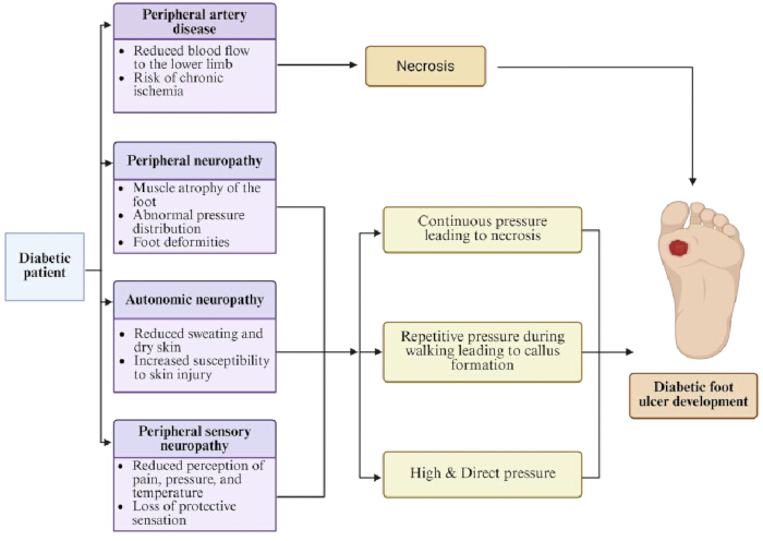
Figure 1: Major pathophysiological mechanisms contributing to diabetic foot ulcer development, including vascular insufficiency, neuropathy, altered plantar pressure, ischemia, and tissue injury, and their roles in ulcer initiation and progression. Please click here to view a larger version of this figure.
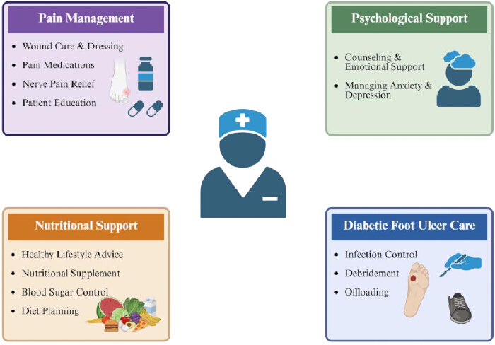
Figure 2: Key nurse-led roles in diabetic foot ulcer care, including pain management, psychological support, nutritional care, wound management, infection control, and offloading. Please click here to view a larger version of this figure.
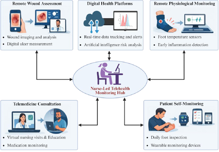
Figure 3: Digital technologies supporting nurse-led diabetic foot ulcer monitoring, including telemedicine, remote wound assessment, wearable sensors, and patient self-monitoring. Please click here to view a larger version of this figure.
Table 1: Clinical characteristics of commonly used wound dressings in diabetic foot ulcers136,137,138,139,140,141,142,143,144,145,146. Dressing selection should be individualized; no dressing type is universally superior. Please click here to download this file.
Table 2: Representative microorganisms identified in diabetic foot ulcers and their potential clinical significance when infection is present147,148,149. Microbial growth alone does not confirm infection, which should be diagnosed clinically. Please click here to download this file.
Disclosures
The authors declare no conflicts of interest. During the preparation of this manuscript, AI-assisted tools were used for language checking and polishing. The figures were created through the integration of BioRender and ChatGPT for conceptual design and visual refinement. All content was reviewed and approved by the authors, who take full responsibility for the final manuscript.
References
- Schmidt AM. Highlighting diabetes mellitus: The epidemic continues. Arterioscler Thromb Vasc Biol. 2018;38(1):e1-e8.
- Dawi J, et al. Diabetic foot ulcers: Pathophysiology, immune dysregulation, and emerging therapeutic strategies. Biomedicines. 2025;13(5):1076.
- McDermott K, Fang M, Boulton AJM, Selvin E, Hicks CW. Etiology, epidemiology, and disparities in the burden of diabetic foot ulcers. Diabetes Care. 2023;46(1):209-221.
- Jeffcoate W, et al. Causes, prevention, and management of diabetes-related foot ulcers. Lancet Diabetes Endocrinol. 2024;12(7):472-482.
- Kumbhar S, Bhatia M. Advancements and best practices in diabetic foot care: A comprehensive review of global progress. Diabetes Res Clin Pract. 2024;217:111845.
- Aditya C, et al. A comprehensive review on diabetic foot ulcer addressing vascular insufficiency, impaired immune response, and delayed wound healing mechanisms. Front Pharmacol. 2025;16:1622055.
- Guo Q, et al. Influencing factors for the recurrence of diabetic foot ulcers: A meta-analysis. Int Wound J. 2023;20(5):1762-1775.
- Jiang C, et al. Impact of multidisciplinary nursing interventions and blood glucose control on diabetic foot ulcer healing, patient emotions, and satisfaction. World J Psychiatry. 2025;15(11):106956.
- Nayeri ND, et al. Experiences of nurses within a nurse-led multidisciplinary approach in providing care for patients with diabetic foot ulcer. J Family Med Prim Care. 2020;9(6):3136-3141.
- Sulistyo AAH, et al. Education program to prevent diabetic foot ulcer in patient with diabetes: A scoping review. Afr J Reprod Health. 2024;28(10 Suppl):397-410.
- Swaminathan N, et al. Early intervention and care for diabetic foot ulcers in low and middle income countries: Addressing challenges and exploring future strategies: A narrative review. Health Sci Rep. 2024;7(5):e2075.
- National Institute for Health and Care Excellence (NICE). Diabetic foot problems: prevention and management. NICE Guideline No. 19. London: National Institute for Health and Care Excellence (NICE); 2023.
- Zamani N, et al. Engaging patients and caregivers to establish priorities for the management of diabetic foot ulcers. J Vasc Surg. 2021;73(4):1388-1395.e4.
- Ojo O, et al. Factors influencing the care and management of diabetic foot ulcers: A scoping review. Endocr Pract. 2025;31(3):380-389.
- Portela Dos Santos O, et al. Effectiveness of educational interventions to increase skills in evidence-based practice among nurses: The EDITcare systematic review. Healthcare (Basel). 2022;10(11):2204.
- Drovandi A, Seng L, Golledge J. Effectiveness of educational interventions for diabetes-related foot disease: A systematic review and meta-analysis. Diabetes Metab Res Rev. 2024;40(3):e3746.
- Monami M, et al. Effects of a short educational program for the prevention of foot ulcers in high-risk patients: A randomized controlled trial. Int J Endocrinol. 2015;2015:615680.
- Aalaa M, et al. Diabetic foot workshop: A strategy for improving the knowledge of diabetic foot care providers. Diabetes Metab Syndr. 2022;16(7):102543.
- Everett E, Mathioudakis N. Update on management of diabetic foot ulcers. Ann N Y Acad Sci. 2018;1411(1):153-165.
- Lydecker AD, et al. Chlorhexidine vs routine foot washing to prevent diabetic foot ulcers: A randomized clinical trial. JAMA Netw Open. 2025;8(2):e2460087.
- Armstrong DG, Boulton AJM, Bus SA. Diabetic foot ulcers and their recurrence. N Engl J Med. 2017;376(24):2367-2375.
- Armstrong DG, Tan TW, Boulton AJM, Bus SA. Diabetic foot ulcers: A review. JAMA. 2023;330(1):62-75.
- Lane KL, et al. Glycemic control and diabetic foot ulcer outcomes: A systematic review and meta-analysis of observational studies. J Diabetes Complications. 2020;34(10):107638.
- Miranda C, Da Ros R, Marfella R. Update on prevention of diabetic foot ulcer. Arch Med Sci Atheroscler Dis. 2021;6:e123-e131.
- Baker CJ, et al. Exercise training for people with diabetes-related foot ulcers: A systematic review of glycemia, fitness, and wound-healing outcomes. Can J Diabetes. 2025;49(3):164-173.e1.
- Alshammari L, O'Halloran P, McSorley O, Doherty J, Noble H. The effectiveness of foot care educational interventions for people living with diabetes mellitus: An umbrella review. J Tissue Viability. 2023;32(3):406-416.
- Ritonga SH, Gea IN, Harahap MA, Sagala NS, Napitupulu NF. Family experience in caring family members with diabetic foot ulcer: Phenomenological study. Enfermería Clínica. 2021;31:556-559.
- Wantonoro W, Komarudin K, Imania DR, Harun S, Nguyen TV. The influence of 6-month interdisciplinary accompaniment on family caregivers' knowledge and self-efficacy regarding diabetic wound care. SAGE Open Nurs. 2023;9:23779608231167801.
- Abbott CA, et al. Innovative intelligent insole system reduces diabetic foot ulcer recurrence at plantar sites: A prospective, randomised, proof-of-concept study. Lancet Digit Health. 2019;1(6):e308-e318.
- Boulton AJ, et al. Comprehensive foot examination and risk assessment: A report of the task force of the foot care interest group of the American Diabetes Association, with endorsement by the American Association of Clinical Endocrinologists. Diabetes Care. 2008;31(8):1679-1685.
- Jones PJ, Rowlands AV, Davies MJ, Bus SA. Diabetes foot ulcer prevention: A review of footwear width assessment for at-risk feet. J Foot Ankle Res. 2025;18(3):e70071.
- Ismail A, et al. Doppler sonographic characteristics and clinical outcomes of diabetic foot syndrome: A 5-year audit from a tertiary hospital in northern Nigeria. J West Afr Coll Surg. 2024;14(2):127-133.
- Khan J, et al. Accuracy of ultrasound for the diagnosis of peripheral vascular disease in diabetic foot: A systematic review. Semin Ultrasound CT MR. 2026;47(1):54-66.
- Mennes OA, Van Netten JJ, Slart R, Steenbergen W. Novel optical techniques for imaging microcirculation in the diabetic foot. Curr Pharm Des. 2018;24(12):1304-1316.
- Anderson M, Arvidson C. Nurse practitioners and barriers to diabetic foot treatment. J Am Assoc Nurse Pract. 2020;32(12):795-799.
- GBD 2021 Diabetes Collaborators. Global, regional, and national burden of diabetes from 1990 to 2021, with projections of prevalence to 2050: A systematic analysis for the Global Burden of Disease Study 2021. Lancet. 2023;402(10397):203-234.
- Zhou J, Zhou L. Comprehensive nursing model for diabetic foot ulcers: A strategy to improve prognosis and quality of life. Medicine (Baltimore). 2024;103(26):e38674.
- Goswami S, Eckert L, Sturgill M, Ramos R, Bridgeman MB. Patient education on self-administration of insulin via pen: An interprofessional team-based educational initiative. J Interprof Educ Pract. 2023;32:100663.
- Rismayanti IDA, et al. Early detection to prevent foot ulceration among type 2 diabetes mellitus patient: A multi-intervention review. J Public Health Res. 2022;11(2):2752.
- Bus SA, et al. Guidelines on offloading foot ulcers in persons with diabetes (IWGDF 2023 update). Diabetes Metab Res Rev. 2024;40(3):e3647.
- Lazzarini PA, et al. Effectiveness of offloading interventions for people with diabetes-related foot ulcers: A systematic review and meta-analysis. Diabetes Metab Res Rev. 2024;40(3):e3650.
- Armstrong DG, et al. Off-loading the diabetic foot wound: A randomized clinical trial. Diabetes Care. 2001;24(6):1019-1022.
- Ababneh A, Finlayson K, Edwards H, Lazzarini PA. Factors associated with adherence to using removable cast walker treatment among patients with diabetes-related foot ulcers. BMJ Open Diabetes Res Care. 2022;10(1):e002640.
- Schaper NC, et al. Practical guidelines on the prevention and management of diabetes-related foot disease (IWGDF 2023 update). Diabetes Metab Res Rev. 2024;40(3):e3657.
- Van Netten JJ, et al. Prevention of foot ulcers in persons with diabetes at risk of ulceration: A systematic review and meta-analysis. Diabetes Metab Res Rev. 2024;40(3):e3652.
- Bus SA, et al. Effect of custom-made footwear on foot ulcer recurrence in diabetes: A multicenter randomized controlled trial. Diabetes Care. 2013;36(12):4109-4116.
- Crews RT, et al. Role and determinants of adherence to off-loading in diabetic foot ulcer healing: A prospective investigation. Diabetes Care. 2016;39(8):1371-1377.
- Wang Q, et al. Mechanisms of microbial infection and wound healing in diabetic foot ulcer: Pathogenicity in the inflammatory-proliferative phase, chronicity, and treatment strategies. Front Endocrinol (Lausanne). 2025;16:1657928.
- Wang R, et al. Diabetic wound repair: From mechanism to therapeutic opportunities. MedComm (2020). 2025;6(10):e70406.
- Dayya D, et al. Debridement of diabetic foot ulcers: Public health and clinical implications - a systematic review, meta-analysis, and meta-regression. BMJ Surg Interv Health Technol. 2022;4(1):e000081.
- Vas PRJ, Edmonds ME. Approach to a new diabetic foot ulceration. In: The Foot in Diabetes. 2020:481-493.
- Anzali BC, et al. Surgical sharp debridement alongside maggot debridement therapy (MDT) for the treatment of diabetic foot ulcers (DFUs): A systematic review of case reports. Surg Pract Sci. 2025;20:100270.
- Amadeh A, Mohebbi N, Amadeh Z, Jamshidbeigi A. Comparative efficacy of autolytic and collagenase-based enzymatic debridement in chronic wound healing: A comprehensive systematic review. Int Wound J. 2025;22(4):e70177.
- Wang L, et al. Research progress on the application of novel wound healing dressings in different stages of wound healing. Pharmaceutics. 2025;17(8).
- Chuangsuwanich A, Rachata P. Wound dressings: Principles and modern applications. n: De Francesco F, Turksen K, eds. Management and Strategies for Wound Healing. Cham: Springer Nature Switzerland; 2026:439-461.
- Ko A, Liao C. Hydrogel wound dressings for diabetic foot ulcer treatment: Status-quo, challenges, and future perspectives. BMEMat. 2023;1(3):e12037.
- Kamińska MS, et al. Effectiveness of hydrocolloid dressings for treating pressure ulcers in adult patients: A systematic review and meta-analysis. Int J Environ Res Public Health. 2020;17(21).
- Yousefian F, et al. Antimicrobial wound dressings: A concise review for clinicians. Antibiotics (Basel). 2023;12(9).
- Simões D, et al. Recent advances on antimicrobial wound dressing: A review. Eur J Pharm Biopharm. 2018;127:130-141.
- Tang Y, et al. Negative pressure wound therapy promotes wound healing of diabetic foot ulcers by up-regulating PRDX2 in wound margin tissue. Sci Rep. 2023;13(1):16192.
- Hasan MY, Teo R, Nather A. Negative-pressure wound therapy for management of diabetic foot wounds: A review of the mechanism of action, clinical applications, and recent developments. Diabet Foot Ankle. 2015;6:27618.
- Zeng Y, Tu J, Qin J. The mechanism by which negative pressure wound therapy promotes wound healing. J Tissue Viability. 2026;35(1):100982.
- Debels H, Hamdi M, Abberton K, Morrison W. Dermal matrices and bioengineered skin substitutes: A critical review of current options. Plast Reconstr Surg Glob Open. 2015;3(1):e284.
- Haxho F, et al. Wound care - part II: Tissue-engineered skin substitutes & other advanced wound therapies. J Am Acad Dermatol. 2025;95(3):605-620.
- Ahmed AB, et al. Platelet-rich plasma (PRP) and recombinant growth factor therapies in cutaneous wound healing: Mechanisms, clinical applications, and future directions. J Clin Med. 2025;14(23):8583.
- Armstrong DG, Lipsky BA. Diabetic foot infections: Stepwise medical and surgical management. Int Wound J. 2004;1(2):123-132.
- Senneville É, et al. IWGDF/IDSA guidelines on the diagnosis and treatment of diabetes-related foot infections (IWGDF/IDSA 2023). Diabetes Metab Res Rev. 2024;40(3):e3687..
- Senneville É, et al. Diagnosis of infection in the foot of patients with diabetes: A systematic review. Diabetes Metab Res Rev. 2024;40(3):e3723.
- Lavery LA, et al. The infected diabetic foot: Re-evaluating the Infectious Diseases Society of America diabetic foot infection classification. Clin Infect Dis. 2020;70(8):1573-1579.
- Lavery LA, Armstrong DG, Murdoch DP, Peters EJ, Lipsky BA. Validation of the Infectious Diseases Society of America's diabetic foot infection classification system. Clin Infect Dis. 2007;44(4):562-565.
- Singer M, et al. The third international consensus definitions for sepsis and septic shock (Sepsis-3). JAMA. 2016;315(8):801-810.
- Huang Y, et al. A comparison of tissue versus swab culturing of infected diabetic foot wounds. Int J Endocrinol. 2016;2016:8198714.
- Demetriou M, et al. Tissue and swab culture in diabetic foot infections: Neuropathic versus neuroischemic ulcers. Int J Low Extrem Wounds. 2013;12(2):87-93.
- Calderwood MS, et al. Strategies to prevent surgical site infections in acute-care hospitals: 2022 update. Infect Control Hosp Epidemiol. 2023;44(5):695-720.
- Kavitha KV, et al. Choice of wound care in diabetic foot ulcer: A practical approach. World J Diabetes. 2014;5(4):546-556.
- Boulton AJ. The diabetic foot: A global view. Diabetes Metab Res Rev. 2000;16 Suppl 1:S2-S5.
- Alexiadou K, Doupis J. Management of diabetic foot ulcers. Diabetes Ther. 2012;3(1):4.
- Wang YB, et al. Clinical comprehensive treatment protocol for managing diabetic foot ulcers: A retrospective cohort study. World J Clin Cases. 2024;12(17):2976-2982.
- Ghadeer A, et al. Diabetic foot ulcer related pain and its impact on health-related quality of life. J Tissue Viability. 2025;34(2):100856.
- Price R, et al. Oral and topical treatment of painful diabetic polyneuropathy: Practice guideline update SHORT ABSTRACT: Report of the AAN Guideline Subcommittee. Neurology. 2022;98(1):31-43.
- Ren Y, et al. Assessment and management of pain during dressing change in patients with diabetic foot ulcers: A best practice implementation project. JBI Database System Rev Implement Rep. 2019;17(10):2193-2201.
- Fitridge R, et al. The intersocietal IWGDF, ESVS, SVS guidelines on peripheral artery disease in people with diabetes and a foot ulcer. Diabetes Metab Res Rev. 2024;40(3):e3686.
- Szeto CC, et al. Non-steroidal anti-inflammatory drug (NSAID) therapy in patients with hypertension, cardiovascular, renal or gastrointestinal comorbidities: Joint APAGE/APLAR/APSDE/APSH/APSN/POA recommendations. Gut. 2020;69(4):617-629.
- American Geriatrics Society Beers Criteria Update Expert Panel. American Geriatrics Society 2023 updated AGS Beers Criteria® for potentially inappropriate medication use in older adults. J Am Geriatr Soc. 2023;71(7):2052-2081.
- Frescos N, Copnell B. Podiatrists' views of assessment and management of pain in diabetes-related foot ulcers: A focus group study. J Foot Ankle Res. 2020;13(1):29.
- Woo K, González CVS, Amdie FZ, De Gouveia Santos VLC. Exploring the effect of wound related pain on psychological stress, inflammatory response, and wound healing. Int Wound J. 2024;21(7):e14942.
- Zhang D, Lee EKP, Mak ECW, Ho CY, Wong SYS. Mindfulness-based interventions: An overall review. Br Med Bull. 2021;138(1):41-57.
- Pan WJ, Wang SF. Understanding patients' emotional needs to strengthen therapeutic relationships: A deep insight into narrative nursing. World J Psychiatry. 2025;15(3):103093.
- Zhu X, et al. Healing through empowerment and active listening: Experience-based co-design of a nurse-led personalised self-care support intervention for primary care patients with diabetic foot ulcers. Health Expect. 2025;28(4):e70386.
- Nakao M, Shirotsuki K, Sugaya N. Cognitive-behavioral therapy for management of mental health and stress-related disorders: Recent advances in techniques and technologies. Biopsychosoc Med. 2021;15(1):16.
- Cederholm T, et al. GLIM criteria for the diagnosis of malnutrition - a consensus report from the global clinical nutrition community. Clin Nutr. 2019;38(1):1-9.
- Yuan Z, et al. Effectiveness of Global Leadership Initiative on Malnutrition and Subjective Global Assessment for diagnosing malnutrition and predicting wound healing in patients with diabetic foot ulcers. Br J Nutr. 2024;132(1):21-30.
- Ran Q, et al. Risk factors for malnutrition in patients with diabetic foot ulcer and its association with prolonged length of hospitalization. Nutr Diabetes. 2024;14(1):26.
- Apergi K, Dimosthenopoulos C, Papanas N. The role of nutrients and diet characteristics in the management of diabetic foot ulcers: A systematic review. Int J Low Extrem Wounds. 2025;24(3):525-541.
- Fawzy El-Sayed KM, Cosgarea R, Sculean A, Doerfer C. Can vitamins improve periodontal wound healing/regeneration? Periodontol 2000. 2024;94(1):539-602.
- Saeg F, Orazi R, Bowers GM, Janis JE. Evidence-based nutritional interventions in wound care. Plast Reconstr Surg. 2021;148(1):226-238.
- Du F, Alhaskawi A, Zou X, Zhou W, Lu H. Advanced applications of vitamin B complex in plastic and cosmetic surgery: Mechanisms and therapeutic benefits. Int J Vitam Nutr Res. 2025;95(4):39087.
- Lin PH, et al. Zinc in wound healing modulation. Nutrients. 2017;10(1):16.
- McDaniel JC, Belury M, Ahijevych K, Blakely W. Omega-3 fatty acids effect on wound healing. Wound Repair Regen. 2008;16(3):337-345.
- Alexander JW, Supp DM. Role of arginine and omega-3 fatty acids in wound healing and infection. Adv Wound Care (New Rochelle). 2014;3(11):682-690.
- Moore ZE, Corcoran MA, Patton D. Nutritional interventions for treating foot ulcers in people with diabetes. Cochrane Database Syst Rev. 2020;7(7):CD011378.
- Chen P, et al. Guidelines on interventions to enhance healing of foot ulcers in people with diabetes (IWGDF 2023 update). Diabetes Metab Res Rev. 2024;40(3):e3644.
- Armstrong DG, et al. Effect of oral nutritional supplementation on wound healing in diabetic foot ulcers: A prospective randomized controlled trial. Diabet Med. 2014;31(9):1069-1077.
- Van Den Berg G, et al. Factors influencing the delivery of nutritional care by nurses for hospitalised medical patients with malnutrition: A qualitative study. J Clin Nurs. 2023;32(15-16):5147-5159.
- Cangelosi G, et al. Nutritional support in the prevention and treatment of pressure ulcers in healthy aging: A systematic review of nursing interventions in community care. Geriatrics (Basel). 2025;10(1):17.
- Ajibade B. Assessing the patient's needs and planning effective care. Br J Nurs. 2021;30(20):1166-1171.
- Vudathaneni VKP, et al. The impact of telemedicine and remote patient monitoring on healthcare delivery: A comprehensive evaluation. Cureus. 2024;16(3):e55534.
- Choudhary P, et al. The challenge of sustainable access to telemonitoring tools for people with diabetes in Europe: Lessons from COVID-19 and beyond. Diabetes Ther. 2021;12(9):2311-2327.
- Maxant G, Pastrav M, Gogeneata I, Bajcz C, Bertaux AC. Clinical and medico-economic benefits of remote monitoring of chronic wounds. Int Wound J. 2025;22(3):e70140.
- Dardari D, et al. Hospital stays and costs of telemedical monitoring versus standard follow-up for diabetic foot ulcer: An open-label randomised controlled study. Lancet Reg Health Eur. 2023;32:100686.
- Blake H. Digital wound management: How it works and its potential benefits in wound care practice. Nurs Stand. 2025;40(6):61-66.
- Lazarus J, et al. Data-driven digital health technologies in the remote clinical care of diabetic foot ulcers: A scoping review. Front Clin Diabetes Healthc. 2023;4:1212182.
- Cay G, et al. Towards a remote patient monitoring platform for comprehensive risk evaluations for people with diabetic foot ulcers. Sensors (Basel). 2024;24(10):2979.
- Hazenberg C, Aan De Stegge WB, Van Baal SG, Moll FL, Bus SA. Telehealth and telemedicine applications for the diabetic foot: A systematic review. Diabetes Metab Res Rev. 2020;36(3):e3247.
- Smith-Strøm H, et al. The effect of telemedicine follow-up care on diabetes-related foot ulcers: A cluster-randomized controlled noninferiority trial. Diabetes Care. 2018;41(1):96-103.
- Cheng J, et al. Recent progress in flexible wearable sensors for real-time health monitoring: Materials, devices, and system integration. Micromachines (Basel). 2025;16(10):1124.
- Orlando G, Prior Y, Reeves ND, Vileikyte L. Patient and provider perspective of smart wearable technology in diabetic foot ulcer prevention: A systematic review. Medicina (Kaunas). 2021;57(12):1359.
- Wang Q, et al. A wireless, self-powered smart insole for gait monitoring and recognition via nonlinear synergistic pressure sensing. Sci Adv. 2025;11(16):eadu1598.
- Tamura T. Advanced wearable sensors technologies for healthcare monitoring. Sensors (Basel). 2025;25(2):322.
- Zhu Z, et al. PEDOT:PSS-based wearable flexible temperature sensor and integrated sensing matrix for human body monitoring. ACS Appl Mater Interfaces. 2024;16(41):56082-56094.
- Frykberg RG, et al. Feasibility and efficacy of a smart mat technology to predict development of diabetic plantar ulcers. Diabetes Care. 2017;40(7):973-980.
- Bus SA, et al. Effectiveness of at-home skin temperature monitoring in reducing the incidence of foot ulcer recurrence in people with diabetes: A multicenter randomized controlled trial (DIATEMP). BMJ Open Diabetes Res Care. 2021;9(1):e002392.
- Anisuzzaman DM, et al. Image-based artificial intelligence in wound assessment: A systematic review. Adv Wound Care (New Rochelle). 2022;11(12):687-709.
- Madić M, Vitković N, Damnjanović Z, Stojanović S. Machine learning applications for venous ulcer assessment and wound care: A review. Diagnostics (Basel). 2026;16(3):373.
- Basiri R, Saleh A, Khan SS, Popovic MR. Temporal machine learning framework for diabetic foot ulcer healing trajectory prediction. Biomed Eng Online. 2026;25(1):41.
- Wang Y, et al. Establishing a clinical prediction model for diabetic foot ulcers in type 2 diabetic patients with lower extremity arteriosclerotic occlusion using machine learning. Sci Rep. 2025;15(1):11737.
- Twilt M. mHealth applications are they the future or another burden for our patients? Mhealth. 2023;9:9.
- Ruissen MM, et al. Clinical impact of an integrated e-health system for diabetes self-management support and shared decision making (POWER2DM): A randomised controlled trial. Diabetologia. 2023;66(12):2213-2225.
- Wang Y, et al. Effectiveness of mobile health interventions on diabetes and obesity treatment and management: Systematic review of systematic reviews. JMIR Mhealth Uhealth. 2020;8(4):e15400.
- Kwon SY, Moon JS. Advances in continuous glucose monitoring: Clinical applications. Endocrinol Metab (Seoul). 2025;40(2):161-173.
- Lanzola G, et al. Remote blood glucose monitoring in mHealth scenarios: A review. Sensors (Basel). 2016;16(12):1983.
- Zivkovic J, et al. Transitioning from self-monitoring of blood glucose to continuous glucose monitoring in combination with a mHealth app improves glycemic control in people with type 1 and type 2 diabetes. Diabetes Technol Ther. 2025;27(1):10-18.
- Mumtaz H, et al. Current challenges and potential solutions to the use of digital health technologies in evidence generation: A narrative review. Front Digit Health. 2023;5:1203945.
- Kuznetsova M, et al. Implementation of a continuous patient monitoring system in the hospital setting: A qualitative study. Jt Comm J Qual Patient Saf. 2024;50(4):235-246.
- Luo G, Liu Y, Wang A. Practical guidelines for the prevention and management of diabetic foot disease in China. Burns Trauma. 2025;13:tkaf064.
- Hargis A, Yaghi M, Bermudez NM, Gefen A. Foam dressings for wound healing. Curr Dermatol Rep. 2024;13(1):28-35.
- Nasra S, Patel M, Shukla H, Bhatt M, Kumar A. Functional hydrogel-based wound dressings: A review on biocompatibility and therapeutic efficacy. Life Sci. 2023;334:122232.
- Dumville JC, Deshpande S, O'Meara S, Speak K. Hydrocolloid dressings for healing diabetic foot ulcers. Cochrane Database Syst Rev. 2012;(2):CD009099.
- Tashkandi H. Honey in wound healing: An updated review. Open Life Sci. 2021;16(1):1091-1100.
- Feketshane Z, et al. Dissolvable sodium alginate-based antibacterial wound dressing patches: Design, characterization, and in vitro biological studies. Int J Biol Macromol. 2023;232:123460.
- Mahheidari N, et al. Biological macromolecules alginate-carboxymethyl cellulose (ALG-CMC) hydrogel loaded with botulinum toxin type A for skin wound treatment and functional tissue regeneration. Int J Biol Macromol. 2025;316(Pt 2):144668.
- Sun D, et al. Silicone elastomer gel impregnated with 20(S)-protopanaxadiol-loaded nanostructured lipid carriers for ordered diabetic ulcer recovery. Acta Pharmacol Sin. 2020;41(1):119-128.
- Gwak HC, et al. Efficacy of a povidone-iodine foam dressing (Betafoam) on diabetic foot ulcer. Int Wound J. 2020;17(1):91-99.
- Haynes JS. A clinical evaluation of a charcoal dressing to reduce malodour in wounds. Br J Nurs. 2018;27(6):S36-S42.
- Liang K, Liu Y, Jiang F. Analysis of therapeutic effect of silver-based dressings on chronic wound healing. Int Wound J. 2024;21(8):e70006.
- Xu H, et al. Preparation and characterization of a polyurethane-based sponge wound dressing with a superhydrophobic layer and an antimicrobial adherent hydrogel layer. Acta Biomater. 2024;181:235-248.
- Maity S, et al. A systematic review of diabetic foot infections: Pathogenesis, diagnosis, and management strategies. Front Clin Diabetes Healthc. 2024;5:1393309.
- Chai W, et al. The profile of microbiological pathogens in diabetic foot ulcers. Front Med (Lausanne). 2021;8:656467.
- Holmes CL, Anderson MT, Mobley HLT, Bachman MA. Pathogenesis of gram-negative bacteremia. Clin Microbiol Rev. 2021;34(2):10-128.