Models of bacterial sepsis in preterm neonates are essential, yet existing rodent models lack clinical relevance. A highly translational model of neonatal sepsis was developed using preterm piglets infected with live Staphylococcus epidermidis.
Method Article
A Translational Preterm Piglet Model For Neonatal Sepsis
* These authors contributed equally
In This Article
Summary
Abstract
Preterm infants are particularly vulnerable to bacterial infections that may rapidly progress to life-threatening sepsis, a state of excessive inflammation and organ dysfunction. To investigate disease mechanisms and advance therapeutic development, robust translational animal models that replicate the complex pathophysiology and clinical presentation of neonatal sepsis are critically needed. This protocol presents a clinically relevant preterm piglet model of neonatal sepsis, induced via intra-arterial infusion of live Staphylococcus epidermidis shortly after birth or on postnatal day three. Piglets are delivered at 90% gestation by cesarean section and cared for under neonatal intensive care conditions. These include temperature- and oxygen-controlled incubators and an indwelling umbilical arterial catheter, which enables delivery of parenteral nutrition and serial blood sampling. Following infection with S. epidermidis, piglets are monitored for 24–48 h. The incidence and severity of sepsis can be modulated by the dose of bacteria and the supply of parenteral glucose. Higher provision of parenteral glucose accelerates pro-inflammatory immune responses, leading to clinical deterioration. Lethal sepsis and humane endpoints are defined by arterial blood gas parameters (pH ≤ 7.1) combined with clinical signs of hemodynamic compromise such as lethargy, skin discoloration, and tachypnea. This model provides a unique platform to explore how energy metabolism and immunomodulatory interventions affect bacterial clearance, systemic inflammation, and tissue injury during neonatal infection. It holds significant potential to inform clinical strategies for the prevention and treatment of neonatal sepsis.
Introduction
Neonatal infection is a leading cause of mortality and long-term morbidity worldwide, particularly among preterm infants, but also in term infants with low birth weight or perinatal complications1,2. Early diagnosis remains challenging due to non-specific clinical signs, immature and highly variable immune responses, and a lack of sensitive and specific biomarkers capable of distinguishing infection from non-infectious inflammation3,4. As a result, clinicians frequently initiate empirical broad-spectrum antibiotic therapy, often before microbiological confirmation, contributing to antibiotic overuse, disruption of early-life microbial colonization, and the development of antimicrobial resistance5.
Neonatal sepsis is a life-threatening condition caused by a dysregulated host response to infection that leads to organ dysfunction4. Although infection and sepsis represent distinct biological and clinical entities, the terms are often used interchangeably in neonatology literature. Clinically, neonatal sepsis is commonly classified as early- or late-onset sepsis (EOS, LOS), occurring before or after 72 h of life, respectively1. EOS is typically caused by pathogens transferred during birth, such as Escherichia coli or Group B Streptococcus. In contrast, LOS frequently presents with non-specific symptoms, may be culture-negative, and is associated with risk factors such as invasive procedures, parenteral nutrition, and prolonged hospitalization. The most common causative organisms include coagulase-negative staphylococci, particularly Staphylococcus epidermidis, as well as Gram-negative bacteria, although fungal and polymicrobial infections also occur1,2,6. Regardless of etiology, neonatal infections can converge on a common sepsis phenotype1,2. Importantly, preterm infants exhibit developmental immaturity of both innate and adaptive immunity, together with limited metabolic reserves, which may affect responsiveness to infection7.
Progress in diagnosis and treatment has been hindered by limitations of existing experimental systems. In vitro approaches allow for mechanistic studies of immune pathways but fail to capture the integrated physiology of complex immune responses. Rodent models, while valuable for genetic and molecular studies into neonatal immune responses, differ substantially from humans in immune ontogeny, metabolism, and size8,9. The clinical management of sepsis is difficult to study in these models due to limited opportunities for intensive monitoring, repeated blood sampling, and testing of clinically realistic supportive or therapeutic care.
Large animal models, particularly preterm newborn piglets, offer a powerful complementary approach. Pigs share close anatomical, physiological, and immunological similarities with humans, including comparable immune cell populations, pattern-recognition receptor signaling, and metabolic regulation10,11,12,13,14. Importantly, pigs are widely used as models for adult sepsis13,15. The physical scale of pigs permits the use of clinically relevant instrumentation, continuous physiological monitoring, repeated bio-sampling, and standardized intensive care interventions such as parenteral nutrition, fluid therapy, antibiotics, and respiratory support. Experimental infection can also be studied in the context of developmental immaturity, enabling investigation of disease trajectories, host responses, and treatment effects under conditions that closely resemble clinical practice.
This paper describes a standardized neonatal sepsis model in preterm piglets designed as a flexible translational platform. The model allows for the controlled induction of a systemic bacterial infection and longitudinal assessment of clinical, immunological, and metabolic responses, which can be used to explore the pathophysiology of neonatal sepsis and to evaluate the efficacy of novel preventive and therapeutic interventions.
Protocol
All procedures detailed were conducted in accordance with the EU directive 2010/63/EU for animal experiments and approved by the Danish Animal Experiments Inspectorate (license number: 2020-15-0201-00520).
1. Procedures surrounding birth
- Perform elective cesarean section to deliver preterm piglets at 90% of gestation (gestational days 106–107).
- Obtain a pregnant sow (Landrace × Large White, third or fourth parity) from a commercial Specific Pathogen Free farm.
NOTE: The sow must be impregnated via artificial insemination (× Duroc) to ensure the exact day of conception is known. - Fast the sow overnight (minimum 12 h) with ad libitum access to water prior to surgery. Induce sedation via intramuscular injection of a cocktail containing atropine (2 mL per sow), butorphanol (2 mL/100 kg body weight; 10 mg/mL), and Zoletil 50 Vet (3.3 mL/100 kg body weight; tiletamine 25/mL and zolazepam 25 mg/mL).
- Once sedated, insert an ear vein catheter and induce general anesthesia with propofol (10 mg/mL), titrated until the loss of the palpebral reflex and jaw tone is achieved.
- Perform endotracheal intubation under direct laryngoscopy to secure the airway. Maintain a surgical plane of anesthesia throughout the cesarean section using vaporized isoflurane delivered in 100% oxygen (1–2 L/min) via a calibrated vaporizer.
- Initiate at 5% and maintain at 1.5–2.5%, titrating the concentration to the lowest effective dose based on the heart rate and peripheral oxygen saturation.
NOTE: If apnea occurs, initiate mechanical ventilation to ensure adequate oxygenation. - Position the sow in right lateral recumbency. Perform an aseptic preparation of the surgical site. Administer a total volume of 120 mL lidocaine (10 mg/mL) via subcutaneous and intramuscular infiltration along the intended left flank incision line.
- Perform a left flank laparotomy using a scalpel to expose and exteriorize the uterine horns. Incise the uterus horns along the greater curvature to deliver individual piglets.
- Prior to transecting the umbilical cord, milk the cord 2–3 times toward the piglet to increase hemoglobin concentration and iron stores. Place a navel clamp labeled A–Z around the umbilical cord together with gauze to prevent bleeding, and transect the cord.
- Immediately after delivery, place each piglet in a polythene bag, leaving the head exposed. The bag minimizes heat loss, thereby reducing the risk of hypothermia.
- Administer a fixed volume of 0.1 mL of doxapram (20 mg/mL) and 0.1 mL of flumazenil (0.1 mg/mL) intramuscularly in the neck. Doxapram stimulates spontaneous respiration, whereas flumazenil, a benzodiazepine antagonist, partially reverses anesthesia transferred from the sow during the cesarean section.
- Transfer the piglets to a neonatal intensive care unit.
- Obtain a pregnant sow (Landrace × Large White, third or fourth parity) from a commercial Specific Pathogen Free farm.
- Resuscitation, randomization, and catheterization
- Weigh and determine the sex of each piglet upon transfer to the neonatal intensive care unit.
- For piglets unable to breathe spontaneously, perform resuscitation on an individual basis. Repeat the doxapram and/or flumazenil doses, and provide positive-pressure ventilation as needed to stabilize respiration.
- Once the piglets are physiologically stable and while still anesthetized from maternally transferred anesthesia, use aseptic technique to insert an umbilical arterial catheter (4 Fr PVC feeding tube) into one of the arteries of the transected umbilical cord. Advance the catheter tip to the dorsal aorta. Secure the catheter to the skin with sutures and connect it to an infusion line for the provision of parenteral nutrition.
NOTE: In the preterm pig, intra-arterial administration is the established standard over the umbilical vein to avoid potential hepatic necrosis resulting from the high risk of the catheter tip malpositioning within the liver parenchyma rather than the ductus venosus21. - Stratify piglets by birth weight and sex and randomly assign them to treatment groups.
- Housing conditions
- House each piglet individually in a heated, padded incubator with controlled ventilation.
- Provide supplemental oxygen (1–2 L/min) for up to 12 h after birth to prevent hypoxia.
- Monitor rectal temperature during the first 12 h after birth. Adjust heating conditions as needed to maintain a core temperature of 38–39 °C (target: 38.5 °C).
NOTE: A brief period of hypothermia is expected immediately after birth and during catheter placement.
2. Passive immunization via plasma infusion
NOTE: Since pigs do not transfer immunoglobulins transplacentally, provide partial passive immunization for experiments lasting longer than 24 h to prevent spontaneous infections.
- Collect maternal blood from the sow following cesarean section into a sterile heparin-coated container. Centrifuge the blood in sterile Falcon tubes at 4 °C, 2500 × g, for 10 min, and repeat once. After separation, carefully collect the plasma into new sterile Falcon tubes and store at 4 °C until use.
- Within 12 h after birth, administer the plasma at a dose of 8 mL/kg as a 30 min continuous intra-arterial infusion using an infusion pump.
3. Induction of experimental infection
NOTE: The experimental infection can be induced either 3–4 h after birth or on postnatal day 3.
- Preparation of the bacterial suspension
- Thaw the frozen S. epidermidis stock vial (WT-1457, 15–25% glycerol) at room temperature. Vortex briefly (2–3 s) to obtain a homogenous suspension.
NOTE: According to the risk group classification for infectious agents, S. epidermidis is classified as a WHO Risk Group 1 microorganism. WHO Risk Group 1 microorganisms are unlikely to cause human disease and do not require special considerations. - Prepare an overnight culture by inoculating 20 mL of brain heart infusion (BHI) broth with 5 µL of the thawed S. epidermidis stock in a sterile 100 mL Erlenmeyer flask. Incubate at 37 °C with orbital shaking at 180 rpm for ~16 h, allowing the culture to reach the stationary phase. Include uninoculated BHI broth control under identical conditions to verify medium sterility.
- Prepare a new culture by inoculating 20 mL of BHI broth with 200 µL of the overnight culture (1:100 dilution) in a sterile 100 mL Erlenmeyer flask. Prepare sufficient culture to infect all piglets (see calculations in steps 3.1.4 – 3.1.6). Incubate at 37 °C with orbital shaking at 180 rpm for 2.5 h, until bacteria reach the early-logarithmic growth phase (Supplementary Figure 1A).
- Estimate the total culture volume required to infect all piglets based on the titrated target dose, average body weight, number of piglets, and culture density.
- Assume the culture will reach an optical density at 600 nm (OD) of ≥1 (Supplementary Figure 1B) after the 2.5 h incubation and use an established conversion rate of OD of 1 = 3 × 10⁸ colony-forming units (CFU)/mL.
- Include 15% extra volume to account for handling losses. Calculate the estimated culture volume using the formula:
VEstimate = [Target dose (CFU/kg BW) × BW) × BW × N × 1.15] / 3 × 10⁸ CFU/mL (1)
where:
VEstimate = estimated total culture volume required (mL)
BW = average body weight of piglet (kg)
N = total number of piglets - Following incubation, determine the OD of the culture. Dilute the sample as necessary to keep readings within the linear range of the spectrophotometer.
- Calculate the final culture volume required to achieve the target CFU concentration using the formula:
VFinal = OD1 · VEstimate / Measured OD (2)
where:
VFinal = final culture volume required (mL) - Transfer the calculated VFinal into 50 mL Falcon tubes and centrifuge at 3,000 × g for 10 min at room temperature.
- Carefully discard the supernatant.
- Resuspend the bacterial pellet in 3 x VFinal of sterile 0.9% saline to prepare for intra-arterial administration (3 mL/kg body weight over 3 min). Ensure that the solution is free of aggregates.
- Prepare 50 mL infusion syringes with the bacterial suspension.
- Prepare a separate 50 mL infusion syringe with sterile 0.9% saline for uninfected controls.
- Thaw the frozen S. epidermidis stock vial (WT-1457, 15–25% glycerol) at room temperature. Vortex briefly (2–3 s) to obtain a homogenous suspension.
- Infection procedure
- Administer the bacterial suspension or sterile saline to each piglet through the umbilical arterial catheter using an infusion pump set to deliver the target dose over 3 min. Maintain a 5-min interval between consecutive piglets.
- Immediately after infection, start the continuous infusion of parenteral nutrition to flush the catheter and ensure delivery of the entire dose.
- Confirm the dose by saving the bacterial suspension at 4 °C. Later, this can be diluted and plated out on blood agar plates to determine the bacterial density (see step 8)
4. Parenteral nutrition
- Provide total parenteral nutrition (TPN) to piglets throughout the duration of the experiment. For example, use a three-chamber human TPN bag, such as Kabiven 1900 kcal, modified to meet the macronutrient requirements of newborn piglets.
- Prepare the parenteral nutrition solution under sterile conditions.
- Remove 300 mL from the glucose chamber.
- Remove 70 mL from the lipid chamber.
- Add 155 mL of 50% glucose to the glucose chamber.
- Add 214 mL of Vamin to the glucose chamber.
- Add 150 mL of sterile water to the glucose chamber.
- Add 0.1 mL of heparin (5000 IE/mL) to the glucose chamber.
- Mix the contents of all three chambers thoroughly.
NOTE: This results in a parenteral nutrition solution with the macronutrient composition shown in Table 1. Use this formulation as the standard control diet when evaluating the effects of alternative nutritional regimens on infection responses. - Store the parenteral nutrition at 4 °C until use. Prepare the solution in batches, and do not use it beyond 48 h after preparation.
NOTE: The model can be modified to evaluate the effects of enteral feeding and gut stimulation by fitting piglets with orogastric feeding tubes.
- Delivery of parenteral nutrition
- Fill 50 mL sterile infusion syringes with parenteral nutrition and infuse continuously through the intra-arterial catheter using an infusion pump.
- Set the infusion rate to 6 mL/kg/h. Weigh the piglets daily and adjust the dose according to the updated body weight.
NOTE: For longer-term studies, if peripheral edema (overhydration) is observed, reduce the infusion rate to 4 mL/kg/h. - Monitor the infusion pumps regularly to ensure that the syringes do not run empty. Refill the syringes as needed.
5. Clinical monitoring
- Assess piglets regularly throughout the study to determine if action is needed. After bacterial infusion, animals should be monitored every 1–2 h by experienced personnel until planned euthanasia.
NOTE: Clinical deterioration, the progression from no symptoms to humane endpoints, can occur rapidly, within 30–60 min.- Observe symptoms related to respiration, including tachypnea, respiratory distress, cyanosis, and agonal breathing.
NOTE: In cases of hypoxia (oxygen saturation < 80%), oxygen supply should be initiated/increased. - Observe symptoms related to circulation, including discoloration, cold extremities, prolonged capillary refill time, tachycardia, and bradycardia.
- Observe symptoms related to activity, including lethargy, inability to stand or walk (relevant from postnatal day 3 onward), and reactivity to pain stimulus.
- Observe symptoms related to coagulation, such as ecchymosis or petechial bleeding in the skin. Apply gentle pressure to discolored areas; if the redness does not blanch, classify the finding as skin bleeding.
- Observe symptoms related to respiration, including tachypnea, respiratory distress, cyanosis, and agonal breathing.
- If clinical symptoms are observed, perform blood gas measurements to assess pH, lactate, oxygen saturation, and partial pressure of carbon dioxide relative to baseline values (see step 6.3).
NOTE: Continuous clinical monitoring (of e.g., blood pressure, heart rate, and respiratory rate) may be used but is not routinely employed in these experiments.
6. Blood sampling
NOTE: Longitudinal blood sampling can be performed in piglets via the intra-arterial catheter or by jugular venipuncture. The total sampled blood volume should not exceed 7.5% of the estimated total blood volume (~7% of body weight). All blood sampling procedures must adhere to local ethical guidelines.
- Intra-arterial catheter blood sampling
- Using clean, gloved hands, carefully access the catheter port. Disinfect the access site with ethanol prior to opening. Avoid contamination of the infusion line.
- Withdraw a minimum of 2 mL of blood and set it aside, as this volume may be contaminated with parenteral nutrition from the infusion line. Maintain sterility of the withdrawn sample.
- Collect the required blood volume for analysis. For example, blood for hematology analyses should be collected in EDTA-coated tubes, while samples for blood gas analyses should be obtained using heparin-coated syringes.
- After sampling is completed, reinfuse the initially withdrawn 2 mL of blood into the piglet and carefully reconnect the catheter to the parenteral nutrition infusion line.
- Jugular puncture blood sampling
- Remove the piglet from its incubator and place it in dorsal recumbency to expose the neck region. Securely restrain the piglet to prevent movement and allow safe access to the jugular vein.
NOTE: Jugular venipuncture is associated with potential risks, including hemorrhage, hematoma formation, and airway compromise. The decision to perform this procedure should be carefully weighed against the scientific importance of the outcomes. - Identify the approximate anatomical location of the jugular vein and disinfect the puncture site with ethanol.
- Perform jugular venipuncture using a Vacutainer system and collect the required blood volume. For example, collect 0.2 mL of blood and store the sample at 4 °C until bacterial plating.
- Apply pressure to the venipuncture site until hemostasis is achieved. Then return the piglet to its incubator.
- Continue monitoring the piglet for signs of renewed bleeding. If significant hemorrhage occurs, euthanize the piglet. Also monitor for subcutaneous hematoma formation or internal bleeding that could compromise the airway.
- Remove the piglet from its incubator and place it in dorsal recumbency to expose the neck region. Securely restrain the piglet to prevent movement and allow safe access to the jugular vein.
- Blood gas measurements
- Inspect the blood sample in the heparinized syringe for air bubbles. If bubbles are present, gently expel or dislodge them by tapping the syringe to avoid altering gas measurements.
- Transport the sample to the blood gas analyzer and follow the manufacturer’s instructions. Ensure the sample is processed promptly to maintain accuracy.
7. Sepsis criteria and humane endpoints
- Euthanize piglets upon reaching predefined humane endpoints indicative of sepsis.
NOTE: For other pathologies or iatrogenic injuries, refer to local ethical guidelines for humane endpoints.- Use blood pH as the main indicator for sepsis and the primary humane endpoint. Euthanize piglets with a blood pH ≤ 7.1 immediately. For piglets with a blood pH ≤ 7.2, repeat blood gas measurements every 15–30 min and monitor for secundary humane endpoints.
- As secondary humane endpoints, euthanize piglets regardless of blood pH upon manifestation of disseminated intravascular coagulation, including ecchymosis or petechial bleeding, respiratory arrest, profound lethargy, and/or lack of responsiveness to pain stimuli (confirmed by absence of withdrawal reflex following a gentle pinch of the interdigital space with anatomical forceps).
- Anesthetize the piglet by intramuscular injection of a Zoletil-based mixture (0.1 mL/kg).
NOTE: Reconstitute one vial of Zoletil 50 Vet (tiletamine 125 mg, zolazepam 125 mg) to 10 mL using 6.25 mL xylazine (20 mg/mL), 1.25 mL ketamine (100 mg/mL), and 2.5 mL butorphanol (10 mg/mL). Final concentrations are 12.5 mg/mL each of tiletamine, zolazepam, xylazine, ketamine, and 2.5 mg/mL butorphanol. - Confirm adequate depth of anesthesia by the absence of a withdrawal reflex in response to a painful stimulus (see step 7.1.2).
- Perform euthanasia by intracardiac injection of an overdose of sodium pentobarbital (3 mL/kg; 400 mg/mL).
NOTE: Intracardiac blood sampling may be performed prior to euthanasia using a Vacutainer system.
8. Bacterial plating and quantification
- Quantify blood bacterial density using jugular venipuncture samples collected during the experiment and cardiac puncture samples collected at euthanasia.
NOTE: Do not use blood drawn from the arterial catheter, as it may contain residual bacteria from the initial experimental infection, which could confound quantifications.- Prepare a sheep blood agar plate for each blood sample, mark quadrants 0, -1, -2, and -3 on the bottom, and label the plate with the animal ID and timepoint.
- Using 50 µL of blood from each sample, prepare 10-fold dilutions of 10-1, 10-2, and 10-3 in 450 µL of sterile PBS in 1.5 mL microcentrifuge tubes.
- Spot each plate with three 20 µL drops of the corresponding dilution in the assigned quadrant, spacing drops to avoid contact or carryover and preventing aerosolization.
- Incubate plates overnight at 37 °C, then store plates at 4 °C until counting.
- Count all bacterial colonies in the dilution containing approximately 10 colonies per spot. Include only colonies of S. epidermidis, which are typically white, circular, and smooth-edged. Carefully inspect and record any colonies with altered morphology, as these may indicate contamination, e.g., from sampling. Calculate CFU/mL using the following equation:
(CFU/mL) = [(colony count · dilution factor)/(3 · 20 μL)] ·[1000μL/mL] (3)
Results
A dose–response experiment was performed in preterm piglets reared until postnatal day 5 (Figure 1). On postnatal day 3, piglets received an infusion of either a low dose of S. epidermidis (109 CFU/kg, n = 9), a high dose of S. epidermidis (5 × 10⁹ CFU/kg, n = 9), or saline as a control (n = 3), and were monitored for 44 h.
Following S. epidermidis infusion, the blood bacterial burden remained higher throughout the 44 h follow-up period in piglets receiving the high dose compared with the low-dose group (Figure 2A, P < 0.05). Only piglets in the high-dose group (3/9) developed sepsis, with onset occurring approximately 12–15 h after the infection (Figure 2B). Consistent with this, piglets receiving the high S. epidermidis dose exhibited a lower blood pH at 12 h (Figure 2C, P < 0.001), accompanied by a marked increase in partial pressure of carbon dioxide (pCO₂) (Figure 2D, P < 0.01), compared with the low-dose group.
During the acute phase of infection (6 h), both infected groups showed lower base excess and higher lactate levels compared with saline controls, with more pronounced changes that persisted until 12 h in the high-dose S. epidermidis group (Figure 2E, F, P < 0.01 – 0.05). Blood glucose levels did not differ between groups (Figure 2G).
At 6 h, both infected groups also showed robust blood neutrophil depletion compared with uninfected controls (Figure 3A, both P< 0.001). Later in the course of infection, neutrophil replenishment was observed in both infected groups, but was greater in the low-dose S. epidermidis group (Figure 3A, P < 0.05). Both infected groups developed thrombocytopenia during the experiment; however, thrombocyte counts declined earlier and more markedly in the high-dose S. epidermidis group compared with both the low-dose group and uninfected controls (Figure 3B, P < 0.01–0.05). Plasma levels of TNF-α, IL-6, and IL-10 were elevated in both infected groups at 6 and 12 h after infection compared with controls (Figure 3C, P < 0.001–0.05). At 24 h, plasma TNF-α and IL-10 levels remained elevated only in the high-dose S. epidermidis group compared with uninfected controls (P < 0.05), whereas by 44 h, only TNF-α levels remained elevated (P < 0.05).
At euthanasia, when piglets either reached humane endpoints or were euthanized at the scheduled time point of 44 h after infection, plasma levels of negative acute-phase reactants and markers of organ injury were measured (Figure 4A–G). Among these, only cholesterol (a negative acute phase reactant) was significantly lower in piglets infected with the high dose of S. epidermidis compared with the low-dose group (Figure 4B, P < 0.05). Mean levels of liver injury markers, including alanine aminotransferase and aspartate aminotransferase, as well as kidney injury markers such as creatinine and blood urea nitrogen, were numerically higher in the high-dose S. epidermidis group than in uninfected controls (Figure 4D–G).
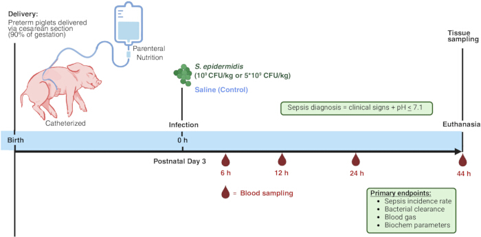
Figure 1: Experimental design. Preterm piglets were delivered via cesarean section at 90% of gestation. On postnatal day 3, piglets received an intra-arterial infusion of live S. epidermidis at either 109 or 5 × 10⁹ CFU/kg, or saline as a control. Following infection, piglets were monitored for 44 h, with longitudinal blood sampling performed at defined time points. Figure created with Biorender.com. Please click here to view a larger version of this figure.
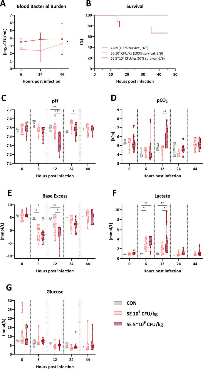
Figure 2: Blood bacterial burden, survival, and blood gas parameters in preterm piglets following arterial infusion with saline (CON) or infection with live S. epidermidis. (A) Density of S. epidermidis in blood samples collected at 6, 24, and 44 h post-infection. (B) Survival curves showing time to humane endpoint or scheduled euthanasia at 44 h post-infection. (C–G) Arterial blood gas parameters at 0, 6, 12, 24, and 44 h post-infection. Data are presented as mean ± SD. (A), survival curves (B), or violin dot plots including median (solid line) and IQR (dotted lines) (C–G). n = 9 for each infected group; n = 3 for the uninfected control. Data were analyzed using a linear mixed-effects model including group × time interactions (A, C–G) or a Cox proportional hazard model (B). *P < 0.05, **P < 0.01, ***P < 0.001. Abbreviations; CON = control; pCO2 = partial pressure of CO2; SE = S. epidermidis. Please click here to view a larger version of this figure.
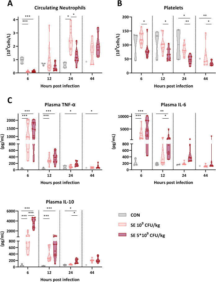
Figure 3: Circulating inflammatory markers in preterm piglets following arterial infusion with saline (CON) or infection with live S. epidermidis. (A, B) Circulating neutrophil and platelet counts at 6, 12, 24, and 44 h post-infection. (C) Plasma levels of TNF-α, IL-6, and IL-10 at 6, 12, 24, and 44 h post-infection. Data are presented as violin dot plots including median (solid line) and IQR (dotted lines) (A–C). n = 9 for each infected group; n = 3 for the uninfected control. Data were analyzed using a linear mixed-effects model including group × time interactions (A–C). *P < 0.05, **P < 0.01, ***P < 0.001. Abbreviations; CON = control; IL = interleukin; SE = S. epidermidis; TNF = tumor necrosis factor. Please click here to view a larger version of this figure.
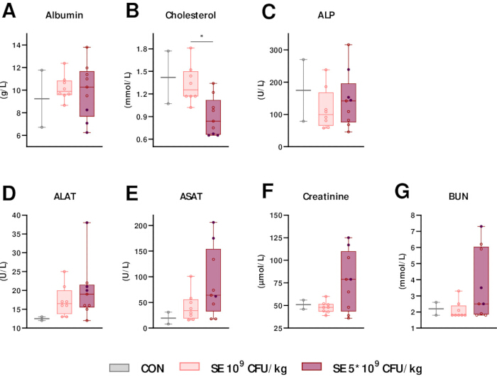
Figure 4: Plasma biochemical parameters in preterm piglets following arterial infusion with saline (CON) or infection with live S. epidermidis (109 or 5 × 10⁹ CFU/kg). (A–G) Plasma levels of albumin, cholesterol, ALP, ALAT, ASAT, creatinine, and BUN at the humane endpoint or at scheduled euthanasia at 44 h post-infection. Data are presented as whisker plots showing the full range (minimum to maximum) (A–G). Individual data points in dark purple indicate piglets that developed sepsis. n = 9 for each infected group; n = 3 for the uninfected control. Data were analyzed using a multiple linear regression model (A–G). *P < 0.05. Abbreviations; ALAT = alanine aminotransferase; ALP = alkaline phosphatase; ASAT = aspartate aminotransferase; BUN = blood urea nitrogen; CON = control; SE = S. epidermidis. Please click here to view a larger version of this figure.
| Total Amount | Concentration | |||
| Energy | 7762 | kJ | 3525 | kJ/L |
| Macronutrients | ||||
| Amino Acids | 92 | g | 42 | g/L |
| Fat | 66 | g | 30 | g/L |
| Glucose | 221 | g | 100 | g/L |
| Micronutrients | ||||
| Na | 64 | mmol | 29 | mmol/L |
| K | 48 | mmol | 22 | mmol/L |
| Mg | 8 | mmol | 4 | mmol/L |
| Ca | 4 | mmol | 2 | mmol/L |
| PO4 | 18 | mmol | 8 | mmol/L |
| SO4 | 8 | mmol | 4 | mmol/L |
| Cl | 93 | mmol | 42 | mmol/L |
| Acetate | 78 | mmol | 35 | mmol/L |
Table 1: Composition of modified total parenteral nutrition. The total amounts and concentrations of energy, macronutrients (amino acids, fat, and glucose), and micronutrients in the parenteral nutrition solution.
Supplementary Figure 1: Growth kinetics of S. epidermidis WT-1457. Bacteria were cultured in brain heart infusion broth at 37 °C with orbital shaking at 180 rpm. Optical density was measured at 600 nm over time. Data are shown on a logarithmic (A) and linear scale (B) (n = 3 independent experiments).Please click here to download this file.
Discussion
A standardized neonatal sepsis model in preterm piglets was designed to study a systemic bacterial infection under clinically relevant conditions of neonatal immaturity. The model combines control of gestational age, resuscitation, and intensive care support, timing of infection, and longitudinal blood sampling. This approach aims to bridge the gap between rodent and in vitro experimental systems and the complex physiology encountered in infected newborn infants. Importantly, this model has been used to confirm the known association of sex, low birthweight, and gestational age, with sepsis sensitivity observed in human preterm infants16,17,18.
Pigs are a highly relevant large-animal model for infection research due to substantial similarities with humans in immune function and physiology. Key components of the innate immune system, including leukocyte subsets, pattern-recognition receptor signaling, and cytokine responses, show close correspondence between pigs and humans. The main immunological differences between newborn piglets and humans lie within adaptive immune responses. Since there is no transplacental transfer of immunoglobulin G (IgG) during porcine gestation, newborn piglets are born naïve in this regard19. Likewise, whereas B cells of infants do not undergo isoform shift from IgM to IgG before 3–6 months of age, porcine B cells are capable of mounting IgG responses shortly after birth20.
In this model system, these differences are mitigated by inducing infection shortly after birth, before adaptive immune responses can occur, and by supplying maternal immunoglobulins via plasma transfusion. This approach results in circulating IgG levels similar to newborn very preterm infants subject to interrupted transplacental IgG transfer due to preterm birth17. In addition, the comparatively large body size at birth (~800 g) further strengthens the model, compared to newborn mice (~3 g). As a result, environmental influences such as temperature, humidity, and oxygen tension more closely resemble those experienced by human neonates. This scale also enables the use of clinical-grade equipment, invasive monitoring, and supportive care modalities, such as vascular access, respiratory support, and continuous physiological monitoring, which are not feasible in neonatal rodent models. Specifically, vascular access for infusions, such as parenteral nutrition, in the preterm pig model is achieved via an umbilical arterial catheter. While this contrasts with the standard intravenous route used in clinical neonatology, it is necessitated by the anatomical constraints of preterm piglets, where umbilical vein catheters rarely traverse the ductus venosus spontaneously and instead malposition within the liver parenchyma21. Intra-arterial delivery provides a reliable and reproducible route for continuous infusion without the high risk of hepatic necrosis associated with umbilical vein malplacement. Likewise, this allows arterial blood to be sampled for blood gas analysis.
Systemic infection in this model is induced by intra-arterial infusion of live S. epidermidis, a leading cause of late-onset sepsis in neonatal intensive care units1,2. However, a diverse set of pathogens causes neonatal sepsis, and coagulase-negative staphylococci, such as S. epidermidis, are likewise known to cause less severe infections and a lower sepsis risk than other, more virulent pathogens, such as E. coli1,2. While the model uses a low-virulence pathogen, the observed clinical deterioration, inflammatory activation, and physiological instability all reflect key hallmarks of neonatal sepsis. The uninfected control group (n = 3) in this study serves as a baseline reference to demonstrate the physiological state of “healthy” preterm piglets under identical intensive care conditions. While this results in a design imbalance, these comparisons are meant to represent the host response to infection. As shown in Figure 2, there was a clear dose–response relationship with increasing S. epidermidis doses, consistent with previous findings in piglets infected on day 1 of life22. Only piglets receiving the highest S. epidermidis dose deteriorated to the humane endpoints requiring euthanasia. However, both infected groups developed metabolic acidosis, while a substantial respiratory component (elevated pCO₂) was observed only in the high-dose S. epidermidis group. Similarly, both groups exhibited marked neutropenia at 6 h post-infection, followed by neutrophil replenishment at 24 h, which was greater in the low-dose group. Thrombocytopenia was also present in both groups, accompanied by increases in plasma cytokines and organ injury markers, with more pronounced responses in piglets receiving the highest S. epidermidis dose.
In preterm piglets, S. epidermidis infection on postnatal day 1 induces the same paraclinical signs of neonatal sepsis (metabolic acidosis, leukopenia, thrombocytopenia, inflammation), as well as lethargy, coagulopathy, and complement consumption17,22,23. The infection also causes systemic disturbances in energy metabolism, most notably reduced mitochondrial oxidative phosphorylation, along with alterations in the plasma proteome and metabolome similar to those reported in neonatal sepsis patients24,25,26. In addition, preterm piglets (90% gestation) are much more likely to develop sepsis than piglets delivered at full term17. Nutritional composition further affects clinical and immunological responses. Glucose-based TPN drives glycolytic metabolism, which amplifies pro-inflammatory immune responses, while impairing disease tolerance in peripheral tissues24,26. The combination of these responses is associated with organ injury and progression towards the sepsis phenotype27. Modulation of TPN composition, either by reducing glucose content or substituting glucose with alternative macronutrients such as galactose, glucogenic amino acids, or β-hydroxybutyrate28,29, attenuates the inflammatory response and preserves tissue tolerance to disease. High glucose supplementation via TPN also results in hyperglycemia (≈10–20 mM). However, it remains unclear to what extent the observed glycolysis-linked inflammatory responses are directly mediated by elevated blood glucose levels.
Experimental observations indicate clear differences in clinical responses depending on the postnatal day of infection. Both the risk of developing sepsis and the severity of clinical symptoms are highest when infection is induced shortly after birth. Infection induction on postnatal day 3 leads to a milder sepsis phenotype with sickness behaviors, respiratory and metabolic acidosis during the first 24 h post-induction, followed by clinical recovery despite elevated markers of organ injuries. The mechanisms underlying these differences between early and late induction are currently unknown and represent an area of ongoing investigation. However, glucose homeostasis following initiation of parenteral nutrition improves over the first three postnatal days in preterm piglets, resulting in reduced hyperglycemia despite similar glucose intake. In all, varying the postnatal day of infection and glucose provision allows the model to capture features of both acute severe and life-threatening sepsis or a milder phenotype. Together, these adjustments enable testing of targeted interventions tailored to specific disease phenotypes. The clinical applicability of this model could be further strengthened by aligning it with established sepsis-related neonatal scoring systems, such as the neonatal Sequential Organ Failure Assessment (nSOFA). A real-time nSOFA-based scoring system was not implemented in the current study due to the infeasibility of continuous arterial blood pressure monitoring and immediate platelet counts required for cardiovascular and hematologic scoring. Future refinements of the protocol could incorporate an adapted nSOFA framework to better categorize disease severity.
Despite its translational strengths, this model has several important limitations. These experiments are resource-intensive, requiring specialized facilities and highly trained personnel to perform cesarean delivery, neonatal resuscitation, postnatal housing, and care of preterm piglets. Infection experiments further require continuous, hands-on monitoring by two to three experienced staff members to ensure animal welfare and reliable data collection, and long-term follow-up increases costs. Compared with rodent models, the availability of species-specific reagents is more limited, particularly for immunohistochemistry and flow cytometry, although many human antibodies can be used successfully following appropriate validation. In addition, the outbred nature of pigs results in considerable within- and between-litter biological variation, necessitating larger group sizes to detect modest differences in infection responses. While this variability increases experimental complexity, it also more closely reflects the heterogeneity observed in clinical neonatal populations, thereby enhancing the model's translational relevance. In conclusion, this validated protocol for neonatal sepsis will be a useful tool for future investigations in neonatology and for testing novel preventive and therapeutic interventions.
Disclosures
The authors have no conflicts of interest to disclose.
Acknowledgements
This work was supported by the staff at the group of Comparative Pediatrics, University of Copenhagen, and funded by the Novo Nordisk Foundation (NNF23OC0085457 and NNF22OC0078747).
Materials
| Name | Company | Catalog Number | Comments |
|---|---|---|---|
| 0.1–10 μL pipette tips “Optifit” | Sartorius | 790010 | |
| 1 mL disposable syringes (sterile) | e-power | 150001S | |
| 1 mL syringe with 25 G X 16 mm needle (sterile) | B. Braun Melsungen AG | 9161465V | |
| 1.5 mL tubes | Eppendorf | 30125215 | |
| 10 mL disposible syringe (sterile) | e-power | 150010S | |
| 100 mL Erlenmeyer flasks | Duran Wheaton Kimble | 212162403 | |
| 1000 μL pipette | Sartorius | 46880171 | |
| 12 G “Intraflon 2” needles (sterile) | Vygon | 122.27 | |
| 16 G X 40 mm “Microlance 3” needles (sterile) | Becton Dickinson | 300637 | |
| 18 G X 40 mm “Microlance 3” needles (sterile) | Becton Dickinson | 303262 | |
| 19 G X 40 mm “Sterican” needles (sterile) | B. Braun Melsungen AG | 466 5112 | |
| 2 mL disposible syringe (sterile) | e-power | 150002S | |
| 2-0 70 cm suture “ Vicryl plus” (sterile) | Johnson & Johnson | VCP453 | |
| 20 G X 25 mm “Microlance 3” needles (sterile) | Becton Dickinson | 304827 | |
| 20 mL disposable syringe (sterile) | e-power | 150020S | |
| 20 μL pipette | Sartorius | 46886095 | |
| 200 μL pipette | Sartorius | 39486615 | |
| 20G Venflon "Pro" 1.1 x 32 mm | Beckton Dickinson | 393255 | |
| 21 G x 25 mm Vacutainer blood collection needles (sterile) | Becton Dickinson | 301746 | |
| 21 G X 40 mm “Sterican” needles (sterile) | B. Braun Melsungen AG | 4665643 | |
| 22 G X 50 mm “Microlance 3” needles (sterile) | Becton Dickinson | 300094 | |
| 23 G X 25 mm “Medoject” needles (sterile) | Chirana T. Injecta | CH23100 | |
| 25 G X 16 mm “Microlance 3” needles (sterile) | Beckton Dickinson | 300600 | |
| 25 G X 25 mm “Sterican” needles (sterile) | B. Braun Melsungen AG | 9186158 | |
| 25 mL plastic pipettes (sterile) | LP Italian Spa | 162510 | |
| 30 mL disposible syringes (sterile) | e-power | 150030S | |
| 4L plastic freezer bags | Cater.Line | ABN-105004 | |
| 5–350 μL pipette tips “Optifit” | Sartorius | 790350 | |
| 5 mL disposible syringe (sterile) | e-power | 150005S | |
| 5 mL syringe (sterile) | B. Braun Melsungen AG | 4617053V | |
| 50–1200 μL pipette tips “Optifit” | Sartorius | 791200 | |
| 50 mL Luer Lock Original Perfusor Syringe | B. Braun Melsungen AG | 8728844F-06 | |
| 50 mL tubes (sterile) | Sarstedt AG and Co. KG | 62.547.254 | |
| 82% ethanol + 0.5% chlorhexidine disinfection wipes | Mediq | 527497 | |
| Atropine "ATROpin" 1 mg/mL | Amgros | 741124 | |
| Bandage scissors, stainless steel, 180 mm | B. Braun Melsungen AG | BC849R | |
| Blood gas machine ABL90 | Radiometer | 994-667 (service manual) | |
| Brain Heart Infusion broth (BHI/B) | Produced in-house | N.A. | Commercially available equivalent product: Brain Heart Infusion Broth (Merck, Cat. No. 53286) |
| Butorphanol "Butomidor vet" 10 mg/mL | VetViva Richter | 53 19 43 | |
| Cell incubator “HeraCell 150i” | Thermo Fisher Scientific | 51032719 | |
| Centrifuge, Rotina 380 R | Hettich | 1701 | |
| Clinical chemistry system “Advia 2120i” | Siemens Healthcare GmbH | 10488923 | |
| Closing cones “Combi-Stopper” (sterile) | B. Braun Melsungen AG | 4495101 | |
| Columbia agar + 5% sheep blood plates | BioRad | 63784 | |
| Disposable soft underpads, Abri-Soft classic | Abena | 4115 | |
| Doxapram-V 20 mg/mL | Dechra | 34144/G | |
| EDTA 3 mL collection tubes (sterile) | Becton Dickinson | 368856 | |
| Endotracheal tube (12 mm x 55 cm, 150 cc), Silicone, cuffed | Mila | GVPmL-METC955 | |
| Enteral feeding tube 04Fr-L.40cm - PVC (sterile) | Vygon | 310.04 | Used as umbilical arterial catheter |
| Ethanol 70% | VWR | 83801.36 | |
| Extension line “Q-Style” (sterile) | Becton Dickinson | 385151 | |
| Flowmeter for oxygen | Dameca | P/N 32018-00 | |
| Flumaznil 0.1 mg/mL | Hamelm pharmacy gmbh | 33659 | |
| Gloves, “Gentle Skin” (sterile) | Meditrade | 9021 | |
| Glucose 500 mg/mL | Fresenius Kabi | 469480 | |
| Heparin 5000 IE/mL | Panpharma | 482480 | |
| Incubator shaker “New Brunswick Innova 40” | Eppendorf | M1299-0092 | |
| Infusion line “Connecta PE Line” (sterile) | Merit Medical | 682192 | |
| Infusion line “Original Infusomat Line” | B. Braun Melsungen AG | 8700036T | |
| Infusion pump A: Perfusor Space | B. Braun Melsungen AG | 8713032U | |
| Infusion pump B: Perfusor Secura FT | B. Braun Melsungen AG | 871822/9 | |
| Infusion pump C: Perfusor Compact | B. Braun Melsungen AG | 87139112 | |
| Infusion pump, Alaris Guardrails plus GH, care fusion | Becton Dickinson | 80023UN00-G | |
| Isoflurane "Attane Vet" 1000 mg/mL (250 mL) | ScanVet | 55226 | |
| Ketamine, “Ketiminol Vet”100 mg/mL | Merck & Co., Inc. | 511519 | |
| Kidney trays, stainless steel, reusabe | B. Braun Melsungen AG | JG504R | |
| Laryngoscope "home made set" | N.A. | N.A. | Scoopula, stainless steel, 37 cm length, with taped flashlight "Coast, G20" within divet and light facing towards wider edge |
| Lidocaine "Xylocain" 10 mg/mL | Aspen | 44 31 22 | |
| Lithium Heparin 2 mL collection tubes (sterile) | Becton Dickinson | 368494 | |
| Lube | Equivet | 180544 | |
| Luer access split septum “Q-style” (sterile) | Becton Dickinson | 385100 | |
| Multidirectional stopcock, “Discofix” (sterile) | B. Braun Melsungen AG | 4095111 | |
| Nitrile gloves, “x-soft” large | ImteX | 1013 | |
| Nitrile gloves, “x-soft” medium | ImteX | 1012 | |
| Nitrile gloves, “x-soft” small | ImteX | 1011 | |
| Nonwoven swab, “Mesoft” | Mölnlycke | 156340 | |
| Operation scissors, stainless steel, 14 cm curved | Medshop.dk | 933007 | |
| Operation scissors, stainless steel, 14 cm straight | Medshop.dk | 933006 | |
| Parenteral nutrition, Kabiven 1900 kcal 2053mL | Fresenius Kabi | 831222221 | |
| Pasteur pipette “Pastette” | Alpha Laboratories Limited | LW4111 | |
| Pentobarbital “Euthananimal” 400 mg/mL | Alfasan | 88672 | |
| Phosphate buffered solution pH 7.4 (sterile filtered) | gibco | 10010-023 | |
| Pipet controller “Stripettor, Ultra” | Corning | 4099 | |
| Pregnant Sow at 90% gestation of Landrace X Large White X Duroc cross | Commercially supplied 2 weeks prior to scheduled c-section | n.a | |
| Propfol "B. Braun" 10 mg/mL (50 mL) | B. Braun Melsungen AG | 17492 | |
| R statistical software | R Foundation for Statistical Computing | Version 4.5.0 | |
| Re-breathing bag, 0.5 Litre | Kruuse | 271600 | |
| Resuscitator mask | McCulloch Medical | 55340X | |
| Rochester pean, stainless steel, 14 cm straight | Kruuse | 140220 | |
| S/5 Avance Anaesthesia System | General Electric (Datex-Ohmeda) | 1009-3206-000 | |
| Scale, max capacity 7.5 kg | Kern | FOB-NL-4mL | |
| Sensor-cassette for blood gass machine (300 tests) | Radiometer | 946-010 | |
| Sodium Chloride 9 mg/mL (50 mL) | B. Braun Melsungen AG | 420079 | |
| Staphylococcus epidermidis stock | Gothenburg University, Sweden | WT-1457 , PMID 28572323 | Generous donation from Xiaoyang Wang, isolated from a septic patient |
| Sterile water | Valby Trekroner Apotek | 805541 | |
| Surgical covers "BUSTER" (120 x 120 cm) | Kruuse | 141840 | |
| Surgical disposable scalpel “Aesculap” (sterile) (size 11) | B. Braun Melsungen AG | 5518040 | |
| Surgical disposable scalpel (sterile) (size 23) | Kruuse | 140964 | |
| Surgical forceps, stainless steel, 15 cm | Kruuse | 140732 | |
| Suture holder, stainless steel, 15 cm | My Medical A/S | 300540 | |
| Suture scissors, stainless steel, 13 cm | Medshop.dk | 933008 | |
| T-piece resuscitation, “Lullaby Resus Plus” | General Electric | 2070100-001 | |
| Tape “Durapore” | 3M | 1538-1 | |
| Thermometer | Geratherm Medical AG | GT-195-1 rapid | |
| Umbilical cord clamps | Kruuse | 142131 | |
| Vamin 18g N/l electrolyte free | Fresenius Kabi | 15977 | |
| Ventilated neonatal incubator | Arrowmight | Custom made (no longer available) | Key specifications and performance parameters: Enclosed design (60 x 40 x 28 cm) for humidity and temperature control, integrated oxygen supply system, and a forced-air circulation system (via dedicated inlet/outlet ports) to ensure continuous atmospheric renewal and a steady rotating airflow |
| Visible spectrophotometer, Genesys 30 | Thermo Fisher Scientific | 840-277300 | |
| Vortex “lab dancer S040” | VWR | 444-0020 | |
| Xysol Vet 20 mg/mL | CP Pharma | CP 054899 | |
| Zoletil “50 Vet” ,Tiletamine 25 mg/mL, Zolazepam 25 mg/mL | Virbac | 83046806 |
References
- Shane, A. L., Sánchez, P. J., Stoll, B. J. Neonatal sepsis. The Lancet. 390 (10104), 1770-1780 (2017).
- Strunk, T., Molloy, E. J., Mishra, A., Bhutta, Z. A. Neonatal bacterial sepsis. The Lancet. 404 (10449), 277-293 (2024).
- Wynn, J. L., et al. Time for a neonatal-specific consensus definition for sepsis. Pediatric critical care medicine : a journal of the Society of Critical Care Medicine and the World Federation of Pediatric Intensive and Critical Care Societies. 15 (6), 523-528 (2014).
- Schlapbach, L. J., et al. International Consensus Criteria for Pediatric Sepsis and Septic Shock. JAMA. 331 (8), 665-674 (2024).
- Fleiss, N., Hooven, T. A., Polin, R. A. Can we back off using antibiotics in the NICU?. Seminars in Fetal and Neonatal Medicine. 26 (3), 101217 (2021).
- Strunk, T., et al. Impaired Cytokine Responses to Live Staphylococcus epidermidis in Preterm Infants Precede Gram-positive, Late-onset Sepsis. Clinical infectious diseases : an official publication of the Infectious Diseases Society of America. 72 (2), 271-278 (2021).
- Collins, A., Weitkamp, J. H., Wynn, J. L. Why are preterm newborns at increased risk of infection?. Archives of Disease in Childhood - Fetal and Neonatal Edition. 103 (4), F391-F394 (2018).
- Kowalski, G. M., Bruce, C. R. The regulation of glucose metabolism: implications and considerations for the assessment of glucose homeostasis in rodents. Am J Physiol Endocrinol Metab. 307 (10), E859-E871 (2014).
- Bailey, M., Christoforidou, Z., Lewis, M. C. The evolutionary basis for differences between the immune systems of man, mouse, pig, and ruminants. Veterinary Immunology and Immunopathology. 152 (1-2), 13-19 (2013).
- Mair, K. H., et al. The porcine innate immune system: An update. Developmental & Comparative Immunology. 45 (2), 321-343 (2014).
- Ezquerra, A., et al. Porcine myelomonocytic markers and cell populations. Developmental & Comparative Immunology. 33 (3), 284-298 (2009).
- Gerner, W., Käser, T., Saalmüller, A. Porcine T lymphocytes and NK cells – An update. Developmental & Comparative Immunology. 33 (3), 310-320 (2009).
- Meurens, F., Summerfield, A., Nauwynck, H., Saif, L., Gerdts, V. The pig: a model for human infectious diseases. Trends in Microbiology. 20 (1), 50-57 (2012).
- Pabst, R. The pig as a model for immunology research. Cell and Tissue Research. 380 (2), 287-304 (2020).
- Goldfarb, R. D., Dellinger, R. P., Parrillo, J. E. Porcine models of severe sepsis: emphasis on porcine peritonitis. Shock (Augusta, Ga). 24 Suppl 1 (SUPPL. 1), 75-81 (2005).
- Bæk, O., Ren, S., Brunse, A., Sangild, P. T., Nguyen, D. N. Impaired neonatal immunity and infection resistance following fetal growth restriction in preterm pigs. Frontiers in Immunology. 11, 1808 (2020).
- Bæk, O., et al. Diet Modulates the High Sensitivity to Systemic Infection in Newborn Preterm Pigs. Frontiers in Immunology. 11, 1019 (2020).
- Bæk, O., et al. Sex-Specific Survival, Growth, Immunity, and Organ Development in Preterm Pigs as Models for Immature Newborns. Frontiers in Pediatrics. 9, (2021).
- Porter, P. Transfer of immunoglobulins IgG, IgA, and IgM to lacteal secretions in the parturient sow and their absorption by the neonatal piglet. Biochimica et Biophysica Acta (BBA) - Protein Structure. 181 (2), 381-392 (1969).
- Butler, J. E., et al. The piglet as a model for B-cell and immune system development. Veterinary immunology and immunopathology. 128 (1-3), 147-170 (2009).
- Sangild, P. T., et al. The preterm pig as a model in pediatric gastroenterology. Journal of Animal Science. 91, 4713-6359 (2013).
- Brunse, A., Worsøe, P., Pors, S. E., Skovgaard, K., Sangild, P. T. Oral Supplementation with Bovine Colostrum Prevents Septic Shock and Brain Barrier Disruption During Bloodstream Infection in Preterm Newborn Pigs. SHOCK. , 1 (2018).
- Krogh, A. K. H., Brunse, A., Thymann, T., Bochsen, L., Kristensen, A. T. Staphylococcus epidermidis sepsis induces hypercoagulability in preterm pigs. Research in veterinary science. 127, 122-129 (2019).
- Bæk, O., et al. Altered hepatic metabolism mediates sepsis preventive effects of reduced glucose supply in infected preterm newborns. eLife. 13, (2024).
- Wu, Z., et al. Regulation of host metabolism and defense strategies to survive neonatal infection. Biochimica et biophysica acta. Molecular basis of disease. 1870 (8), (2024).
- Muk, T., Brunse, A., Henriksen, N. L., Aasmul-Olsen, K., Nguyen, D. N. Glucose supply and glycolysis inhibition shape the clinical fate of Staphylococcus epidermidis–infected preterm newborns. JCI Insight. 7 (11), (2022).
- Zhong, J., et al. Reduced parenteral glucose supply during neonatal infection attenuates neurological and renal pathology associated with modulation of innate and Th1 immunity. Biochimica et biophysica acta. Molecular basis of disease. 1871 (4), (2025).
- Wu, Z., et al. Harnessing systemic glycolysis-TCA cycle axis to boost host defense against neonatal infection. EMBO Molecular Medicine. , (2026).
- Bæk, O., et al. Rethinking parenteral nutrition as supportive therapy for neonatal sepsis. Med. , (2026).
Reprints and Permissions
This article has been published
Video Coming Soon