This retrospective cohort study evaluated fertility and recurrence outcomes after laparoscopic and laparotomic surgery in 219 patients with endometriosis. Laparoscopy was associated with faster perioperative recovery and higher early spontaneous pregnancy rates, although adjusted analyses did not demonstrate surgical superiority.
A subscription to JoVE is required to view this content. Sign in or start your free trial.
Research Article
Laparoscopic versus Laparotomic Surgery for Endometriosis: Fertility and Recurrencein a Retrospective Cohort
In This Article
Summary
Abstract
Endometriosis is frequently associated with infertility and postoperative recurrence, and fertility-sparing surgery may be considered when symptoms, endometriomas, adhesions, or deep infiltrating disease are unlikely to be adequately managed with medical therapy or assisted reproduction alone. Because the choice of laparoscopic surgery (LS) or laparotomic surgery (LT) is strongly influenced by lesion phenotype and disease burden, pooled surgical comparisons are vulnerable to confounding. This retrospective cohort study evaluated 219 surgically treated patients with pathologically confirmed endometriosis who underwent LS (n = 150) or LT (n = 69) between January 2020 and December 2024. Outcomes were assessed within a 12-month postoperative window. Because comprehensive patient-level, subtype-specific analyses were not performed, findings should be interpreted with caution across heterogeneous endometriosis phenotypes, and any phenotype-specific observations are exploratory. LS was associated with faster perioperative recovery and a higher unadjusted spontaneous clinical pregnancy rate (37.3% versus 23.2%; 42.4% versus 27.1% among patients attempting conception). However, the adjusted odds ratio for any clinical pregnancy within 12 months was not statistically significant (OR = 1.42, 95% CI, 0.80–2.53). Composite recurrence was numerically lower after LS (18.0% versus 29.0%), but the adjusted estimate did not reach statistical significance (HR = 0.63, 95% CI: 0.39–1.03; P = 0.061). These findings support cautious, individualized interpretation in surgically selected patients and should not be interpreted as evidence that LS is superior across all endometriosis phenotypes or all patients with infertility.
Introduction
Endometriosis is a chronic inflammatory gynecologic disorder characterized by the presence of endometrial-like tissue outside the uterine cavity. It is associated with pelvic pain, adhesions, impaired quality of life, and infertility among reproductive-aged women1,2,3. For patients who desire fertility preservation, surgery may be used to remove endometriotic lesions, restore pelvic anatomy, and improve access to natural conception or assisted reproductive technology (ART). However, recurrence remains common after conservative treatment, and postoperative management must balance symptom control with reproductive goals4,5.
Endometriosis is not a single surgical entity. Isolated ovarian endometriomas, adenomyosis-associated disease, superficial peritoneal lesions, and deep infiltrating endometriosis (DIE) differ in pathophysiology, operative strategy, fertility-preservation priorities, and recurrence risk. Therefore, aggregated analyses that pool these phenotypes can be clinically misleading if interpreted as reflecting a uniform treatment effect. A phenotype-aware interpretation is essential, especially because patients with severe adhesions, large endometriomas, or suspected bowel or urinary tract involvement are more likely to be selected for laparotomy.
The choice between laparoscopic and laparotomic surgery is clinically important but methodologically difficult to study. Laparoscopy offers magnified visualization, smaller incisions, reduced perioperative trauma, and faster recovery, whereas laparotomy may still be selected for extensive disease, dense adhesions, suspected bowel or urinary tract involvement, or situations requiring tactile assessment and complex reconstruction6,7,8.
Previous studies suggest that conservative laparoscopic treatment may improve postoperative reproductive efficiency in selected patients, but the independent association between surgical route and fertility or recurrence remains difficult to estimate because patients selected for laparotomy often have more severe disease at baseline9,10. Surgery is not mandatory for every infertile patient with endometriosis; patients with mild lesions, diminished ovarian reserve, advanced reproductive age, or an urgent fertility timeline may reasonably prioritize ART, selected less-invasive alternatives for endometrioma management, or individualized nonoperative management11. Propensity-score methods and multivariable models can improve balance for measured covariates, but they do not create true randomization and cannot eliminate unmeasured confounding from surgeon judgment, patient preference, subtle anatomical complexity, completeness of excision, or postoperative adherence. This study compared short-term fertility and recurrence outcomes after laparoscopic versus laparotomic surgery in surgically treated patients with endometriosis, while limiting interpretation to 12-month, phenotype-aware, association-based findings and aligning clinical interpretation with current guidance on individualized endometriosis management11.
Access restricted. Please log in or start a trial to view this content.
Protocol
The study was conducted in accordance with the Declaration of Helsinki and approved by the Institutional Review Board of Tianjin Central Hospital of Obstetrics and Gynecology (Approval No. CDPK991890). The requirement for written informed consent was formally waived due to the retrospective observational design. All retrieved clinical data were strictly anonymized and de-identified prior to analysis to ensure patient confidentiality and data security. The research tools used in the protocol are listed in the Table of Materials.
1. Patient selection and cohort definition
Electronic medical records were reviewed to identify patients who underwent fertility-sparing surgical treatment for pathologically confirmed endometriosis at Tianjin Central Hospital of Obstetrics and Gynecology between January 2020 and December 2024, thereby establishing a single-center retrospective cohort. Eligible patients were women of reproductive age with intraoperative and pathological confirmation of endometriosis who underwent fertility-preserving surgery with retention of the uterus and at least one ovary.
Patients were required to have complete baseline clinical characteristics, disease severity metrics including revised American Society for Reproductive Medicine (rASRM) staging, lesion phenotype classification, operative details, and at least 12 months of continuous postoperative follow-up. Cases were classified according to the dominant disease phenotype, including isolated ovarian endometrioma, deep infiltrating endometriosis (DIE) with or without endometrioma, adenomyosis-associated disease, and superficial peritoneal or adhesion-dominant disease. These classifications were used to support phenotype-aware interpretation of pooled outcome estimates.
Patients without definitive pathological confirmation of endometriosis, those with concurrent systemic malignancies, those who underwent hysterectomy or bilateral oophorectomy, and those lost to follow-up or lacking primary fertility outcome data were excluded. All reporting was conducted in accordance with the Strengthening the Reporting of Observational Studies in Epidemiology (STROBE) guidelines. The patient screening workflow and final cohort assembly process are shown in Figure 1.
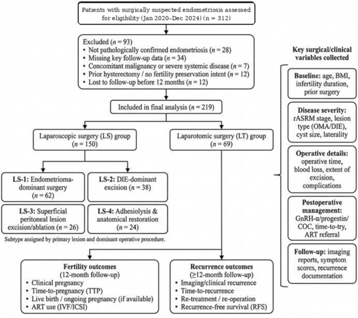
Figure 1: Patient selection flowchart and study design. A total of 312 patients with surgically suspected endometriosis were screened between January 2020 and December 2024. After application of exclusion criteria, 219 patients with pathologically confirmed endometriosis were included in the final analysis and classified into laparoscopic surgery (LS; n = 150) and laparotomic surgery (LT; n = 69) cohorts. The figure also summarizes phenotype classification, collected clinical variables, postoperative management variables, and fertility and recurrence outcomes evaluated during follow-up. Please click here to view a larger version of this figure.
2. Surgical intervention
- Laparoscopic approach
Patients undergoing laparoscopic surgery received general anesthesia and were positioned in the modified lithotomy position. Pneumoperitoneum was established using either a Veress needle or an open Hasson technique at the umbilicus, with intra-abdominal pressure maintained at 12–14 mmHg. A primary 10 mm trocar was inserted for the laparoscope, and three accessory 5 mm trocars were placed in the lower abdomen under direct visualization to facilitate pelvic access and operative triangulation.
The pelvic cavity was systematically inspected to determine the extent of endometriotic disease and to assign the rASRM stage. Adhesiolysis was performed using cold scissors or ultrasonic energy devices to mobilize adherent structures and restore pelvic anatomy. The extent of lesion excision was determined by intraoperative assessment of tissue planes, with priority given to preserving critical neurovascular structures in anatomically distorted regions.
Ovarian endometriomas were managed using a cystectomy stripping technique. The cleavage plane between the cyst capsule and the normal ovarian cortex was identified whenever feasible, and hemostasis was achieved with selective bipolar coagulation, minimizing thermal injury to preserve ovarian tissue and ovarian reserve.
Deep-infiltrating endometriosis (DIE) lesions were resected according to intraoperative anatomical findings and surgical feasibility. When deep nodular involvement was identified, ureterolysis and pararectal space dissection were performed as appropriate. Multidisciplinary surgical collaboration was undertaken when bowel or urinary tract resection was required. - Laparotomic approach
Patients undergoing laparotomic surgery received general anesthesia and underwent either a Pfannenstiel or midline vertical abdominal incision to access the pelvic cavity. A self-retaining retractor was used to optimize visualization of pelvic structures.
Complex pelvic adhesions, particularly in cases with complete cul-de-sac obliteration, were identified and carefully dissected using a combination of sharp and blunt techniques to expose critical neurovascular and ureteral landmarks. Extensive endometriotic lesions and large ovarian cysts were resected using conventional surgical instrumentation. Hemostasis was achieved primarily through meticulous suture ligation, with minimal thermal coagulation. Following completion of the procedure, the pelvic cavity was irrigated with sterile saline, hemostasis was confirmed, and the abdominal wall was closed in anatomical layers.
3. Postoperative management and fertility guidance
Postoperative care was individualized according to clinical recovery and disease characteristics. Analgesia was administered as indicated, and recovery parameters, including time to first flatus and length of hospital stay, were recorded throughout the postoperative period.
Postoperative hormonal suppression therapy was initiated within four weeks after surgery according to lesion severity, residual disease status, reproductive goals, and clinician judgment. Gonadotropin-releasing hormone agonists or oral progestins were administered for three to six months in selected patients with advanced disease phenotypes to suppress residual disease activity and reduce postoperative inflammatory burden.
Comprehensive fertility counseling was provided on the basis of postoperative pelvic anatomy, ovarian reserve, patient age, fertility timeline, and individual reproductive preferences. Natural conception was encouraged when clinically appropriate. Patients with diminished ovarian reserve, advanced reproductive age, severe disease, or prolonged infertility were counseled regarding timely referral for assisted reproductive technology (ART) rather than assuming surgery alone would represent the preferred fertility strategy.
4. Outcome evaluation and follow-up
Patients underwent structured postoperative follow-up assessments at 3, 6, and 12 months after surgery. Reproductive outcomes were documented systematically, including clinical pregnancy (CP) and time to pregnancy (TTP).
Clinical pregnancy was defined by ultrasonographic visualization of an intrauterine gestational sac. Time to pregnancy was calculated from the initiation of conception attempts to the first recognized clinical pregnancy.
Disease recurrence was monitored using structured symptom assessment and pelvic ultrasonography. Suspected recurrence events were independently reviewed by two experienced gynecologists. Imaging recurrence was defined as the detection of newly developed cystic lesions that met the established morphological criteria for ovarian endometrioma. Operational definitions for fertility outcomes and recurrence endpoints are summarized in Table 1.
Table 1: Definitions of fertility outcomes, recurrence endpoints, and follow-up schedule. Clinical definitions, diagnostic criteria, treatment variables, and scheduled follow-up time points applied throughout the analysis are summarized. Abbreviations: ART = assisted reproductive technology; TTP = time to pregnancy; VAS = visual analog scale; MRI = magnetic resonance imaging; GnRH-a = gonadotropin-releasing hormone agonist; COC = combined oral contraceptive; RFS = recurrence-free survival; rASRM = revised American Society for Reproductive Medicine. Please click here to download this Table.
5. Data extraction and statistical analysis
Demographic, surgical, and therapeutic variables were extracted from the institutional hospital database. Key analytical covariates included age, body mass index (BMI), duration of infertility, and preoperative cancer antigen 125 (CA125) levels.
Continuous variables were summarized as means with standard deviations (SDs) or medians with interquartile ranges (IQRs), depending on data distribution. Categorical variables were reported as frequencies and percentages. Missing data were addressed using multiple imputation when appropriate, and sensitivity analyses were conducted to assess the potential impact of incomplete records on the study findings.
Baseline imbalance between surgical groups was quantified using standardized mean differences (SMDs), with values < 0.10 indicating acceptable balance. Multivariable regression served as the primary adjusted analytical approach for the aggregate cohort. Propensity-score matching or inverse probability of treatment weighting (IPTW) was specified only as a sensitivity analysis and was to be reported only if patient-level covariate overlap was adequate, including matched or weighted sample size and post-adjustment SMDs. Because the present submission reports the aggregate multivariable model rather than a matched or weighted cohort, propensity-based findings should not be interpreted unless those diagnostics are supplied.
Adjusted reproductive outcomes were evaluated using multivariable logistic regression models. Time-to-event outcomes, including recurrence-free survival (RFS), were assessed using Kaplan–Meier survival analysis and Cox proportional hazards regression.
All statistical tests were two-sided, and P < 0.05 was considered statistically significant. Subgroup analyses were interpreted as exploratory and hypothesis-generating due to limited subgroup sample sizes and the potential for multiple comparisons. The primary recurrence-free survival analysis was restricted to the prespecified 12-month postoperative period. Any analyses extending beyond 12 months were considered exploratory.
The reported aggregate regression models and survival analyses were performed using R software (version 4.3.0; R Foundation for Statistical Computing). Analytical workflows were verified through standardized code execution to support methodological reproducibility.
Access restricted. Please log in or start a trial to view this content.
Results
A total of 219 surgically treated patients were included, comprising 150 patients in the laparoscopic surgery (LS) group and 69 patients in the laparotomic surgery (LT) group. Demographic and reproductive-history characteristics were broadly similar between groups; however, the cohorts were not fully balanced. Standardized mean differences (SMDs) were added to Table 2 to quantify baseline imbalance more appropriately than P values alone. The LT group had a greater disease burden, including more advanced revised American ...
Access restricted. Please log in or start a trial to view this content.
Discussion
This retrospective cohort study compared fertility and recurrence outcomes after LS and LT among surgically treated patients with endometriosis. The principal finding was not definitive surgical superiority but substantial confounding by indication. LS was associated with faster perioperative recovery, a higher unadjusted spontaneous clinical pregnancy rate, and shorter time to pregnancy. However, the adjusted odds ratio for any 12-month clinical pregnancy was not statistically significant, and the adjusted recurrence es...
Access restricted. Please log in or start a trial to view this content.
Disclosures
The authors declare no competing interests.
Acknowledgements
The authors gratefully acknowledge the clinical and administrative staff of the Department of General Gynecology at Tianjin Central Hospital of Obstetrics and Gynecology for their technical support and assistance with data retrieval. The authors also thank the patients whose clinical records contributed to this retrospective study. This research received no specific grant from any funding agency in the public, commercial, or not-for-profit sectors.
Access restricted. Please log in or start a trial to view this content.
Materials
| Name | Company | Catalog Number | Comments |
|---|---|---|---|
| Advanced bipolar energy device | Hangzhou Kangji Medical Instrument Co., Ltd. | KANGJI disposable bipolar forceps; public hospital laparoscopic surgical-instrument contract lists Kangji, and product catalog examples include 109Y.201/109Y.202, 5 mm x 330 mm; exact study model not specified | Hemostasis during ovarian endometrioma cystectomy while minimizing thermal injury |
| Electronic Medical Record System | Yihui Technology Co., Ltd. | Institutional hospital database; hospital information-system reconstruction/interconnectivity project vendor identified from public tender information; exact EMR module/version not specified | Identification of eligible patients and extraction of demographic, surgical, therapeutic, and follow-up data |
| GraphPad Prism | GraphPad Software | Version 9.0 | Preparation and refinement of summary graphs for reporting |
| Laparoscopic imaging system | Olympus | Olympus endoscope/laparoscope and working handles identified from public hospital equipment-repair notices; exact model/catalog number not specified | Pelvic visualization, magnification, and operative triangulation during laparoscopic surgery |
| Microsoft Excel | Microsoft | Microsoft 365 | Data curation, data checking, and table organization |
| Pathology Reporting System | Tianjin Central Hospital of Obstetrics and Gynecology | Institutional pathology archive | Confirmation of pathologically diagnosed endometriosis |
| R Software | R Foundation for Statistical Computing | Version 4.3.0 | Regression modeling, survival analysis, multiple imputation, sensitivity analysis, and standardized mean difference calculation |
| Self-retaining abdominal retractor | Hangzhou Kangji Medical Instrument Co., Ltd. | KANGJI retractor/reusable surgical-instrument product line identified from public manufacturer pages and registration documents; exact self-retaining abdominal-retractor model not specified | Optimization of pelvic visualization during laparotomic surgery |
| Surgical suturing materials | Ethicon, LLC / Johnson & Johnson MedTech | Absorbable surgical sutures for suture ligation and anatomical layer closure; exact suture type, size, needle type, and catalog number not specified in the manuscript | Hemostasis and abdominal wall closure during laparotomic surgery |
| Surgical trocars | Hangzhou Kangji Medical Instrument Co., Ltd. | KANGJI disposable trocar; public hospital laparoscopic surgical-instrument contract lists Kangji; manuscript specifies one primary 10 mm trocar and three accessory 5 mm trocars, exact catalog number not specified | Laparoscopic abdominal access and operative triangulation |
| Transvaginal Ultrasound Platform | GE HealthCare | GE Voluson 730EXP, P8, E8, E10, and LOGIQ e ultrasound systems identified from public hospital equipment-maintenance survey; exact follow-up unit not specified | Monitoring for suspected recurrence during structured postoperative follow-up |
| Ultrasonic scalpel | Ruishengda (Tianjin) Technology Co., Ltd. | Public procurement source identifies this supplier for the hospital ultrasonic-scalpel handle-cable project; exact ultrasonic energy platform/tip used in the study not specified | Mobilization of adherent pelvic structures and tissue dissection |