CAR T-cell application to solid tumors is limited by complex ex vivo manufacturing and safety concerns. This protocol describes a method for the in situ generation of transient CAR-T cells via LNP–mediated delivery of CAR-encoding mRNA, providing a reproducible platform adaptable to other mRNA-based immune cell engineering applications.
Method Article
In Situ Generation of Claudin18.2-Specific CAR-T Cells via Lipid Nanoparticle–Mediated mRNA Delivery
* These authors contributed equally
In This Article
Summary
Abstract
Chimeric antigen receptor (CAR) T-cell therapy has achieved remarkable clinical success in hematological malignancies; however, its broader application is limited by high manufacturing costs and insufficient efficacy against solid tumors. To overcome these challenges, an in situ CAR-T cell generation strategy was developed based on lipid nanoparticle (LNP)-mediated delivery of CAR mRNA targeting Claudin18.2, enabling the transient production of CAR-T cells directly within tumor tissue. Following LNP transfection, activated T cells exhibited efficient surface expression of CARs. These transient CAR-T cells demonstrated potent cytotoxic activity against Claudin18.2-positive gastric carcinoma cell lines, including NUGC4 and NCI-N87, and produced significant antitumor efficacy in tumor-bearing mouse models. This protocol describes detailed procedures for in vitro transcription of CAR mRNA, LNP formulation, T-cell activation and transfection, CAR expression analysis, and in vitro and in vivo functional evaluation of CAR-T-mediated cytotoxicity. Taken together, this strategy provides a cost-effective and versatile platform for generating transient CAR-T cells and offers a promising approach for treating solid tumors.
Introduction
The incidence and mortality of cancer are rising rapidly worldwide. This poses a threat to human survival and societal development. Gastric cancer is the third leading cause of cancer-related deaths globally and is highly prevalent1. Due to low rates of early diagnosis, a significant proportion of patients are diagnosed at advanced stages. Traditional treatments include surgery, radiotherapy, and chemotherapy. Surgical resection for patients with mid-to-late-stage or complex gastric cancer may be incomplete. Furthermore, the significant surgical trauma and prolonged postoperative recovery can adversely affect patients' quality of life and potentially lead to complications. Radiotherapy and chemotherapy severely compromise normal tissues, generating numerous side effects during treatment. In contrast, targeted and cell therapies offer precision and efficacy. These treatments have emerged as crucial contemporary care modalities and represent the prevailing trend in the selection of future therapeutic strategies.
The advent of chimeric antigen receptor T-cell (CAR-T) therapy has propelled cell therapy toward a landmark breakthrough. It has demonstrated potent efficacy in treating hematologic malignancies2,3. CAR-T therapy utilizes the patient's own T cells, which are genetically modified in vitro by inserting a specific chimeric antigen receptor (CAR). This receptor recognizes specific antigens on the surfaces of tumor cells and activates T cells to attack cancer cells. In recent years, CAR-T therapy research for gastric cancer has also advanced. The selection of therapeutic targets is particularly critical as it determines the targeting precision and efficacy of CAR-T cells. Following HER2, Claudin18.2 has emerged as another prominent therapeutic target in gastric cancer, exhibiting expression rates of 40% to 87% among patients. Claudin18.2 is typically buried within the gastric mucosa. When malignant tumors disrupt the tight junctions responsible for intercellular molecular exchange, Claudin18.2 becomes exposed on the surface of tumor cells. This makes it a specific therapeutic target for these tumors4.
Typically, the standard manufacturing process for CAR-T cells involves cell collection, T-cell selection and enrichment, T-cell activation, CAR gene construction, gene transduction, expansion, quality control, storage and transportation, followed by patient infusion. The entire CAR-T cell preparation process is highly complex, requiring sterile conditions and demanding stringent environmental and equipment specifications. This results in the high cost of CAR-T therapy, limiting its clinical application. To address the complexities of ex vivo CAR-T production, the concept of in vivo CAR-T preparation has been proposed. This approach involves directly programming CAR-T cells within the patient's body, eliminating the need for ex vivo cell collection, modification, and expansion. This strategy aims to reduce costs, enhance safety and accessibility, and enable broader applications of disease treatment5.
The key to in vivo CAR-T preparation lies in selecting an optimal delivery vector. Currently, vectors are categorized as viral or non-viral. Among viral options, lentiviral (LV) and adeno-associated viral (AAV) vectors are predominantly utilized. However, the significant risks and limited payload capacity associated with viral vectors create major obstacles for clinical research6. A primary disadvantage of viral vectors for in vivo delivery is their potential for genomic integration, which may result in genetic toxicity and insertional mutations. Previous cases have documented fatal immune responses or cancer in a small portion of patients following treatment. Moreover, the inherent toxicity and immunogenicity of viruses severely restrict their application for repeated in vivo administration. Another challenge is the limited payload capacity of viral vectors, which prevents the simultaneous delivery of multiple genes.
Non-viral mRNA delivery systems effectively address these shortcomings. They avoid integrating into the host genome and thus circumvent many risks. Their transient action prevents off-target toxicity, enabling short-term, controllable CAR-T cell expression, thereby reducing side effects and safety risks. Among non-viral systems, lipid nanoparticles (LNPs) offer a safer approach. They overcome the size limitations of viral vectors. LNPs also resolve issues of insertion mutations and immunogenicity. This positions them as promising candidates in CAR-T cell therapy, with the potential to reduce toxicity, enhance safety, and lower costs7.
Current research on immunotherapy for solid tumors primarily focuses on LNP formulation optimization, targeted modification, and mRNA sequence engineering across various cancer models, including melanoma8, breast cancer, and colorectal cancer9. Efforts have also been directed toward constructing chimeric antigen receptor macrophages (CAR-M)10 and dendritic cells (CAR-DC)11 via LNP delivery systems to breach the complex physical barriers and immunosuppressive tumor microenvironment (TME) of solid tumors. The platform presented herein provides a versatile, non-viral mRNA delivery system characterized by high modularity. By simply exchanging the mRNA sequence, this platform can be rapidly adapted to solid tumor models targeting other antigens or extended to non-oncology therapeutic areas such as autoimmune, cardiovascular, and fibrotic diseases.
However, a technical gap remains in gastric cancer treatments using these platforms: prior studies have predominantly focused on myeloid cell editing, while strategies for highly efficient in vivo T-cell reprogramming remain largely exploratory. To address this gap, this protocol details a method for generating transient CAR-T cells via LNP-mediated mRNA delivery. This approach aims to overcome the limitations inherent to conventional CAR-T cell therapies, including prohibitive costs, complex manufacturing processes, and systemic toxicities, thereby offering a more efficacious and accessible therapeutic alternative for patients. It is demonstrated that transfection of T cells with an optimized mRNA sequence encoding a specific CAR targeting Claudin18.2 enables robust cell-surface CAR expression, directing the modified T cells to exert targeted cytotoxic activity against specific tumor cells. This protocol is structured into five distinct sections: in vitro transcription of CAR mRNA, LNP preparation and characterization, T-cell activation and transfection, detection of CAR expression, and evaluation of CAR-T cell cytolytic efficacy in vitro and in vivo.
Compared with existing mRNA-LNP delivery technologies, the core methodological innovation of this protocol lies in the kinetic optimization of transient expression. A synergistic optimization was performed on the 5' UTR, 3' UTR, 5' Cap, and a 120-bp poly(A) tail. This tailored design modulates the mRNA half-life within the tumor microenvironment, achieving highly efficient translation within a defined therapeutic window followed by safe degradation. Characterized by rapid and transient CAR generation, this platform serves as an "off-the-shelf" formulation for immediate use, preventing disease progression during the prolonged manufacturing window required for conventional therapies. Furthermore, this transient expression profile offers a controllable safety advantage when targeting antigens with potential "on-target, off-tumor" toxicities, or for patients unable to tolerate severe cytokine release syndrome (CRS) associated with conventional CAR-T therapies.
Despite the profound advantages of in vivo in situ CAR-T cell generation, namely the elimination of complex ex vivo manufacturing and reduced economic burdens, certain limitations persist. In systemic circulation, LNPs are prone to non-specific accumulation in the liver and rapid internalization by macrophages and dendritic cells. Without specific targeting moieties directed toward T cells, the transfection efficiency of the target T-cell population is inevitably compromised. Furthermore, upon entering the tumor tissue, in situ generated CAR-T cells are subjected to the hostile TME; under hypoxic, acidic, and immunosuppressive conditions, these T cells are highly susceptible to immune exhaustion, thereby restricting their cytotoxic potency. Additionally, while the transient nature of mRNA expression enhances safety, it restricts the duration of the therapeutic window. To achieve sustained antitumor efficacy, intermittent and repeated administrations are often required. Consequently, future investigations will focus on addressing these limitations by refining LNP delivery efficiency to achieve precise, lineage-specific targeting.
Protocol
All animal experiments were approved by the Ethics Committee for Laboratory Animals at Shanghai Tenth People’s Hospital (Approval No.: SHDSYY-2025-P0085; Approval Date: January 1, 2025). This study complies with local laws, regulations, and institutional requirements. NSG female mice aged 6–8 weeks were used. The Peripheral Blood Mononuclear Cells (PBMCs) used in this study were obtained from a commercial source. The reagents and equipment used in this study are listed in the Table of Materials.
1. Preparation of CAR mRNA
- Linearized DNA templates preparation
NOTE: The plasmid containing the CAR sequence was linearized using the BspQI restriction enzyme. The selected restriction enzyme should insert the DNA fragment downstream of the promoter region on the right side and contain no recognition sites within the inserted DNA fragment.- Add double-distilled water (ddH2O), 10× buffer, DNA template, and BspQI enzyme in the order specified in Table 1. Incubate at 50 °C for 2 h, then inactivate the enzyme at 80 °C for 20 min. The linearized plasmid can be stored at 4 °C.
- Set up the restriction digestion reaction in a sterile microcentrifuge tube. Add double-distilled water, restriction enzyme buffer, plasmid DNA template, and BspQI restriction enzyme in the specific order and volumes indicated in Table 1.
- Mix the reaction mixture gently by pipetting up and down, then spin down briefly in a microcentrifuge. Incubate the mixture at 50 °C for 2 h in a thermal cycler to allow complete digestion. Inactivate the restriction enzyme by incubating the mixture at 80 °C for 20 min.
NOTE: Optional - Store the linearized plasmid at 4 °C for short-term use or proceed immediately to quality control.
- Prepare a 1% agarose gel by weighing 0.25 g of agarose and dissolving it in 25 mL of 1× Tris–Acetate–EDTA (TAE) buffer. Add 2.5 µL of Gel Green and mix thoroughly. Bring to a boil, then shake vigorously. Repeat this process three times before pouring the mixture into the gel casting tray. Run at 110 V for 10 min, followed by 130 V for 20 min.
- Perform ultraviolet (UV) visualization to verify complete digestion. By comparing the band positions with those of DNA markers and the uncut plasmid, the linearized plasmid showed only two distinct bands corresponding to its predicted molecular weights: the target sequence was approximately 1,791 bp, and the remaining fragment was approximately 2,535 bp.
- Purify the digestion products using DNA isolation beads.
- Vortex the magnetic beads to ensure thorough mixing. Pipette the specified volume of magnetic beads into the DNA sample and mix gently. Incubate at Room Temperature (RT) for 10 min to allow DNA binding to the beads.
- Place the sample on a magnetic stand for 5 min. Carefully remove the supernatant. Add 200 µL of freshly prepared 80% ethanol for washing. Incubate at RT for 30 s and carefully remove the supernatant. Repeat this step twice.
- Keep the sample on the magnetic stand at room temperature with the lid open for 5 min to dry. Remove the sample from the magnetic stand. Add 11 µL nuclease-free ddH2O. Mix thoroughly by pipetting up and down. Incubate at room temperature for 2 min. Allow the solution to settle on the magnetic stand for 5 min until clear.
- Carefully transfer the supernatant to a new nuclease-free centrifuge tube. Use a spectrophotometer to measure the DNA concentration; the resulting DNA must be used immediately for in vitro transcription. Calculate the recovery rate based on the amount of purified DNA divided by the initial amount of DNA added.
- Add double-distilled water (ddH2O), 10× buffer, DNA template, and BspQI enzyme in the order specified in Table 1. Incubate at 50 °C for 2 h, then inactivate the enzyme at 80 °C for 20 min. The linearized plasmid can be stored at 4 °C.
- In Vitro Transcription (IVT)
- Thaw all kit components of a T7 RNA polymerase–based in vitro transcription and RNA capping kit on ice (except for the T7 RNA Polymerase Mix, which should be kept at -20 °C until use). Vortex the buffers briefly and spin down.
- Assemble the IVT reaction mixture in a sterile, nuclease-free microcentrifuge tube at room temperature according to the volumes specified in Table 2. Add the components in the listed order, ending with the T7 enzyme mix.
- Mix the reaction mixture thoroughly but gently by flicking the tube or pipetting up and down. Spin down briefly to collect the liquid at the bottom. Incubate the mixture at 37 °C for 2 h in a thermal cycler.
- Add 2 µL of DNase I enzyme directly to the IVT reaction to degrade the residual DNA template. Mix gently and incubate at 37 °C for an additional 15 min. Proceed immediately to the purification step.
- Purify the resulting RNA product using RNA isolation beads following the same procedure as for DNA digestion product purification.
- Dissolve in 20 µL enzyme-free water. Use a NanoDrop spectrophotometer to measure mRNA concentration and the absorbance (OD) ratio. Use 1 µL of nuclease-free water as a blank control. The OD260/280 ratio should be between 1.8 and 2.1, and the OD260/230 ratio should be between 2.0 and 2.4.
NOTE: If the ratios fall outside these ranges, this indicates contamination, and the sample must be purified again. - Use an automated capillary electrophoresis system for nucleic acid and protein analysis to analyze the length and integrity of the synthesized CAR mRNA. The electrophoresis profile should show a single, sharp peak corresponding to the expected full-length CAR mRNA size (approximately 1800 nt), with no tailing and a flat, clean baseline. No significant low-molecular-weight diffuse bands or multiple peaks should be present; otherwise, this indicates RNA degradation.
- Aliquot the validated CAR mRNA to avoid repeated freeze-thaw cycles and store it at -80 °C for long-term preservation.
- Dissolve in 20 µL enzyme-free water. Use a NanoDrop spectrophotometer to measure mRNA concentration and the absorbance (OD) ratio. Use 1 µL of nuclease-free water as a blank control. The OD260/280 ratio should be between 1.8 and 2.1, and the OD260/230 ratio should be between 2.0 and 2.4.
2. LNP preparation and characterization
- Dissolve SM102, cholesterol, 1,2-Distearoyl-sn-glycero-3-phosphocholine (DSPC), and DMG-PEG2000 in anhydrous ethanol to prepare a 10 mg/mL stock solution. Prepare the organic phase using a 1:50:38.5:10:1.5 ratio.
- Calculate the required mRNA amount based on the 8:1 ratio of positively charged amino N to negatively charged phosphate P groups. Dissolve mRNA in sodium citrate solution to prepare the aqueous phase. Maintain an organic: aqueous phase ratio of 1:3 with flow rates of 1.5 mL/min each. Prepare LNPs using a herringbone mixer microfluidic device.
- First, rinse the tubing and microfluidic chip (herringbone mixer) once with anhydrous ethanol and a sodium citrate solution. After mixing the prepared organic and aqueous phases by purging, draw them up using a 1 mL syringe; the ratio of organic to aqueous phase is 1:3. Load them into the two channels of the syringe pump, respectively.
- Set the flow rates for the organic and aqueous phases. Add 5 mL of sodium citrate buffer (50 mM) to the centrifuge tube connected to the outlet. After flushing out any residual washing solution from the tubing, start the device and collect the eluate.
- Transfer the LNPs dispersed in the sodium citrate solution to a 100 kDa ultrafiltration tube, centrifuge at 4000 × g for 5–10 min, then add 2.5 mL of sodium citrate solution and 2.5 mL of 1× TBS solution to redisperse them, and repeat the centrifugation once.
- Add 1 mL of sodium citrate solution and 4 mL of 1× TBS solution to redisperse the particles, and centrifuge again. Store the concentrated LNPs in 1 mL of TBS solution at 4 °C for a short period.
- Use a fluorescence-based RNA quantification kit to measure the mRNA content in the LNP before and after demulsification; the difference between the two values represents the encapsulation efficiency of the LNP.
- Prepare 1× TE buffer, probe working solution, and 2% Triton X-100. Dilute the mRNA standard (100 µg/mL) to 2 µg/mL using 1× TE buffer, and prepare a standard curve with blank, 10, 20, 100, 500, and 1000 ng/mL concentrations. Vortex 4 µL of LNP solution with 16 µL of 2% Triton X-100 solution, then let stand for 10 min.
- Add 5 µL of the demulsified LNP solution to 95 µL of 1× TE buffer. Add 100 µL of probe working solution to both the standard curve and sample wells, mix well, protect from light, and transfer to a black-bottom 96-well plate.
- After incubation, measure the fluorescence intensity (Ex480/Em520) using a microplate reader. Subtract the absorbance values of the blank wells from those of the standard curve wells and sample wells, respectively, to plot the standard curve.
- Use the standard curve to calculate the mRNA concentrations in the LNP solution before and after demulsification, multiply these values by 200 to obtain the pre-dilution concentrations, and substitute them into the formula for calculation:

- Take 10 µL of LNP and dilute it in 1 mL of TBS solution. Determine the particle size, zeta potential, and Polydispersity Index (PDI) value using a Malvern particle size analyzer.
3. T cell activation and transfection
- T cell sorting
- Take PBMCs at a concentration of 1 × 10⁷/mL, resuspend them at 37 °C, add 10 mL of an immunomagnetic cell separation buffer, mix thoroughly, centrifuge at 500 × g for 8 min, and count.
- Resuspend cells and adjust density to 5 × 10⁷ cells/mL, add 50 µL of Isolation Cocktail per mL of cell suspension and incubate at room temperature for 5 min; add 40 µL of magnetic particles for immunomagnetic cell isolation per mL of cell suspension and mix; place the tube on a magnetic stand and incubate at RT for 10 min.
- Carefully transfer the enriched cell suspension to a new centrifuge tube using a pipette. Remove the tube from the magnet, add 4 mL of fresh immunomagnetic cell separation buffer, and incubate for an additional 5 min for a second separation round.
- Wash cells with T-cell culture medium (RPMI 1640 GlutaMAX + 10% Fetal Bovine Serum + 1% Penicillin/Streptomycin + 100 IU/mL rhIL-2 + 5 ng/mL rhIL-7 + 5 ng/mL rhIL-15), then centrifuge and count. Add T-cell culture medium to adjust cell density to 1 × 10⁶/mL and add 12.5 µL human T cell activator CD3/CD28 magnetic beads. Incubate at 37 °C in a 5% CO₂ incubator for 48 h to activate cells.
- T-cell activation parameter detection
- Transfer 200 µL of T cells activated for 48 h into a 1.5 mL microcentrifuge tube. Remove CD3/CD28 activation magnetic beads, add 1 mL of FACS buffer for washing, gently mix by pipetting, then centrifuge at 500 × g, 4 °C for 5 min. Carefully remove the supernatant and repeat 2–3 times.
- Resuspend cells in 100 µL FACS buffer. Add 1 µL each of anti-human CD3, anti-human CD25, and anti-human CD69 antibodies. Gently mix samples and incubate on ice in the dark for 30 min.
- Add 1 mL of FACS buffer to wash the cells, centrifuge, then carefully remove the supernatant, add 100 µL of FACS buffer and 0.1 µL of LIVE/DEAD fixable dead cell stain, and incubate on ice in the dark for 10 min.
- Resuspend the cells in 1 mL FACS buffer and centrifuge at 500 x g, 4 °C for 5 min, then carefully remove supernatant. Repeat 2–3 times. Discard supernatant and resuspend cells in 200 µL FACS buffer. Analyze cells using flow cytometry.
- Select target cells using FSC-A and SSC-A, isolate single cells using FSC-A and FSC-H, and identify live cells using LIVE/DEAD Cell Discrimination Dye (LIVE/DEAD). Compare the CD25 and CD69 ratios between inactive and activated T cells.
- LNP Transfection of T cells
- Based on the encapsulation efficiency calculation, take the required volume of LNP according to the amount of mRNA. First, centrifuge the LNP dispersed in TBS to separate it, then transfer it to RPMI 1640 medium.
- After 48 h, use a magnetic rack to remove CD3/CD28 activation magnetic beads. Centrifuge at 500 × g for 8 min, count, and add T-cell culture medium to adjust density to 2 × 10⁶ cells/mL. Add 1 µg of LNP-mRNA per 1 × 10⁵ cells. Gently mix by pipetting and incubate at 37 °C in a 5% CO₂ incubator for 4–6 h.
4. CAR expression detection
- 4–6 h post-transfection, gently pipette CAR-T cells to mix thoroughly. Transfer at least 2 × 10⁵ cells to a 1.5 mL microcentrifuge tube.
- Add 1 mL FACS buffer to the 1.5 mL microcentrifuge tube. Pipette to mix, then centrifuge at 500 x g for 5 min at 4 °C. Carefully remove the supernatant. Repeat this step 2–3 times.
- Resuspend the cell pellet in 100 µL of FACS buffer and divide the sample into a control group and an experimental group, each containing 50 µL. Add 1 µL of the CAR detection antibody G4S-linker to the cell suspension in the experimental group, gently mix the sample, and incubate on ice in the dark for 30 min. For the control group, do not add the antibody; incubate on ice.
- Pipette 0.2 µL of LIVE/DEAD fixable dead cell stain (1000×) and dilute it in 100 µL of FACS buffer; then pipette 50 µL into each of the control and experimental groups, gently mix the sample, and incubate on ice in the dark for 10 min.
- Wash cells with 1 mL FACS buffer. Centrifuge at 500 x g for 5 min at 4 °C. Carefully remove supernatant. Repeat 2–3 times.
- Discard supernatant and resuspend cells in 200 µL FACS buffer. Analyze cells using flow cytometry.
- Evaluate CAR expression using the following gating strategy: gate on the target cells using FSC-A and SSC-A plot, gate on the single cells using FSC-A and FSC-H plot, and gate on the live cells using LIVE/DEAD fixable cell stain.
NOTE: Based on the selection of cells stained positive for the G4S-linker antibody in the far-right column of the control group’s live cell population, the percentage shown for the experimental group represents the CAR positivity rate.
5. In vitro cytotoxicity analysis of CAR-T cells
- Digest NUGC4 and NCl-N87 tumor cells expressing luciferase with trypsin. Centrifuge at 300 x g for 5 min. Add 1 mL RPMI 1640 medium, count and adjust the cell density to 5 × 10⁴ cells/mL. Aliquot 100 µL into each well of a 96-well plate. Add an equal volume of Phosphate-Buffered Saline (PBS) to the remaining wells to prevent evaporation. Incubate in a 37 °C incubator with 5% CO₂ for 24 h.
- Calculate the CAR positivity rate based on flow cytometry results. Add CAR-T or T cells at effector-to-target cell ratios of 10:1, 5:1, 3:1, 1:1, 1:3, and 1:5 to wells containing tumor cells (100 µL/well). Incubate at 37 °C with 5% CO₂ for 24 h.
- Using the firefly luciferase reporter gene assay kit, detect the luciferase signal from remaining tumor cells according to the product manual and calculate the kill rate: (1 - (effector cell RLU value/target cell RLU value)) × 100%.
6. In vivo functional validation of CAR-T cells
- After a 1-week acclimatization period prior to inoculation, inject each mouse subcutaneously in the axilla with 5 × 10⁶ NUGC4 cells. Measure the tumor size daily, and calculate the tumor volume using the formula: V (mm3) = L × W2/2 (where L is the longest diameter of the tumor and W is the shortest diameter) until the tumor reached 100 mm3.
- Randomly divide the mice into four groups: three treatment groups receiving different doses and a control group receiving PBS, with three mice in each group.
NOTE: The Blindness Act was not implemented. - After incubating 10 µg of mRNA with 1 × 10⁶ activated T cells for 4 h, prepare dosages of 5 × 10⁵ cells/mouse, 1 × 10⁶ cells/mouse, and 2 × 10⁶ cells/mouse. Administer the mixture of LNP and T cells to the mice via tail vein injection, with each mouse receiving a 100 µL volume of cells or PBS. Doses were administered on days 1, 4, 7, and 10, for a total of four doses.
- Measure the tumor size of the mice on days 0, 2, 4, 6, 8, 10, 12, and 14, calculate the tumor volume, and plot a growth curve. Express the data as mean ± SD. Analyze the differences between groups using two-way analysis of variance (ANOVA) with a statistical and graphing software, combined with Tukey’s multiple comparison test. ns: not statistically significant; *P < 0.05; **P < 0.01; ***P < 0.001; ****P < 0.0001.
Results
A CAR sequence targeting Claudin18.2 was employed, composed of the single-chain variable fragment (scFv) from a humanized anti-Claudin18.2 monoclonal antibody, the CD8 hinge region, the CD28 transmembrane region, the co-stimulatory domain, and the CD3ζ activation domain. A DNA template containing the T7 promoter and poly A tail was constructed based on this sequence for in vitro CAR mRNA transcription (Figure 1A). The CAR mRNA was approximately 1800 bp in length, as verified by capillary electrophoresis (Figure 1B). A single band with a sharp peak and no tailing, along with a straight and clean baseline, indicates that the mRNA has not degraded.
Then, LNPs were prepared using a microfluidic device. LNPs dispersed in TBS remained stable at 4 °C for 1 week. A quantitative assay kit was used to detect mRNA encapsulation efficiency (Figure 2A), and particle size, zeta potential, and PDI were also measured (Figure 2B). Results indicate LNP encapsulation efficiency exceeds 90%, with particle size around 150 nm and PDI below 0.2, demonstrating large size uniformity within the ideal range for intravenous administration. The zeta potential, approaching neutrality with a slight negative bias, prevents non-specific binding and enables prolonged circulation.
Following T cell activation, flow cytometry analysis of activation markers at 48 h revealed significant increases in early activation markers CD69 and CD25 compared to non-activated T cells (Figure 3A). Transfection was performed using the LNP according to the aforementioned protocol, and CAR positivity rates were assessed, with untransfected T cells serving as negative controls. CAR expression was observed to commence at 4 h post-transfection, with the proportion of CAR-T cells peaking at over 40% by 18 h and subsequently declining to below 10% by 72 h (Figure 3B).
Finally, the in vitro cytotoxic function of CAR-T cells was validated using two gastric cancer cell lines overexpressing Claudin18.2, namely NUGC4 and NCI-N87. Following co-culture of LNP-transfected CAR-T cells with target cells, luciferase signaling in tumor cells revealed that LNP-mediated CAR-T cells exhibited significantly greater in vitro killing efficacy against both gastric cancer cell lines compared to T cells alone. Killing rates exceeded 50% across target-to-effector ratios ranging from 10:1 to 1:3 (Figure 4A). Concurrently, in vivo killing experiments in the NUGC4 mouse model demonstrated that the LNP-CAR-T cell group exhibited tumor growth inhibition compared to the PBS group between days 0 and 14. The tumor volume ratio of the three treatment groups versus the PBS group showed statistically significant differences with p-values < 0.01 (Figure 4B).
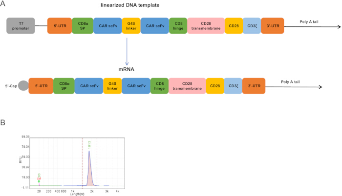
Figure 1: CAR plasmid structure and mRNA quality control. (A) The Claudin18.2 -targeted CAR contains an extracellular Claudin18.2-binding single-chain variable fragment (scFv) derived from a humanized anti-Claudin18.2 monoclonal antibody, a CD8 hinge region, a CD28 transmembrane domain, and an intracellular CD28 co-stimulatory domain followed by the CD3ζ activation domain. The DNA encoding the CAR was flanked by a 5' untranslated region (UTR) and a 3' UTR, followed by a 120-bp poly(A) tail. Transcription of the mRNA was driven by a T7 promoter, and the mRNA was capped at the 5' end. (B) CAR mRNA was detected via capillary electrophoresis as a single band with a size of 1,800 bp, which is consistent with the predicted size. Please click here to view a larger version of this figure.
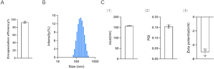
Figure 2: Characterization of lipid nanoparticles. (A) Encapsulation efficiency of CAR-LNPs (n = 3). (B) The size distribution of CAR-LNPs measured by dynamic light scattering (DLS) is shown as a representative. (C1) Size of CAR-LNPs (n = 3). (C2) PDI of CAR-LNPs (n = 3). (C3) Zeta potential of CAR-LNPs (n = 3). Please click here to view a larger version of this figure.
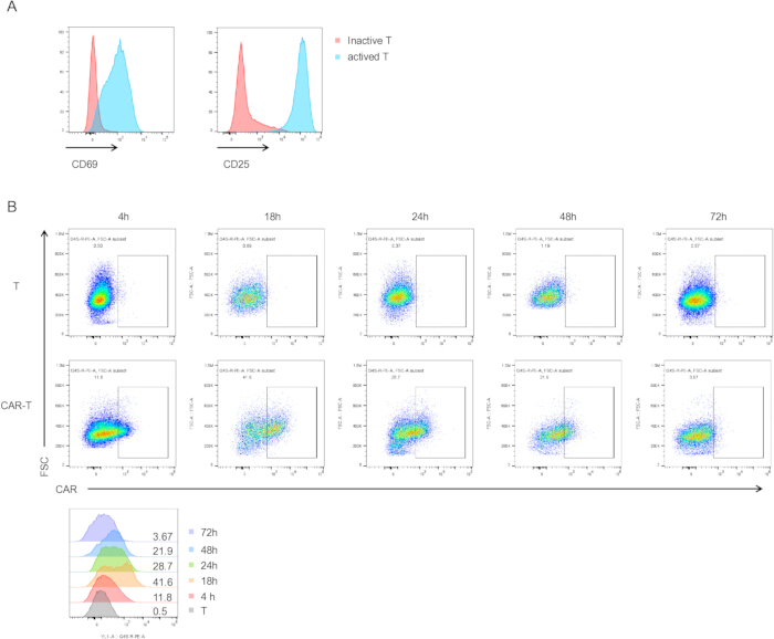
Figure 3: Flow cytometric analysis of T-cell activation markers and CAR expression. (A) Flow cytometric analysis of CD69 and CD25 expression in unactivated and activated T cells. (B) T cells activated by LNP transfection; changes in CAR expression on the cell surface were assessed at 4 h, 18 h, 24 h, 48 h, and 72 h. Please click here to view a larger version of this figure.
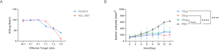
Figure 4: Validation of the in vitro and in vivo cytotoxic activity of CAR-T cells produced using LNP-delivered mRNA. (A) Cytotoxicity curves of CAR-T cells against two target cell types at different effector-to-target ratios. Killing rates are shown as Mean ± SD (n = 3). (B) The line graph shows changes in tumor volume growth curve in the treatment and control groups for the three dosage levels. Data were analyzed using two-way analysis of variance (ANOVA) combined with Tukey’s multiple comparison test. Data are presented as Mean ± SD. ***P = 0.0002, ****P < 0.0001 (n = 3). Please click here to view a larger version of this figure.
| Component | |
| DNA template | 3 µg |
| 10× buffer (r3.1) | 15 µL |
| BspQI | 3 µL |
| ddH2O | To 150 µL |
Table 1: DNA plasmid linearization reaction. This table shows the quantities and volumes of each component in the 150 µL reaction system. A commercially available restriction endonuclease was used to ensure efficient plasmid linearization.
| Component | |
| RNase free H₂O | To 20 µL |
| 1 × Transcription Buffer | 2 µL |
| CTP | 2 µL |
| GTP | 2 µL |
| ATP | 2 µL |
| N-Me-pUTP | 2 µL |
| CAP GAG (100mM) | 2 µL |
| Linearized DNA template | 1 μg |
| T7 RNA Polymerase Mix | 2 µL |
Table 2: mRNA in vitro transcription (IVT) reaction. The concentrations and volumes of each component used in a 20 µL IVT reaction are shown. The UTP was replaced by modified N-Methylated Uridine Triphosphate.
Discussion
This protocol provides a non-viral mRNA delivery system that avoids integration into the host genome, thereby circumventing numerous risks, including immune rejection. Its transient functionality also prevents off-target toxicity, enabling short-term, controllable expression in CAR-T cells to reduce side effects and safety risks.
Optimization of the CAR mRNA sequence design was performed to regulate its translation efficiency and expression duration. The 5'UTR, 3'UTR, 5'-Cap, and poly(A) tail of the mRNA molecule synergistically regulate mRNA translation and stability12. In sequence design, the T7 promoter was employed, which forms a highly specific and efficient transcription system with T7 RNA polymerase13, enabling large-scale in vitro RNA synthesis. A 120-base-pair poly(A) tail was then incorporated14, extending mRNA half-life and prolonging protein expression duration. Concurrently, in vitro transcription added a 5'-cap structure to the mRNA 5' end. The correct positioning and integrity of the 5'-cap relative to the 3'-poly(A) tail are critical for ensuring mRNA translation activity and efficiency15.
mRNA-based cancer immunotherapies hold immense potential. Current studies have successfully utilized mRNA-LNP platforms to produce CAR-macrophages (CAR-Ms) and CAR-T cells in vitro. Compared to electroporation or viral transfection, LNP-generated CAR-T cells exhibit equivalent cancer-killing capabilities16,17,18. LNPs circumvent the integration risks and immune responses associated with viral vectors, avoid the severe cytotoxicity induced by electroporation, and overcome limitations of in vivo in situ application. However, non-specific transfection by LNPs poses significant challenges for in vivo CAR-T production19. Typically, LNPs comprise four components: ionizable cationic lipids, phospholipids, cholesterol, and cycloethylated lipids. Each component plays a critical role in LNP stability, transfection efficiency, and safety20. Thus, effective in vivo mRNA delivery can be achieved by optimizing the ratio or composition of these components. Additionally, modifying the LNP surface by conjugating antibodies or targeting peptides21,22 enhances precise localization, enabling specific organ or cell targeting. Numerous research advances have been reported. Antibody-modified LNPs targeting CD323, CD424, and CD525 have been demonstrated to deliver mRNA to T cells in vivo. Shi et al. described a CD117 antibody-modified LNP capable of efficiently delivering mRNA for editing to hematopoietic stem and progenitor cells (HSPCs) in vivo26.
The approach enables in situ production of small-scale transient CAR-T cells, demonstrated to be effective in both in vitro and in vivo experiments. In situ refers to transient CAR expression induced by mRNA delivery within the tumor-localized T-cell compartment, rather than permanent genetic modification ex vivo. It requires a large initial pool of activated T cells for transfection and rapid utilization. Furthermore, the transient nature of mRNA is a double-edged sword: on the one hand, it allows for precise dose modulation at any time to avoid off-target effects and severe systemic toxicity that may arise during long-term treatment; on the other hand, as we are targeting solid tumors with complex immune microenvironments, this non-persistent mode of administration necessitates an increase in the frequency of dosing. Therefore, in subsequent studies, we will extend the half-life by optimising the mRNA sequence, thereby significantly prolonging the expression period of the CAR protein without increasing the initial dose. At the same time, we will consider replacing linear mRNA with circular RNA to enhance translation efficiency.
Furthermore, another limitation of this approach is the lack of active cell targeting on the LNP surface. During circulation in vivo, serum proteins rapidly adsorb to the LNP surface, forming a “biomolecular crown.” Apolipoprotein E (ApoE) in plasma is one of the core components of the biomolecular crown. It is primarily synthesized by hepatocytes; this endogenous tendency leads to significant non-specific accumulation in the liver. At the same time, the mononuclear-macrophage system rapidly clears these substances, resulting in premature release of the drug encapsulated within the LNPs. To reduce non-specific targeting and clearance, in subsequent studies, the LNP surface was modified with targeted antibodies to enhance active targeting of T cells, thereby improving LNP delivery efficiency for the in vivo generation of CAR-T cells.
Currently, the biggest bottleneck in in situ CAR-T therapy for solid tumors remains the influence of the immune microenvironment on CAR-T cells after they enter the tumor tissue; in a hypoxic, highly acidic, and immunosuppressed environment, T cells become exhausted, and T-cell infiltration is limited. Therefore, to enhance antitumor activity and increase T-cell infiltration, the LNP delivery system can be comprehensively optimized in the future to achieve breakthroughs in three dimensions: First, the physical barrier of the dense stroma in tumor tissue can be overcome by enhancing cell infiltration through ligand modification of chemokine receptors or the co-delivery of enzymes that degrade the extracellular matrix; second, mRNA encoding cytokines (such as IL-7) or transcription factors (such as c-Jun) can be co-delivered to regulate T-cell metabolism; and finally, this approach can be combined with immune checkpoint inhibitors to delay the premature exhaustion of T cells.
Compared to traditional CAR-T preparation methods, the advantages currently demonstrated by LNP technology make it a promising approach for advancing CAR-T cell engineering. LNP-mediated in situ CAR-T reprogramming technology is evolving from functional validation toward clinical translation. This technology holds the promise of transforming CAR-T therapy from an expensive, individually tailored treatment into a universally applicable in vivo regeneration model, providing patients with solid tumors safer, more effective, and more affordable treatment options.
Disclosures
The authors declare no conflicts of interest
Acknowledgements
We would like to express our gratitude to Shanghai University and the Central Laboratory at Shanghai Tenth People’s Hospital for providing a research platform for this study.
Materials
| Name | Company | Catalog Number | Comments |
|---|---|---|---|
| Absolute Ethanol | Sigma-Aldrich | 459836 | Organic phase |
| Agarose | Beyotime | ST004L | Gel electrophoresis |
| anti-G4S linker (B02H1) mAb PE | Hycells | GS-ARPE100 | CAR detection |
| BspQI Restriction Enzyme | NEB | R0712 | Plasmid linearization |
| BV421 Mouse Anti-Human CD25 | BD Biosciences | 564033 | Activation marker |
| CD3/CD28 Activation Beads | Thermo Fisher Scientific | 11161D | T cell activation |
| Cholesterol | Sigma-Aldrich | C8667 | Structural lipid |
| CO2 Cell Culture Incubator | Thermo Fisher / equivalent | 250i | For T cell and tumor cell culture |
| DMG-PEG2000 | Avanti Polar Lipids | 880151 | PEGylated lipid |
| DNA Isolation Magnetic Beads | SYNTHGENE | 10501-005 | DNA purification |
| DNase I (RNase-free) | NEB | M0303L | Removal of DNA template |
| DSPC | Avanti Polar Lipids | 850365 | Phospholipid |
| Dynamic Light Scattering Analyzer | Malvern Panalytical | Zetasizer | For LNP size, PDI, zeta potential |
| EasySep Buffer | STEMCELL Technologies | 20144 | Magnetic separation |
| Fetal Bovine Serum (FBS) | Gibco | 10099-141 | Culture supplement |
| FITC Mouse Anti-Human CD3 | BD Biosciences | 555332 | T cell identification |
| Flow Cytometer | BD Biosciences / equivalent | FACSCelesta / LSRFortessa | For CAR and activation marker |
| GelGreen Nucleic Acid Stain | Biotium | 41005 | DNA visualization |
| LIVE/DEAD Fixable Dye | Invitrogen | L34955 | Viability staining |
| Luciferase Assay Kit | Promega | E1500 | Cytotoxicity readout |
| Magnetic Separation Stand | STEMCELL Technologies / equivalent | 18000 | For PBMC and T cell isolation |
| Nanodrop Spectrophotometer | Thermo Fisher Scientific | NanoDrop 2000 | For DNA/RNA quantification |
| NSG Female Mice (6–8 weeks) | Jackson Laboratory | 5557 | In vivo model |
| PBMCs | Shanghai Saili Biotechnology Company | ||
| PCR thermal cycler | Bio-Rad / equivalent | T100 | For DNA amplification |
| PE Mouse Anti-Human CD69 | BD Biosciences | 557050 | Early activation |
| Penicillin-Streptomycin | Gibco | 15140-122 | Antibiotics |
| Qsep Nucleic Acid Analyzer | BiOptic Inc. | Qsep100 | For mRNA size/integrity analysis |
| Quant-iT RiboGreen RNA Kit | Invitrogen | R11490 | Encapsulation efficiency |
| RapidSpheres | STEMCELL Technologies | 15061 | Magnetic labeling |
| Recombinant Human IL-15 | PeproTech | 200-15 | Memory phenotype support |
| Recombinant Human IL-2 | PeproTech | 200-02 | T cell expansion |
| Recombinant Human IL-7 | PeproTech | 200-07 | T cell survival |
| Refrigerated Centrifuge | Eppendorf / equivalent | 5425R | For LNP concentration and cell processing |
| RNA 6000 Ladder | Thermo Fisher Scientific | AM7152 | RNA characterization |
| RNA Cartridge Kit(1pc) | Bioptic | C105110 | RNA quality control |
| RNA Isolation Magnetic Beads | SYNTHGENE | 10511-005 | mRNA purification |
| RNase-free Water | Thermo Fisher Scientific | AM9937 | IVT and RNA handling |
| RPMI 1640 GlutaMAX | Gibco | 61870-036 | T cell culture |
| SM-102 | MedChemExpress / equivalent | HY-112563 | Ionizable lipid |
| T Cell Isolation Cocktail | STEMCELL Technologies | 15061 | CD3+ T cell isolation |
| T7 Co-Transcription and Cap-Addition Kit | SYNTHGENE | 10111-3011 | IVT mRNA synthesis |
| TBS Buffer | Gibco | 3005 | LNP storage |
| Trypsin-EDTA | Gibco | 25200-056 | Cell detachment |
| UV Gel Imaging System | Syngene / equivalent | NuGenius/NuGenius+ | For DNA digestion verification |
References
- Bray F, Ferlay J, Soerjomataram I, Siegel RL, Torre LA, Jemal A. Global cancer statistics 2018: GLOBOCAN estimates of incidence and mortality worldwide for 36 cancers in 185 countries. CA Cancer J Clin. 2018;68(6):394-424.
- Saez-Ibañez AR, Upadhaya S, Partridge T, Shah M, Correa D, Campbell J. Landscape of cancer cell therapies: trends and real-world data. Nat Rev Drug Discov. 2022;21(9):631-632.
- Dunbar CE, High KA, Joung JK, Kohn DB, Ozawa K, Sadelain M. Gene therapy comes of age. Science. 2018;359(6372):eaan4672.
- Jiang H, Shi Z, Wang P, Li X, Hou W, Zhang H, et al. Claudin18.2-specific chimeric antigen receptor engineered T cells for the treatment of gastric cancer. J Natl Cancer Inst. 2019;111(4):409-418.
- Baker DJ, Arany Z, Baur JA, Epstein JA, June CH. CAR T therapy beyond cancer: the evolution of a living drug. Nature. 2023;619(7971):707-715.
- Raguram A, Banskota S, Liu DR. Therapeutic in vivo delivery of gene editing agents. Cell. 2022;185(15):2806-2827.
- Khawar MB, Afzal A, Si Y, Sun H. Steering the course of CAR T cell therapy with lipid nanoparticles. J Nanobiotechnology. 2024;22(1):380.
- Hamilton AG, Thatte AS, Xu J, Mai D, Billingsley MM, Mitchell MJ, et al. High-throughput in vivo screening using barcoded mRNA identifies lipid nanoparticles with extrahepatic tropism for in situ immunoengineering. Adv Mater. 2026;38(13):e14370.
- Żak MM, Yoo J, Utrero-Rico A, Kim J, Smith R, Wang L, et al. A tumor-selective mRNA system enables precision cancer treatment. Mol Ther. 2026;34(2):1066-1083.
- Han JH, Fagan E, Yeom K, Park JH. In situ chimeric antigen receptor macrophage therapy via co-delivery of mRNA and immunostimulant. ACS Nano. 2025;19(48):40798-40816.
- Chen X, Li X, Yan Y, Wang J, Liu Y, Zhang Q, et al. Splenic dendritic cell-targeting mRNA transfection of H-type ionizable lipid-based LNPs for enhancing tumor immunotherapy. Bioact Mater. 2026;61:429-447.
- Roundtree IA, Evans ME, Pan T, He C. Dynamic RNA modifications in gene expression regulation. Cell. 2017;169(7):1187-1200.
- Ikeda RA, Richardson CC. Interactions of the RNA polymerase of bacteriophage T7 with its promoter during binding and initiation of transcription. Proc Natl Acad Sci U S A. 1986;83(11):3614-3618.
- Holtkamp S, Kreiter S, Selmi A, Simon P, Koslowski M, Huber C, et al. Modification of antigen-encoding RNA increases stability, translational efficacy, and T-cell stimulatory capacity of dendritic cells. Blood. 2006;108(13):4009-4017.
- Tada M, Abe N, Inagaki M, Sato K, Yamaguchi T, Tanaka H, et al. The effect of cap structure and poly(A) positioning on mRNA translation efficiency. Angew Chem Int Ed Engl. 2025;64(50):e202514124.
- Ye Z, Chen J, Zhao X, Li Y, Wang X, Zhang Y, et al. In vitro engineering chimeric antigen receptor macrophages and T cells by lipid nanoparticle-mediated mRNA delivery. ACS Biomater Sci Eng. 2022;8(2):722-733.
- Billingsley MM, Singh N, Ravikumar P, Zhang R, June CH, Mitchell MJ. Ionizable lipid nanoparticle-mediated mRNA delivery for human CAR T cell engineering. Nano Lett. 2020;20(3):1578-1589.
- Billingsley MM, Hamilton AG, Mai D, Singh N, Zhang R, Lee Y, et al. Orthogonal design of experiments for optimization of lipid nanoparticles for mRNA engineering of CAR T cells. Nano Lett. 2022;22(1):533-542.
- Zong Y, Lin Y, Wei T, Cheng Q. Lipid nanoparticle (LNP) enables mRNA delivery for cancer therapy. Adv Mater. 2023;35(51):e2303261.
- Sun D, Lu ZR. Structure and function of cationic and ionizable lipids for nucleic acid delivery. Pharm Res. 2023;40(1):27-46.
- Su FY, Zhao QH, Dahotre SN, Li X, Wang Y, Zhang Z, et al. In vivo mRNA delivery to virus-specific T cells by light-induced ligand exchange of MHC class I antigen-presenting nanoparticles. Sci Adv. 2022;8(8):eabm7950.
- Herrera-Barrera M, Ryals RC, Gautam M, Patel A, Kim S, Wilson J, et al. Peptide-guided lipid nanoparticles deliver mRNA to the neural retina of rodents and nonhuman primates. Sci Adv. 2023;9(2):eadd4623.
- Kheirolomoom A, Kare AJ, Ingham ES, Mahakian LM, Tam SM, Silvestrini MT, et al. In situ T-cell transfection by anti-CD3-conjugated lipid nanoparticles leads to T-cell activation, migration, and phenotypic shift. Biomaterials. 2022;281:121339.
- Tombácz I, Laczkó D, Shahnawaz H, Muramatsu H, Natesan A, Yadegari A, et al. Highly efficient CD4+ T cell targeting and genetic recombination using engineered CD4+ cell-homing mRNA-LNPs. Mol Ther. 2021;29(11):3293-3304.
- Rurik JG, Tombácz I, Yadegari A, Méndez Fernández PO, Shewale SV, Li L, et al. CAR T cells produced in vivo to treat cardiac injury. Science. 2022;375(6576):91-96.
- Shi D, Toyonaga S, Anderson DG. In vivo RNA delivery to hematopoietic stem and progenitor cells via targeted lipid nanoparticles. Nano Lett. 2023;23(7):2938-2944.
Reprints and Permissions
This article has been published
Video Coming Soon