This review examines current methods for bedside sublingual microcirculation monitoring, emphasizing acquisition quality, standardized reporting, validation, and clinical translation. It distinguishes established evidence from author-proposed concepts and outlines practical priorities for reproducibility, safety, and future clinical evaluation.
A subscription to JoVE is required to view this content. Sign in or start your free trial.
Review Article
Current Methods, Quality Control, and Translational Perspectives for Bedside Sublingual Microcirculation Monitoring in Critical Illness
* These authors contributed equally
In This Article
Summary
Abstract
Microcirculatory abnormalities may persist after blood pressure, cardiac output, and other systemic hemodynamic targets have been restored in critically ill patients. The sublingual mucosa provides an accessible site for repeated bedside assessment of the microcirculation, but routine clinical implementation remains limited by acquisition artifacts, operator dependence, image-analysis burden, variable measurement quality, and uncertain relationships with therapeutic decision-making. This narrative review aims to examine current methods for bedside sublingual microcirculation monitoring, evaluate their strengths and limitations, and identify practical priorities for translation into routine clinical practice. The review summarizes established and emerging measurement approaches for assessing vascular morphology, vessel density, blood-cell flow patterns, regional velocity-related signals, tissue oxygenation, and data quality, emphasizing that these complementary measurements are not interchangeable and require rigorous quality control. It further outlines standardized acquisition and reporting considerations, performance metrics, repeatability testing, analytical validation, and benchmark comparisons appropriate for different measurement domains. Finally, an author-proposed modular workflow is presented to illustrate a structured framework for sensing, quality control, data processing, and bedside reporting. This conceptual framework is intended to guide future technical development and clinical validation and does not represent a clinically validated device, diagnostic system, or treatment algorithm.
Introduction
Shock resuscitation is commonly guided by blood pressure, cardiac output, lactate, and other systemic variables. Although these measures are indispensable, they do not always indicate whether blood is reaching individual capillary beds effectively. In sepsis, endothelial injury, glycocalyx disruption, altered blood-cell interactions, and dysregulated vascular control can reduce perfused capillary density and produce heterogeneous blood flow1,2. When systemic hemodynamics improve without a corresponding recovery in tissue perfusion, this dissociation is described as hemodynamic incoherence3,4.
The sublingual mucosa provides a practical and readily accessible site for repeated bedside assessment of the microcirculation. Handheld vital microscopy (HVM) has generated the largest body of bedside evidence in critically ill patients5,6. However, routine clinical implementation remains challenging. Consensus statements and methodological reviews have identified saliva, probe pressure, bubbles, motion, and unstable illumination as common causes of poor image acquisition7. In addition, image analysis is technically demanding and often time-consuming8. Formal image-quality scoring systems and validation studies have further demonstrated that poor-quality videos can substantially influence reported microcirculatory variables9,10.
This review examines the capabilities and limitations of established and emerging methods for bedside sublingual microcirculation monitoring, identifies where the supporting evidence is strongest, and discusses the practical requirements for successful clinical translation. Rather than providing a broad overview of microcirculatory pathophysiology or monitoring technologies, the review focuses on standardized HVM-derived measurements, acquisition quality, recognition of failure modes, minimum reporting requirements, module-level verification, domain-specific analytical validation, and a staged pathway toward clinical evaluation. Throughout the review, established evidence is clearly distinguished from author-proposed design concepts and validation priorities to avoid implying that the proposed framework represents a clinically validated monitoring system.
Access restricted. Please log in or start a trial to view this content.
Review and Perspective
Literature search and scope
The initial targeted search covered PubMed/MEDLINE, Embase, and the Web of Science Core Collection from database inception through 1 January 2026. A focused update search was conducted on June 22, 2026 to identify newly published studies directly relevant to sublingual microcirculation monitoring, standardized handheld vital microscopy metrics, acquisition quality, candidate sensing approaches, automated analysis, module-level validation, and clinical translation. Search terms combined sublingual microcirculation with orthogonal polarization spectral (OPS) imaging, sidestream dark field (SDF) imaging, incident dark field (IDF) imaging, near-infrared spectroscopy (NIRS), laser- and Doppler-based methods, transcutaneous carbon dioxide monitoring, automated image analysis, artificial intelligence, sepsis, shock, and critical illness. Reference lists of relevant reviews, consensus statements, and methodological papers were also examined.
The search was designed to support a narrative and translational synthesis rather than to provide an exhaustive estimate of treatment effect or diagnostic accuracy. Eligible sources included consensus statements, methodological and validation studies, clinical observational and interventional studies, and reporting guidance relevant to early-stage monitoring systems. Animal and non-sublingual studies were included only when they clarified physiological plausibility, technical limitations, or domain-matched benchmarks. The review was limited to English-language publications. Except for the disclosed patent used as an illustrative design example, non-peer-reviewed sources were not used as evidence of measurement performance or clinical effectiveness. No systematic patent-landscape search was undertaken. Formal risk-of-bias assessment and quantitative synthesis were not performed. The author-disclosed patent discussed below was not treated as evidence of analytical validity, safety, feasibility, regulatory readiness, or clinical effectiveness.
Clinical evidence and limits of systemic hemodynamics
Most clinical evidence concerns associations rather than treatment guidance. In septic shock, combined peripheral and sublingual perfusion measures have been studied for prognostic assessment11. Lower perfused vessel density has been associated with organ dysfunction and mortality12, and abnormal sublingual perfusion at intensive care unit (ICU) admission was independently associated with 30-day mortality in patients aged 80 years or older with shock13. These studies support the clinical relevance of sublingual microcirculation assessment but do not demonstrate that acting on a microcirculatory measurement improves patient outcomes.
The main randomized evidence remains cautionary. In a multicenter trial, incorporating sublingual microcirculatory variables into the treatment plan resulted in more changes in fluid and vasoactive therapy but did not reduce 30-day mortality14. Reviews of fluid administration likewise report variable responses: improvement is more likely when cardiac output increases and tissue perfusion is impaired, but benefit cannot be assumed for every patient or every fluid strategy15,16. A broader review of microcirculation-guided resuscitation reached a similar conclusion, namely that the clinical utility of this approach remains unproven17.
Physiological plausibility, prognostic association, measurement validity, bedside feasibility, and clinical utility therefore represent distinct questions. A clinically useful monitoring system must do more than detect an abnormal signal; it must produce reproducible measurements, indicate when data are unreliable, and integrate into a prespecified reassessment process without prompting inappropriate intervention.
Clinical value and limitations of the sublingual site
The sublingual mucosa contains a dense superficial vascular network that can be repeatedly assessed at the bedside. Its vascular morphology has been mapped in healthy volunteers18, and anatomy-guided site selection may improve consistency between acquisitions19. Sublingual capillary rarefaction has also been described in chronic heart failure20. Collectively, these findings support the sublingual mucosa as a feasible observation site, although they do not establish universal reference ranges across diseases or devices.
Sublingual measurements remain regional rather than systemic. A porcine hemorrhagic-shock study demonstrated that a sublingual tonometric signal tracked local microcirculatory changes21, and an ovine study identified parallel changes in conjunctival and sublingual microcirculation under selected septic and hemorrhagic conditions22. Human data are less consistent: intestinal and sublingual microcirculation responded differently to fluid challenge following surgery for abdominal sepsis23. Sublingual monitoring should therefore be interpreted as providing information from an accessible tissue bed rather than serving as a universal surrogate for every organ.
Established and candidate measurement domains
The methods considered in this review can be grouped into four core measurement categories: vascular morphology and density, blood-cell flow or regional velocity, tissue oxygenation, and data quality. The maturity of the available evidence differs substantially. HVM has dedicated standards for sublingual image acquisition and analysis5,6, whereas several other modalities are included as complementary, indirect, emerging, or candidate approaches. Tissue-gas measurements are best considered separately as optional contextual information because they do not directly visualize sublingual vessels or capillary flow. No single method addresses all measurement domains, and outputs derived from different physical principles should not be regarded as interchangeable.
HVM provides the most direct visualization of red blood cells moving through superficial sublingual vessels. OPS imaging first enabled polarized-light visualization of the microcirculation at the bedside24. SDF imaging subsequently improved image contrast through the use of a ring of pulsed light-emitting diodes25, and IDF imaging was later introduced as a handheld technique for postoperative and intensive care studies26. These methods primarily characterize vascular morphology, vessel density, and flow patterns rather than absolute volumetric blood flow.
HVM is particularly sensitive to acquisition quality. Saliva, bubbles, tissue compression, motion, poor focus, and unstable illumination can obscure vessels or alter the apparent flow pattern9,10. Consequently, quality assessment is an integral component of the measurement itself. A clinically interpretable report should indicate the proportion of data accepted for analysis, reasons for rejecting image segments, whether compression or motion artifacts were detected, and whether an acceptable acquisition was achieved.
Near-infrared reflectance imaging and NIRS represent distinct approaches. In this review, near-infrared reflectance imaging refers to a candidate camera-based technique intended to enhance the contrast of superficial vessels. In contrast, NIRS derives a volume-averaged tissue oxygenation signal from light absorption and scattering and does not resolve individual capillaries27. NIRS measurements depend on sampling geometry, tissue composition, probe contact, and device-specific algorithms; therefore, measurements obtained using different systems are not necessarily interchangeable28. Because much of the available evidence is not specific to the sublingual ICU setting, NIRS is considered a complementary oxygenation modality rather than an established substitute for HVM.
Laser Doppler flowmetry provides a local perfusion-related signal29, whereas laser speckle contrast imaging generates spatial perfusion maps30. Although these techniques have broader applications in perfusion assessment, they are not established substitutes for sublingual HVM in intensive care practice. Camera-based photoplethysmography of the ventral tongue is a promising but still exploratory approach that requires further standardization of its derived features31. Likewise, transcutaneous carbon dioxide monitoring may provide additional information regarding gas exchange or peripheral perfusion in selected ICU settings, but it is neither a sublingual imaging technique nor a direct measure of microvascular morphology or capillary flow32.
The practical implication is that a density measure, a Doppler-derived velocity estimate, an oxygenation measurement, and a transcutaneous gas signal address different physiological questions. Although combining these measurements may be advantageous, each signal should retain its own units, limitations, quality indicators, and appropriate reference method. The measurement categories and representative techniques are summarized in Table 1.
| Measurement category | Representative methods and candidate approaches | Principal outputs | Main strengths | Major limitations and artifacts | Evidence maturity and translational role | Representative references |
| Morphology and density | Handheld vital microscopy (HVM; orthogonal polarization spectral [OPS], sidestream dark field [SDF], and incident dark field [IDF] imaging; established sublingual research approach); camera-based near-infrared reflectance imaging (candidate superficial-vessel approach) | HVM: total vessel density (TVD), perfused vessel density (PVD), vessel-size-stratified density, vessel-width distribution, segment continuity, and spatial heterogeneity. Candidate reflectance imaging: superficial-vessel contrast, segment continuity, and visible-vessel density. | HVM directly visualizes red blood cell-filled microvessels and has the most developed standards for sublingual image acquisition and analysis. Camera-based reflectance imaging may support superficial-vessel mapping and embedded image analysis. | HVM is sensitive to probe pressure, motion, saliva, bubbles, focus, and illumination. Definitions and measured values depend on vessel-size thresholds, clip selection, software, and analysis rules. Near-infrared reflectance imaging is not equivalent to HVM and may not resolve capillary red blood cell flow. | Established: HVM morphology and density assessment at the sublingual ICU site. Candidate: Reflectance imaging as a superficial-vessel input requiring independent construct and criterion validation. | 5, 6, 8–10, 24–26 |
| Flow and velocity | HVM perfusion and flow scoring (PPV, MFI, and flow heterogeneity index), image-derived red blood cell velocity, laser Doppler flowmetry, laser speckle contrast imaging, camera-based photoplethysmography (emerging), and regional Doppler sensing (candidate input) | Proportion of perfused vessels (PPV), semiquantitative microvascular flow index (MFI), flow heterogeneity index, image-derived red blood cell velocity, perfusion-related flux or maps, pulsatility-related features, and candidate regional velocity-related signals. | Provides complementary information on perfusion, spatial flow heterogeneity, velocity, and pulsatile changes. HVM offers standardized semiquantitative flow assessment. | MFI is a semiquantitative flow score rather than an absolute measure of red blood cell velocity. Depending on the modality, outputs may be relative, regional, or depth dependent. Device characteristics, analytical algorithms, vessel-size thresholds, motion, calibration, sampling depth, acoustic coupling, and insonation angle all affect comparability. | Established (limited to HVM): Sublingual perfusion and flow scoring. Limited/indirect: Laser-based perfusion methods. Emerging: Ventral-tongue photoplethysmography. Candidate: Regional Doppler sensing requiring domain-matched validation. | 5, 6, 8, 29–31, 33, 40 |
| Oxygenation | Near-infrared spectroscopy (NIRS); calibrated multispectral or spectroscopic approaches (optional) | Tissue oxygenation-related estimates and dynamic responses during controlled physiological perturbation. | Provides noninvasive trending and complementary information regarding tissue oxygenation. | Signals are volume averaged and influenced by sampling depth, tissue optical properties, probe pressure, ambient light, and device-specific calibration. Individual microvessels are not resolved. | Limited/indirect at the sublingual ICU site. Suitable as an optional complementary oxygenation module following calibration and physiological validation; not a substitute for vessel morphology or flow assessment. | 27, 28 |
| Data quality | Consensus acquisition standards, image-quality scoring systems, and manual or expert quality review | Accepted-data proportion, rejection reasons, focus, motion, pressure, illumination, or secretion-related quality flags, acquisition failure rate, and repeatability indicators. | Makes measurement reliability, uncertainty, and reasons for data exclusion explicit. | Quality thresholds are modality specific. Any automated quality-gating system requires independent validation against a prespecified reference standard. | Established: Cross-cutting HVM acquisition and analysis standards. Future application: Automated quality gating requiring independent validation. | 5–10 |
| Optional contextual tissue-gas measurements | Transcutaneous carbon dioxide monitoring (contextual; not a sublingual imaging method) | Transcutaneous carbon dioxide tension, transcutaneous-to-arterial carbon dioxide difference, and temporal trends. | Provides continuous, noninvasive contextual information regarding ventilation and may reflect hemodynamic impairment when interpreted alongside arterial measurements. | Does not visualize sublingual microvascular morphology or blood-cell flow. Accuracy depends on sensor temperature, equilibration, calibration, skin perfusion, and local tissue conditions. | Indirect contextual adjunct: Not a sublingual monitoring method or core platform module. | 32 |
Table 1: Established and candidate measurement categories for bedside sublingual microcirculation assessment. The table summarizes representative measurement techniques, principal outputs, major strengths, limitations and artifacts, evidence maturity, translational roles, and representative references. Physiological measurement categories (morphology and density, flow and velocity, and oxygenation) are presented separately from data quality, which is a cross-cutting requirement, and optional contextual tissue-gas measurements. Note. Morphology and density, flow and velocity, and oxygenation represent physiological measurement categories. Data quality is a cross-cutting requirement applicable to all measurement domains. Optional tissue-gas monitoring is presented separately as contextual information rather than as a core sublingual measurement category. A single technique may contribute to more than one category; therefore, these categories are not mutually exclusive. HVM currently has the most developed standards for sublingual ICU image acquisition and analysis. NIRS, laser-based methods, camera-based reflectance imaging, photoplethysmography, regional Doppler sensing, and tissue-gas monitoring are included as limited, indirect, emerging, candidate, or contextual approaches and should not be interpreted as established substitutes for sublingual HVM. Findings established using one measurement modality should not be extrapolated directly to another without domain-matched validation. Abbreviations: HVM, handheld vital microscopy; ICU, intensive care unit; IDF, incident dark field; MFI, microvascular flow index; NIRS, near-infrared spectroscopy; OPS, orthogonal polarization spectral; PPV, proportion of perfused vessels; PVD, perfused vessel density; SDF, sidestream dark field; TVD, total vessel density.
From visualization to quality-controlled analysis
Consensus recommendations have standardized site selection, image acquisition, and reporting of density- and flow-related HVM variables5,6. Subsequent methodological guidance further clarified how video selection and image quality influence quantitative analysis8,9. Automated tools, such as MicroTools, can quantify capillary density and red blood cell velocity while reducing manual workload33. Although automation improves analytical efficiency, it does not eliminate the need for quality gating, agreement testing against expert review, and evaluation under real-world bedside conditions.
Within HVM, commonly reported variables include total vessel density (TVD), perfused vessel density (PVD), the proportion of perfused vessels (PPV), the semiquantitative microvascular flow index (MFI), and indices of spatial flow heterogeneity5,6. TVD and PVD quantify the length of visible and perfused vessels per unit image area, respectively. PPV represents the proportion of visible vessels classified as perfused, whereas MFI grades the predominant flow within predefined image regions and should not be interpreted as an absolute measure of red blood cell velocity. Heterogeneity indices summarize spatial maldistribution of flow across image fields. Reports should prespecify the anatomical site, vessel-size strata and diameter cutoffs, the number and duration of video clips, clip-selection and quality criteria, software version, and whether image analysis was performed manually, semiautomatically, or automatically5,6,8,9. Values generated using different devices, analytical algorithms, vessel-size thresholds, or quality criteria should not be assumed to be interchangeable.
Author-proposed modular framework
The framework described below is author-proposed and informed in part by the author-disclosed patent. It is intended to organize development and validation questions rather than imply superiority, completeness, or clinical readiness. A practical monitoring system does not need to begin as a single integrated device. Instead, its principal functions can be developed as separate modules, including signal acquisition, stabilization of the mucosal interface, signal-quality assessment, data processing, and bedside reporting. This modular approach allows each function to be evaluated independently before system integration introduces additional sources of error. The author-proposed architecture is summarized in Figure 1.
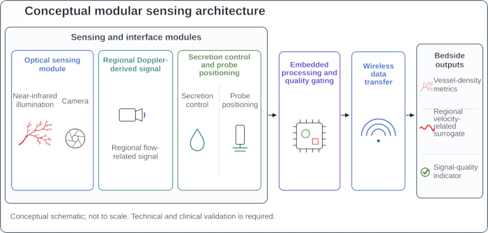
Figure 1. Author-proposed modular sensing architecture for bedside sublingual microcirculation monitoring. Conceptual modular sensing architecture showing optical sensing, regional Doppler-derived sensing, secretion control and probe positioning, embedded processing and quality gating, wireless data transfer, and bedside outputs. Arrows indicate functional links and do not imply a validated serial signal pathway. The schematic is not drawn to scale and does not represent a clinically validated all-in-one device. The framework is informed in part by the disclosed patent, which is cited solely as an illustrative design example and not as evidence of performance or clinical validity34. Please click here to view a larger version of this figure.
One publicly disclosed patent describes near-infrared illumination, camera-based imaging, an ultrasound probe, saliva removal, embedded processing, wireless transmission, and terminal display34. The patent is cited solely as an illustrative design example and is not presented as evidence of analytical validity, safety, feasibility, regulatory readiness, treatment benefit, or clinical effectiveness.
Within this conceptual framework, near-infrared camera imaging would be evaluated as a superficial-vessel input rather than as a substitute for HVM or as a measure of tissue oxygenation. Similarly, an ultrasound component would most likely provide a regional vascular signal rather than direct measurement of capillary red blood cell velocity. Interpretation of such signals would depend on sampling depth, acoustic coupling, tissue motion, insonation angle, output definition, and spatial registration with the optical field.
Testing should begin at the individual module level. Optical evaluation would require endpoints addressing vessel contrast, illumination stability, field-of-view repeatability, and resistance to saliva- and pressure-related artifacts. Evaluation of velocity-related components would require a predefined sampling volume, stable measurement units, assessment of angle sensitivity and signal-to-noise ratio, and comparison with an appropriate calibrated flow reference. Interface and reporting modules would require separate evaluation of secretion control, contact pressure, user comfort, contamination risk, processing delay, synchronization, data loss, and presentation of quality information.
Bedside workflow and minimum report
The proposed workflow shown in Figure 2 comprises four stages. Acquisition would obtain the intended optical signal and, when available, regional Doppler-derived or calibrated oxygenation inputs. Stabilization and quality control would identify saliva, blur, tissue compression, bubbles, motion, and inadequate illumination. Only signals meeting prespecified quality criteria would proceed to analysis. Reporting would then present the measurement, its temporal trend, and an explicit statement of uncertainty or acquisition failure.
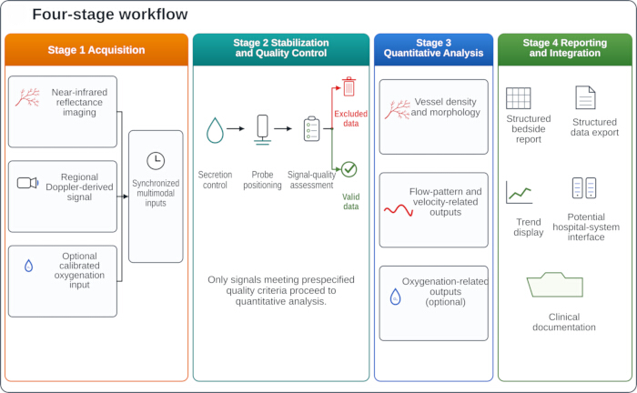
Figure 2. Author-proposed bedside workflow for sublingual microcirculation monitoring. Proposed four-stage workflow linking acquisition, stabilization and quality control, quantitative analysis, and reporting and integration. The optional calibrated oxygenation input is shown as a complementary input and is not considered a required component of the core sensing architecture. Displayed outputs represent candidate measurement categories rather than validated diagnostic indices. Please click here to view a larger version of this figure.
The initial bedside report should be deliberately concise. A reasonable starting set includes one density-related measure, one perfusion- or flow-pattern measure when flow is resolvable, a regional velocity-related estimate only when its interpretation has been validated, the proportion of accepted data, the reason for any failed acquisition, and a time-stamped within-patient trend. Early studies should emphasize changes within the same patient rather than applying universal thresholds across devices or patient populations.
Measurement outputs should remain distinct from treatment recommendations. An abnormal measurement should first prompt confirmation of signal quality and repeat acquisition, when feasible. If the abnormality persists, it may prompt reassessment of systemic hemodynamics, current vasoactive drug dose, hemoglobin concentration, oxygenation, temperature, ventilation, and potential local artifacts. It should not automatically trigger fluid administration or changes in vasoactive therapy.
Performance metrics and statistical considerations
The endpoints described below are proposed for future evaluation and should not be interpreted as validated acceptance thresholds. Morphological studies may assess visible or perfused vessel density, vessel-width distribution, segment continuity, and spatial heterogeneity. Flow studies may evaluate prespecified flow categories, calibrated image-based velocity, or clearly defined Doppler-derived surrogates. Oxygenation endpoints are appropriate only when a calibrated multispectral or NIRS component is incorporated. Across all measurement domains, quality reporting should include the proportion of usable data, reasons for data rejection, acquisition time, artifact flags, and missing segments or segments that cannot be spatially co-registered.
Reliability analysis requires a prespecified unit of analysis. Frames and vessel segments are nested within acquisitions, and repeated acquisitions are nested within participants; therefore, treating all observations as independent would produce artificially narrow estimates of uncertainty. The intraclass correlation coefficient model, agreement definition, and rater structure should be specified in advance. Within-subject coefficients of variation, test–retest bias, and Bland–Altman limits of agreement should be estimated using methods that account for repeated observations within each participant. The smallest detectable change quantifies measurement error and should not be interpreted as a clinically important change unless the corresponding clinical threshold has been established independently.
Benchmark comparisons should be matched to the specific signal under evaluation. Morphology- and flow-pattern outputs may be compared with expert-reviewed HVM analyses or consensus annotations. Velocity estimates require flow phantoms, controlled Doppler reference methods, or calibrated image-based tracking. Oxygenation measurements require an appropriate tissue-oxygenation protocol with controlled physiological perturbation. Optional tissue-gas measurements require a suitable gas-tension reference. Data-transfer and reporting modules should additionally be evaluated for timestamp integrity, access control, data loss, auditability, and interoperability. These proposed benchmark comparisons and interpretation considerations are summarized in Table 2.
| Author-proposed module and primary function | Current principle or limitation | Design consideration | Candidate reportable outputs | Quality and safety checks | Validation and repeatability endpoints | Domain-matched benchmark and interpretation caution |
| Surface-vessel imaging (morphology and density) | Handheld vital microscopy (HVM) directly visualizes red blood cell-filled microvessels but is sensitive to probe pressure, motion, saliva, bubbles, focus, and illumination. Camera-based near-infrared reflectance imaging remains a candidate superficial-vessel approach. | Controlled illumination, reproducible field of view, positioning support, explicit quality gating, and spatial registration only when multimodal fusion is intended. | Visible-vessel density; perfused-vessel density only when flow is directly resolved; vessel-width distribution; segment continuity; spatial heterogeneity. | Focus, illumination, motion, compression, secretion burden, accepted data proportion, and missing segments or segments that cannot be spatially co-registered. | Optical resolution; illumination stability; field-of-view reproducibility; registration error when multimodal fusion is intended; agreement with expert-reviewed HVM; robustness to artifacts; and acquisition failure rate. | Expert-reviewed HVM or consensus annotation. Associations established using HVM should not be extrapolated to near-infrared reflectance imaging without independent construct and criterion validation. |
| Regional Doppler-derived sensing (flow and velocity) | Regional Doppler sensing is a candidate velocity-related input rather than an established sublingual microvascular measurement and is influenced by insonation angle, sampling depth, acoustic coupling, and motion. | Standardized probe geometry, angle and depth metadata, synchronized acquisition, and visible signal quality feedback. | Regional velocity-related surrogate; pulsatility-related variables; temporal variability. | Angle and depth validity, signal-to-noise ratio, coupling quality, motion flag, spectral completeness, and synchronization error. | Flow-phantom linearity; angle sensitivity; sampling-depth stability; agreement with controlled Doppler or calibrated image-based velocity references; synchronization performance; and acquisition failure rate. | Calibrated flow phantom, reference Doppler method, or calibrated image-based velocity method. Regional Doppler signals should be interpreted as regional and depth dependent rather than as direct measures of capillary red blood cell velocity. |
| Optional calibrated oxygenation module (oxygenation) | Near-infrared spectroscopy (NIRS) provides volume-averaged oxygenation-related signals but is influenced by sampling depth, tissue optical properties, probe pressure, ambient light, and device calibration. Individual microvessels are not resolved. | Optional calibrated multispectral or NIRS module maintained analytically separate from camera-based reflectance imaging. | Tissue oxygenation-related value; recovery slope or recovery area during a controlled physiological perturbation; within-patient trend. | Optical coupling, ambient-light control, pressure flag, calibration status, signal drift, and physiological plausibility. | Optical-phantom calibration; controlled physiological perturbation; agreement with an appropriate reference tissue-oxygenation method; response time; signal drift; and calibration stability. | Reference tissue-oxygenation protocol or accepted physiological perturbation. Oxygenation measurements are complementary and should not be interpreted as direct measures of capillary morphology or blood-cell flow. |
| Mucosal-interface stabilization and acquisition (data quality and safety) | Freehand probe contact, variable site selection, oral secretions, and uncontrolled pressure can produce tissue compression, motion, contamination, and repeatability artifacts. | Secretion control, atraumatic contact interface, positioning and pressure support, anatomy-guided site selection, reacquisition prompts, and validated single-use barriers or validated cleaning and disinfection procedures. | Accepted data proportion; acquisition duration; reason for acquisition failure; contact-pressure indicator; secretion burden; site identifier. | Pressure and site confirmation, mucosal injury assessment, oral-access status, barrier integrity, and contamination-control status. | Usable-data yield; time to acceptable acquisition; user comfort; mucosal adverse events; cleaning or disinfection validation; cross-contamination testing; same-site reacquisition reproducibility; and learning-curve assessment. | Standardized HVM acquisition protocol or another prespecified acquisition standard. Improvements in acquisition quality and safety should be demonstrated rather than assumed. |
| Embedded analytics and quality gating (processing and uncertainty) | Offline or semiautomated analysis may be delayed and influenced by subjective clip selection, algorithm choice, and nontransparent data exclusion. | On-device or edge processing with explicit quality gates, visible rejection reasons, modular algorithms, uncertainty display, version control, and an auditable record. | Quality-approved physiological measurements; accepted and rejected data fractions; uncertainty indicator; rejection reason; algorithm version. | Prespecified quality thresholds, failure flag, missing-data handling, override logging, version tracking, and an out-of-distribution flag when artificial intelligence is used. | Agreement with an expert or domain-specific reference; sensitivity and specificity for unacceptable data at prespecified thresholds; processing latency; robustness to artifacts; failure-mode analysis; reanalysis consistency; calibration stability; and software-version comparability. | Expert-reviewed HVM, annotated datasets, or other domain-specific reference standards. Faster computation alone does not establish analytical validity or clinical readiness. |
| Reporting, interoperability, and cybersecurity (workflow integration) | Manual data export and delayed documentation can result in isolated values, incomplete metadata, inconsistent units, and limited traceability. | Structured bedside report with time-stamped trends, quality and uncertainty indicators accompanying each value, standardized data export, controlled access, encryption, and an audit trail. | Current value; prespecified baseline or reference measurement; absolute and relative within-patient change; time since the previous measurement; quality flag; uncertainty indicator; reason for acquisition failure; structured data export. | Timestamp integrity, unit and metadata completeness, data-loss testing, encryption, access control, auditability, and export validation. | End-to-end latency; export accuracy; data integrity; interoperability; human-factors testing; consistency of display across devices or sites; and user-comprehension testing. | Clinical information-system and human-factors requirements. Reporting should support structured reassessment and documentation rather than autonomous treatment recommendations. |
| Integrated bedside workflow (clinical utility and safety) | Physiological relevance and prognostic association do not establish treatment benefit, and responses to fluid or vasoactive interventions remain heterogeneous. | Clinician-supervised confirm-repeat-reassess pathway with prespecified triggers for repeat measurement or reassessment, escalation and stopping rules, and documentation of clinician overrides. | Early phase: completion of repeat measurement or reassessment, management changes, prespecified potentially inappropriate interventions, protocol deviations, and device-related adverse events. Later phase: patient-centered outcomes. | Protocol adherence, completion of repeat measurements, reasons for clinician override, protocol deviations, adverse-event surveillance, and data completeness. | Prospective feasibility and human-factors studies followed, when justified, by decision-triggered comparative evaluation; workflow fidelity; training effects; reproducibility across patient groups and failure scenarios; and prespecified progression criteria. | Usual care or standard hemodynamic assessment. No treatment threshold or outcome claim is justified until prospective clinical utility and safety have been demonstrated. |
Table 2: Author-proposed modular framework for development and validation of bedside sublingual microcirculation monitoring. The table summarizes author-proposed monitoring modules, current measurement principles or limitations, design considerations, candidate reportable outputs, quality and safety checks, validation and repeatability endpoints, and domain-matched benchmark methods with interpretation cautions. The framework distinguishes established measurement principles from author-proposed components and separates early technical, process, and safety outcomes from subsequent evaluation of clinical utility. This framework is conceptual and has not been clinically validated. Note. This author-proposed framework has not been clinically validated. The disclosed patent is cited solely as an illustrative design example and is not evidence of analytical validity, safety, feasibility, regulatory readiness, or clinical effectiveness. All candidate outputs, quality and safety assessments, validation endpoints, repeatability targets, and progression criteria require empirical evaluation. Reliability analyses should prespecify the unit of analysis and account for nested and repeated observations. Abbreviations: HVM, handheld vital microscopy; ICC, intraclass correlation coefficient; NIRS, near-infrared spectroscopy.
Validation and reproducibility
The proposed pathway adapts established principles of verification, analytical validation, and clinical validation35. The first stage would verify optical resolution, illumination stability, signal-to-noise performance, Doppler angle sensitivity, registration error, processing delay, battery performance, data integrity, and relevant electrical and acoustic safety. The second stage would evaluate acquisition time, user comfort, secretion control, reasons for acquisition failure, and intraoperator and interoperator repeatability in simulation studies or controlled volunteer investigations. The staged validation pathway is summarized in Figure 3.
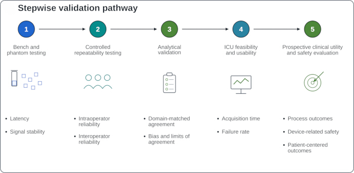
Figure 3. Author-proposed staged validation pathway for bedside sublingual microcirculation monitoring. Conceptual validation pathway progressing from bench and phantom testing and controlled repeatability testing to domain-matched analytical validation, intensive care unit feasibility and usability, and prospective evaluation of clinical utility and safety. Selected endpoints are provided as illustrative examples and do not constitute an exhaustive validation checklist. Abbreviation: ICU, intensive care unit. Please click here to view a larger version of this figure.
The third stage would determine whether each measurement output agrees with an appropriate reference method and responds as expected during controlled physiological changes. In this context, bias, limits of agreement, heteroscedasticity, repeatability, and failure rates are more informative than correlation alone. Evidence obtained using HVM should not be transferred automatically to near-infrared camera imaging, NIRS, or Doppler-derived signals.
The fourth stage would evaluate feasibility in the ICU, including performance during mechanical ventilation, edema, shock, limited oral access, excessive secretions, and patient movement. Failed acquisitions and mucosal adverse events should be reported rather than excluded from performance analyses. The fifth stage would evaluate clinical utility and safety. Initial investigations may focus on process outcomes, inappropriate interventions, and device-related events before larger trials assess patient-centered outcomes. Progression criteria should be specified before data analysis.
When artificial intelligence (AI) is used to accept or reject acquisitions, extract variables, or generate clinician-facing outputs, early studies should clearly describe the intended role of the AI system, its inputs and outputs, the intended clinical setting, the degree of human interaction, and known failure modes36. Likewise, trials evaluating AI- or device-triggered interventions should describe the mechanisms of human oversight and explain how system outputs were translated into clinical actions37.
Potential clinical applications
The most plausible near-term application is targeted reassessment when systemic hemodynamic variables and tissue perfusion appear discordant. In septic shock, repeated sublingual measurements may help characterize persistent perfusion abnormalities after restoration of blood pressure. Fluid responsiveness remains heterogeneous, however, and a favorable average response should not be assumed for individual patients15,16. Accordingly, reviews of microcirculation-guided resuscitation regard bedside monitoring as promising but not yet suitable to direct routine treatment decisions17.
In trauma or hemorrhagic shock, direct microcirculatory assessment has been investigated as an adjunct to conventional systemic resuscitation endpoints21,38. These studies do not establish validated treatment thresholds. Similarly, in perioperative and cardiac critical care, repeated IDF measurements have been used to characterize sublingual microcirculatory changes during postoperative recovery following cardiac surgery26. At present, this application should be regarded as exploratory rather than an established basis for clinical decision-making.
Across all clinical settings, a persistent abnormality is best interpreted as a prompt to reassess both the patient and the measurement rather than as a stand-alone indication for intervention. This approach preserves clinical judgment and reduces the risk of overinterpreting a technically plausible signal before its measurement error and clinical significance are fully understood.
Challenges and future directions
Conditions encountered in the ICU are substantially less controlled than those in bench experiments or volunteer studies. Secretions, restricted oral access, edema, patient movement, and variable contact pressure can all compromise signal quality. Integrating optical imaging, ultrasound coupling, mucosal cleaning, and stable positioning within a compact oral probe is particularly challenging because these components have different physical requirements and may interfere with one another. Consequently, failure rates and reasons for failed acquisitions are as important as average performance among successful measurements.
Device readiness also encompasses safety and information governance. Feasibility studies should evaluate mucosal biocompatibility, pressure injury, cleaning or single-use barriers, cross-contamination, electrical and acoustic exposure, and compatibility with ICU infection-control practices. Wireless communication and reporting functions additionally require assessment of encryption, access control, timestamp integrity, audit trails, data-loss resilience, and secure integration with clinical information systems.
Future development can proceed according to three practical priorities. First, retain only outputs with clear physiological meaning and acceptable repeatability. Second, display rejection reasons and measurement uncertainty alongside every reported result. Third, integrate individual modules only after each has demonstrated independent feasibility. Camera-based photoplethysmographic estimates depend on illumination and signal-extraction conditions31,39, whereas Doppler-derived velocity estimates depend strongly on sampling geometry and insonation angle29,40. These limitations should remain explicit after system integration.
Technical sophistication alone will not ensure clinical usefulness. A monitoring system may still fail if it is uncomfortable, slow, difficult to position, or challenging to interpret. Conversely, a limited set of carefully validated measurements may provide meaningful clinical value when acquisition is rapid, failure is clearly identified, repeatability is well characterized, and results are incorporated into a structured reassessment process.
Limitations of this review
This review used targeted literature searches, including a focused update before final manuscript submission, rather than a systematic review protocol and therefore may not have identified all relevant studies. Because formal risk-of-bias assessment and quantitative synthesis were not performed, the review cannot compare measurement modalities on the basis of pooled diagnostic accuracy or treatment effect. The conceptual workflow incorporates one publicly disclosed patent as an illustrative example of system integration; however, no original evidence of performance, usability, safety, regulatory readiness, or clinical benefit is presented for that architecture. Several proposed endpoints and progression criteria represent author-generated recommendations that require empirical validation. Accordingly, this review should be interpreted as a structured synthesis of current measurement approaches and validation priorities rather than as evidence that an integrated monitoring system is clinically effective.
Access restricted. Please log in or start a trial to view this content.
Conclusions
Sublingual microcirculation monitoring addresses an important limitation of conventional hemodynamic assessment: restoration of systemic variables does not necessarily indicate recovery of tissue-level perfusion. Current and emerging monitoring approaches provide complementary, but incomplete, information regarding vascular morphology, vessel density, blood-cell flow, regional velocity, and tissue oxygenation, and the interpretation of these measurements depends fundamentally on acquisition quality and appropriate qualit...
Access restricted. Please log in or start a trial to view this content.
Disclosures
Zhejiang University holds intellectual property related to the sublingual microcirculation monitoring architecture discussed in this manuscript, including United States Patent No. US 11,877,886 B2, entitled Sublingual Microcirculation Detection Device, Sublingual Microcirculation Detection System and Processing Method Thereof. Siyi Jiang is a named inventor on this patent34. The patent is cited solely as an illustrative design example and is not presented as evidence of analytical validity, safety, feasibility, regulatory readiness, clinical performance, or clinical effectiveness. The authors declare no other financial or personal relationships that could be construed as potential conflicts of interest.
Artificial intelligence disclosure: ChatGPT (OpenAI) was used during manuscript revision to assist with language editing, restructuring of author-prepared text, and format checking. It was not used as a source of scientific evidence or scientific content. The authors independently evaluated the literature, verified the scientific content, references, and patent information, reviewed and approved the final manuscript, and accept full responsibility for its content.
Acknowledgements
This work was supported by the Basic Public Welfare Research Project of Zhejiang Province (Project No. LGF22H150003). The authors thank their clinical and engineering colleagues for providing feedback on bedside workflow requirements for sublingual microcirculation monitoring.
Access restricted. Please log in or start a trial to view this content.
References
- Lundy DJ, Trzeciak S. Microcirculatory dysfunction in sepsis. Crit Care Nurs Clin North Am. 2011;23:67-77.
- Miranda M, Balarini M, Caixeta D, Bouskela E. Microcirculatory dysfunction in sepsis: pathophysiology, clinical monitoring, and potential therapies. Am J Physiol Heart Circ Physiol. 2016;311:H24-H35.
- González R, Urbano J, López-Herce J. Resuscitating the macro- vs. microcirculation in septic shock. Curr Opin Pediatr. 2024;36:274-281.
- Ince C. Hemodynamic coherence and the rationale for monitoring the microcirculation. Crit Care. 2015;19(Suppl 3):S8.
- De Backer D, et al. How to evaluate the microcirculation: report of a round table conference. Crit Care. 2007;11:R101.
- Ince C, et al. Second consensus on the assessment of sublingual microcirculation in critically ill patients: results from a task force of the European Society of Intensive Care Medicine. Intensive Care Med. 2018;44:281-299.
- Dilken O, Ergin B, Ince C. Assessment of sublingual microcirculation in critically ill patients: consensus and debate. Ann Transl Med. 2020;8:793.
- Massey MJ, Shapiro NI. A guide to human in vivo microcirculatory flow image analysis. Crit Care. 2016;20:35.
- Massey MJ, et al. The microcirculation image quality score: development and preliminary evaluation of a proposed approach to grading quality of image acquisition for bedside videomicroscopy. J Crit Care. 2013;28:913-917.
- Damiani E, et al. Impact of microcirculatory video quality on the evaluation of sublingual microcirculation in critically ill patients. J Clin Monit Comput. 2017;31:981-988.
- Pan P, et al. Role of combining peripheral with sublingual perfusion on evaluating microcirculation and predicting prognosis in patients with septic shock. Chin Med J (Engl). 2018;131:1158-1166.
- Hernandez G, et al. Severe abnormalities in microvascular perfused vessel density are associated to organ dysfunction and mortality and can be predicted by hyperlactatemia and norepinephrine requirements in septic shock patients. J Crit Care. 2013;28:538.e9-538.e14.
- Bruno RR, et al. Sublingual microcirculatory assessment on admission independently predicts the outcome of old intensive care patients suffering from shock. Sci Rep. 2024;14:25668.
- Bruno RR, et al. Direct assessment of microcirculation in shock: a randomized controlled multicenter study. Intensive Care Med. 2023;49:645-655.
- Cusack R, O'Neill S, Martin-Loeches I. Effects of fluids on the sublingual microcirculation in sepsis. J Clin Med. 2022;11:7277.
- Dubin A. Effects of fluids on sublingual microcirculation: a point-of-view review. Ann Intensive Care. 2025;15:178.
- Damiani E, et al. Microcirculation-guided resuscitation in sepsis: the next frontier? Front Med (Lausanne). 2023;10:1212321.
- Güven G, et al. Morphologic mapping of the sublingual microcirculation in healthy volunteers. J Vasc Res. 2022;59:199-208.
- Uz Z, et al. Identifying a sublingual triangle as the ideal site for assessment of sublingual microcirculation. J Clin Monit Comput. 2023;37:639-649.
- Wadowski PP, et al. Sublingual functional capillary rarefaction in chronic heart failure. Eur J Clin Invest. 2018;48:e12869.
- Palágyi P, et al. Monitoring microcirculatory blood flow with a new sublingual tonometer in a porcine model of hemorrhagic shock. Biomed Res Int. 2015;2015:847152.
- Hessler M, et al. Monitoring of conjunctival microcirculation reflects sublingual microcirculation in ovine septic and hemorrhagic shock. Shock. 2019;51:479-486.
- Kanoore Edul VS, et al. Dissociation between sublingual and gut microcirculation in the response to a fluid challenge in postoperative patients with abdominal sepsis. Ann Intensive Care. 2014;4:39.
- Groner W, et al. Orthogonal polarization spectral imaging: a new method for study of the microcirculation. Nat Med. 1999;5:1209-1212.
- Goedhart PT, et al. Sidestream dark field imaging: a novel stroboscopic LED ring-based imaging modality for clinical assessment of the microcirculation. Opt Express. 2007;15:15101-15114.
- Uz Z, et al. Recruitment of sublingual microcirculation using handheld incident dark field imaging as a routine measurement tool during the postoperative de-escalation phase: a pilot study in post-ICU cardiac surgery patients. Perioper Med (Lond). 2018;7:18.
- Marin T, Moore J. Understanding near-infrared spectroscopy: an update. Crit Care Nurs Clin North Am. 2024;36:41-50.
- Steenhaut K, et al. Evaluation of different near-infrared spectroscopy technologies for assessment of tissue oxygen saturation during a vascular occlusion test. J Clin Monit Comput. 2017;31:1151-1158.
- Do Amaral Tafner PF, et al. Recent advances in bedside microcirculation assessment in critically ill patients. Rev Bras Ter Intensiva. 2017;29:238-247.
- Heeman W, Steenbergen W, van Dam GM, Boerma EC. Clinical applications of laser speckle contrast imaging: a review. J Biomed Opt. 2019;24:080901.
- Uribe Acevedo R, et al. Non-invasive assessment of sublingual microcirculation using flow derived from green light PPG: evaluation and reference values. J Biomed Opt. 2024;29:017001.
- Mari A, et al. Transcutaneous PCO₂ monitoring in critically ill patients: update and perspectives. J Thorac Dis. 2019;11(Suppl 11):S1558-S1567.
- Hilty MP, et al. MicroTools enables automated quantification of capillary density and red blood cell velocity in handheld vital microscopy. Commun Biol. 2019;2:217.
- Jiang S, Zheng Y, Li C, inventors; Zhejiang University, assignee. Sublingual microcirculation detection device, sublingual microcirculation detection system and processing method thereof. United States patent US 11,877,886 B2. January 23, 2024.
- Goldsack JC, et al. Verification, analytical validation, and clinical validation (V3): the foundation of determining fit-for-purpose for Biometric Monitoring Technologies (BioMeTs). NPJ Digit Med. 2020;3:55.
- Vasey B, et al. Reporting guideline for the early-stage clinical evaluation of decision support systems driven by artificial intelligence: DECIDE-AI. Nat Med. 2022;28:924-933.
- Liu X, et al. Reporting guidelines for clinical trial reports for interventions involving artificial intelligence: the CONSORT-AI extension. Nat Med. 2020;26:1364-1374.
- Lee YLL, et al. Evaluation of microvascular perfusion and resuscitation after severe injury. Am Surg. 2015;81:1272-1278.
- Verkruysse W, Svaasand LO, Nelson JS. Remote plethysmographic imaging using ambient light. Opt Express. 2008;16:21434-21445.
- Saad AA, Loupas T, Shapiro LG. Computer vision approach for ultrasound Doppler angle estimation. J Digit Imaging. 2009;22:681-688.
Access restricted. Please log in or start a trial to view this content.