This protocol describes CRISPR-Cas9-mediated disruption of the nodC locus in Mesorhizobium ciceri using homology-directed repair and GFP-based screening to generate symbiosis-defective mutants for functional genomics studies in chickpea-associated rhizobia.
Method Article
CRISPR-Cas9 Genome Editing in Mesorhizobium ciceri Through nodC Disruption for Chickpea Symbiosis Studies
* These authors contributed equally
In This Article
Summary
Abstract
Mesorhizobium ciceri, a nitrogen-fixing symbiont of chickpea (Cicer arietinum), remains genetically challenging to manipulate using conventional homologous recombination approaches, which are labor-intensive and often leave undesirable selection markers. In this protocol, we describe a streamlined genome-editing strategy using a broad-host-range Clustered Regularly Interspaced Short Palindromic Repeats (CRISPR)-associated protein 9 (Cas9) system adapted for M. ciceri. We demonstrate the efficacy of this method by targeting nodC, which encodes the N-acetylglucosaminyltransferase required for chitin backbone synthesis in Nod factors, the primary signaling molecules involved in symbiotic molecular communication. This protocol details the systematic design of single-guide RNAs (sgRNAs) and the construction of a homology-directed repair (HDR) template. The HDR template was designed to facilitate site-specific integration of a green fluorescent protein (GFP) reporter flanking the nodC cleavage site. Following delivery of the Cas9/sgRNA/HDR construct through biparental mating, putative mutants were identified using a fluorescence-based screening approach. Successful disruption of the 1.3 kb nodC locus within the nodulation (nod) cassette was initially screened by visualization of GFP expression in mutant colonies using fluorescence microscopy. The disruption was further validated by restriction digestion, amplification of the integrated cassette from genomic DNA, and Sanger sequencing. GFP expression was additionally quantified by reverse transcription quantitative polymerase chain reaction. To validate the functional impact of the mutation, chickpea infection assays were performed, demonstrating impaired nodulation in plants inoculated with ΔnodC M. ciceri compared with the wild-type strain. Overall, this protocol provides an efficient and reproducible framework for precise gene disruption and functional genomics studies in Mesorhizobium.
Introduction
The Clustered Regularly Interspaced Short Palindromic Repeats (CRISPR)-associated protein 9 (Cas9) nuclease system, originally identified as an adaptive immune mechanism in prokaryotes, has been adapted into an effective and reliable tool for genome editing1,2. The system uses a synthetic guide RNA (sgRNA) to identify target DNA sequences through Watson-Crick base pairing. The sgRNA comprises 20 nucleotides complementary to the genomic region adjacent to the protospacer adjacent motif (PAM) sequence NGG. Cas9 generates a double-stranded break (DSB) at a specified site within the target sequence, generally 3 base pairs (bp) upstream of the PAM site1. The resulting DSBs can be repaired either by non-homologous end joining or by homology-directed repair (HDR) using a donor template3,4. This strategy has been widely applied for gene disruption, knock-in, and knock-out approaches in bacteria5,6,7,8. The primary objective of this protocol is to develop an efficient genome-editing method for Mesorhizobium ciceri using a broad-host-range CRISPR-Cas9 system.
M. ciceri is a Gram-negative soil bacterium that functions as an essential nitrogen-fixing endosymbiont of chickpea (Cicer arietinum)9. One strain, M. ciceri CC1192, exhibits high nodulation efficiency, and its genome has been sequenced10. Despite its agricultural significance, genetic manipulation of Mesorhizobium species remains limited. CRISPR-based systems have previously been implemented in other rhizobia, including CRISPR interference (CRISPRi) in Rhizobium etli11, genome editing in Sinorhizobium meliloti12, and base editing in S. meliloti13. However, a robust genome-editing platform for M. ciceri has not been reported. Conventional approaches rely on suicide-plasmid-mediated homologous recombination, which is labor-intensive, requires multiple rounds of selection, and frequently leaves antibiotic-resistance markers as genomic scars14.
To address these limitations, the pCasPP vector system, originally developed for Paenibacillus polymyxa, was adapted for use in M. ciceri15. The pCasPP framework utilizes a Streptococcus pyogenes Cas9 codon-optimized for high-GC actinomycetes and contains a lacZ cassette for blue-white screening of sgRNA cloning under the constitutive gapdh promoter8. Biparental mating was performed between the donor Escherichia coli strain and the recipient Mesorhizobium strain to deliver the editing vector16. To enhance screening efficiency, the green fluorescent protein (GFP) variant bjGFP, originally codon-optimized for Bradyrhizobium diazoefficiens, was incorporated into the HDR donor template16. This fluorescence-based screening strategy was applied to target the essential nodC locus, which is required for the early stages of symbiotic signaling between the bacterium and chickpea17,18.
This protocol provides a precise and efficient alternative to random chemical or ultraviolet (UV) mutagenesis approaches. In validation experiments, over 80% of recovered colonies displayed GFP fluorescence, and representative edited colonies were further confirmed by polymerase chain reaction (PCR) and Sanger sequencing. The resulting ΔnodC M. ciceri mutants provide a useful resource for studying symbiotic signaling and host-microbe interactions. Overall, this framework offers the rhizobium research community an efficient and scalable approach for functional genomics studies in Mesorhizobium.
Protocol
All experimental protocols involving plant growth, microbial inoculation, and the handling of genetically modified bacteria were conducted in accordance with institutional guidelines and regulations. Plant cultivation and bacterial inoculations were performed under controlled growth-chamber conditions. The M. ciceri strain used in this study is classified as a Biosafety Level 1 (BSL-1) microorganism. Because all experiments were conducted under contained laboratory conditions without field release, formal institutional biosafety approval was not required in accordance with institutional biosafety policies. All biological waste generated during the study was autoclaved prior to disposal.
1. Competent Cell Preparation
Chemically competent E. coli DH5α and E. coli S17-1λpir cells were prepared and validated according to the procedure described in Supplementary File 1.
2. Guide RNA Design
- Design of Guide RNAs
- Open the Benchling platform (https://benchling.com/). Sign in to an existing Benchling account or create a new account.
- Create a new project and open the CRISPR Guide Design Tool.
- Upload or paste the complete target DNA sequence for guide RNA design. Select the “Single Guide” design option and set the guide length to 20 bp.
- Select the genome reference from the available options. For guide RNA design targeting M. ciceri, select Bradyrhizobium diazoefficiens USDA 110 as the reference genome. Use NGG as the protospacer adjacent motif (PAM) sequence required for Streptococcus pyogenes Cas9 (SpCas9) activity.
- Use the Hsu et al.19 algorithm to calculate predicted off-target cutting frequencies. Maintain a threshold score of ≥50 to minimize nonspecific cleavage across the reference genome background.
- Use the Doench-Root algorithm20 to calculate predicted on-target efficiency scores. Set a target-selection threshold of ≥60 to identify guides with favorable predicted Cas9 cleavage efficiency at the target locus.
- Click “Finish” to generate the list of candidate guide RNAs.
- Selection of Guide RNAs
- Open the guide RNA output panel.
- Prioritize candidate sgRNAs predicted to generate a double-strand break within 100 bp of the translational start codon (ATG), increasing the likelihood of producing a non-functional protein.
- Evaluate the off-target and on-target scores. Prioritize a higher off-target score over a slightly higher on-target score to reduce the risk of unintended off-target cleavage.
- Select one or two candidate single-guide RNAs (sgRNAs) that satisfy all selection criteria.
- Preparation of gRNA Oligonucleotides
- Generate the reverse-complement sequence of the selected guide RNA.
- Add the sequence 5′-ACGC to the 5′ end of the guide RNA and add the sequence 5′-AAAC to the 5′ end of the reverse-complement strand.
- Order both oligonucleotides with 5′ phosphorylation and PAGE purification at a 25 nmol synthesis scale.
- Annealing of gRNA Oligonucleotides
- Prepare 30 mM HEPES buffer by dissolving 7.15 g HEPES free acid in approximately 800 mL distilled water. Adjust the pH to 7.8 using sodium hydroxide and bring the final volume to 1 L.
- Resuspend each phosphorylated oligonucleotide in nuclease-free water to a final concentration of 100 µM. Combine 5 µL of each oligonucleotide with 90 µL of 30 mM HEPES buffer in a sterile microcentrifuge tube.
- Vortex briefly and centrifuge to collect the liquid. Anneal the oligonucleotides in a thermocycler by incubating at 95°C for 5 min and cooling to 4°C at a ramp rate of 0.1°C/s.
- Dilute the annealed oligonucleotides 10-fold using nuclease-free water.
- Assembly of gRNA into pCasPP Vector
- Obtain the pCasPP vector described previously by Rütering et al.15.
- The plasmid sequence was provided by Jochen Schmid, and the construct used in this study was obtained through Dr. Syed Shams Yazdani (ICGEB).
- Assemble a Golden Gate reaction containing 100 ng pCasPP vector, 0.3 µL annealed oligonucleotide insert, 2 µL 10× T4 DNA ligase buffer, 1 µL T4 DNA ligase, 1 µL BbsI restriction enzyme, and nuclease-free water to a final volume of 20 µL.
- Prepare a negative-control reaction by replacing the insert with nuclease-free water.
- Mix the reactions gently and briefly centrifuge.
- Golden Gate Assembly Cycling Conditions
- Load the reaction tubes into a thermocycler. Perform nine cycles consisting of 37°C for 10 min followed by 16°C for 10 min.
- Incubate the reactions at 50°C for 5 min and then at 65°C for 20 min. Hold the reactions at 4°C until use.
- Transformation of Assembly Reactions
- Thaw 50 µL competent Escherichia coli DH5α cells on ice for 10–15 min.
- Add 3 µL of each Golden Gate assembly reaction to the competent cells. Process the negative-control reaction in parallel. Incubate the mixtures on ice for 30 min.
- Heat shock the cells at 42°C for 45 s and immediately return the tubes to ice for 2 min. Add 950 µL Super Optimal broth with Catabolite repression (SOC) medium.
- Prepare SOC medium containing 0.5% yeast extract, 2% tryptone, 10 mM NaCl, 2.5 mM KCl, 10 mM MgCl₂, 10 mM MgSO₄, and 20 mM glucose. Filter-sterilize the medium using a 0.4 µm syringe filter under aseptic conditions.
- Incubate the cultures at 37°C and 180 rpm for 1 h.
- Selection of Positive Transformants
- Prepare LB agar plates supplemented with 50 µg/mL neomycin, 0.2 mM isopropyl β-D-1-thiogalactopyranoside (IPTG), and 32 mg/L 5-bromo-4-chloro-3-indolyl β-D-galactopyranoside (X-gal). Plate 100 µL of the recovery culture onto the selective plates. Incubate the plates at 37°C for 12–16 h.
- Select white colonies and discard blue colonies. Verify that the negative-control plate contains fewer than five colonies.
- Screening and Expansion of Positive Clones
- Patch three to four independent white colonies onto fresh selective LB agar plates.
- Incubate the plates at 37°C for 12–16 h. Inoculate individual colonies into 10 mL LB medium supplemented with 50 µg/mL neomycin.
- Process each guide RNA construct independently throughout the downstream workflow.
- Validation of pCasPP-gRNA Constructs
- Harvest bacterial cultures by centrifugation. Isolate plasmid DNA using a silica-column plasmid purification kit.
- Digest the isolated plasmid DNA with XbaI and EcoRI in the appropriate reaction buffer for 1 h at 37°C.
- Resolve the digestion products on a 1% agarose gel prepared in 1× Tris-acetate-EDTA (TAE) buffer containing ethidium bromide at a final concentration of 0.5 µg/mL.
- Run the gel at 90–110 V until the dye front migrates across approximately 70–80% of the gel length. Visualize the DNA fragments using a UV transilluminator.
- Confirm positive clones by detecting a 1,280 bp fragment, and identify empty-vector controls by detecting a 1,630 bp fragment. The empty pCasPP vector is approximately 9,663 bp and contains a lacZ gene; insertion of the 24 bp gRNA oligonucleotide removes the lacZ fragment, producing a smaller gRNA-containing pCasPP construct. The XbaI and EcoRI restriction sites flanking the lacZ region are used for diagnostic confirmation of gRNA cloning.
- Preparation of Glycerol Stocks
- Grow a confirmed positive clone in selective LB medium.
- Prepare a glycerol stock by mixing 700 µL log-phase culture with 300 µL sterile 50% glycerol. Transfer the mixture into sterile cryovials. Store the glycerol stocks at −80°C.
NOTE: Avoid repeated freeze–thaw cycles. Prepare multiple aliquots to maintain plasmid stability and bacterial viability. The completed Golden Gate assembly reaction may be stored at 4°C overnight or at −20°C for 1–2 weeks before transformation.
3. Assembly of the Homology-Directed Repair Template
- Preparation of the green fluorescent protein (GFP) Insert
- Obtain the plasmid pRJPaph-bjGFP containing the fluorescent reporter cassette16 (Table 1)10,15,16,21,22,23. The plasmid was provided by Prof. Hans-Martin Fischer (ETH Zurich, Switzerland) and is available through Addgene (Plasmid #67032).
- Excise the gfp cassette from pRJPaph-bjGFP by SacI and KpnI restriction digestion as described in Supplementary File 2 (Step 3.1). The gfp sequence in pRJPaph-bjGFP is a synthetic bjGFP gene codon-optimized for Bradyrhizobium diazoefficiens.
- Purify the expected 1081 bp gfp fragment.
- Construction of pUC18-gfp
- Obtain the pUC18 backbone (Addgene plasmid #50004)21.
- Linearize pUC18 using SacI and KpnI as described in Supplementary File 2 (Step 3.2).
- Ligate the purified gfp fragment into the linearized pUC18 vector as described in Supplementary File 2 (Step 3.2).
- Transform competent E. coli DH5α cells using the transformation procedure described in Step 2.7, substituting the ligation mixture generated in Step 3.2.3 for the Golden Gate assembly reaction.
- Select positive transformants and verify gfp insertion by colony polymerase chain reaction (PCR) as described in Supplementary File 2 (Step 3.2).
- Amplification of Homology Arms
- Design primers to amplify upstream (UHA) and downstream (DHA) homology arms of approximately 400–500 bp from M. ciceri genomic DNA (Table 2).
- Incorporate SacI/EcoRI restriction sites into the UHA primers and XbaI/KpnI restriction sites into the DHA primers. Use primers F_nodC_Up_EcoRI and R_nodC_Up_SacI for amplification of the UHA and primers F_nodCdown_KpnI and R_nodCdown_XbaI for amplification of the DHA (Table 2).
- Isolate M. ciceri genomic DNA according to Weerakkody et al.25.
- Amplify and purify the UHA and DHA fragments as described in Supplementary File 2 (Step 3.3).
- Construction of pUC18-UHA-gfp
- Clone the UHA fragment into pUC18-gfp using EcoRI and SacI as described in Supplementary File 2 (Step 3.4).
- Confirm correct plasmid assembly by restriction digestion with XbaI and KpnI. Successful assembly should generate fragments of approximately 500 and 3725 bp.
- Proceed with one verified clone.
- Construction of pUC18-UHA-gfp-DHA
- Clone the DHA fragment into pUC18-UHA-gfp using KpnI and XbaI as described in Supplementary File 2 (Step 3.5).
- Confirm correct assembly by restriction digestion with SacI and EcoRI. Successful assembly should generate fragments of approximately 400 and 4271 bp.
- Verify 5–6 candidate clones by Sanger sequencing using primers F_nodC_Up_EcoRI, BjGFP_BP_F1, and R_nodCdown_XbaI (Table 2), which together cover the majority of the Homology-Directed Repair (HDR) cassette sequence.
- Amplification and Purification of the Final HDR Template
- Amplify the complete HDR cassette using primers F_FL_nodCgfp_XbaI and R_nodCdown_XbaI (Table 2).
- Purify the expected 2012 bp HDR fragment and digest the purified PCR product with XbaI as described in Supplementary File 2 (Step 3.6).
- Preparation of pCasPP-gRNA Vectors
- Retrieve the pCasPP-gRNA1 and pCasPP-gRNA2 plasmids generated in Step 2.
- Linearize the plasmids with XbaI and dephosphorylate the vector backbone as described in Supplementary File 2 (Step 3.7).
- Cloning of the HDR Template into pCasPP-gRNA Vectors
- Ligate the HDR cassette into XbaI-linearized pCasPP-gRNA1 or pCasPP-gRNA2 as described in Supplementary File 2 (Step 3.8).
- Transform competent E. coli DH5α cells and select transformants on LB agar containing 50 µg/mL neomycin.
- Screening of HDR-Containing Clones
- Screen transformants for GFP fluorescence using a fluorescence stereomicroscope equipped with a standard GFP filter set (excitation 470–488 nm; emission 507–510 nm).
- Classify colonies as GFP-positive based on the presence of distinct, uniform green fluorescence that clearly exceeds the weak autofluorescence observed in non-edited negative-control colonies.
- Expand GFP-positive clones and isolate plasmid DNA.
- Validation of HDR Vector Assembly
- Confirm HDR insertion by XbaI restriction digestion.
- Verify successful assembly by release of the expected 2012 bp HDR fragment together with an approximately 9,300 bp pCasPP-gRNA vector backbone.
NOTE: Following heat inactivation of the restriction enzymes and phosphatase, reaction mixtures may be stored at 4°C overnight or at −20°C for up to 1 month. Store confirmed plasmid constructs at −20°C for long-term preservation.
| Strain or plasmid | Description | Reference | ||||
| Mesorhizobium ciceri CC1192 | Recipient strain used for CRISPR-Cas9 genome editing and chickpea nodulation assays | Haskett et al.10 | ||||
| pCasPP | CRISPR-Cas9 genome-editing vector used for guide RNA cloning and HDR template delivery | Rütering et al.15 | ||||
| pRJPaph-bjGFP | GFP reporter plasmid used as the source of the GFP cassette (Addgene plasmid #67032) | Ledermann et al.16 | ||||
| pUC18 | General cloning vector used for HDR template assembly (Addgene plasmid #50004) | Norrander et al.21 | ||||
| Escherichia coli S17-1λpir | Donor strain used for biparental conjugation | Simon et al.22 | ||||
| Escherichia coli DH5α | Cloning host used for plasmid propagation | Hanahan et al.23 | ||||
Table 1: Bacterial strains and plasmids used in this study. This table lists the bacterial strains and plasmids used for plasmid propagation, CRISPR-Cas9-mediated genome editing, homology-directed repair (HDR) template assembly, biparental mating, and fluorescent labeling of Mesorhizobium ciceri CC1192.
| Category | Name | Sequence (5′  3′) or description 3′) or description |
| Bacterial strain | Escherichia coli DH5α | Cloning host strain |
| Bacterial strain | Escherichia coli S17-1λpir | Donor strain for biparental mating |
| Cloning vector | pUC18 | Cloning backbone vector |
| CRISPR vector | pCasPP | Broad-host-range CRISPR-Cas9 vector |
| Reporter plasmid | pRJPaph-bjGFP | GFP reporter plasmid |
| Primer | F_guide1_BbsI | pACGCTGTTTACAGAGGCATGCAAG |
| Primer | R_guide1_BbsI | pAAACCTTGCATGCCTCTGTAAACA |
| Primer | F_guide2_BbsI | pACGCGCTTGTTCGGATCCGACCGT |
| Primer | R_guide2_BbsI | pAAACACGGTCGGATCCGAACAAGC |
| Primer | F_nodC_Up_EcoRI | AAAGAATTCCTCATTGCAAGCCAGCAGT |
| Primer | R_nodC_Up_SacI | AAAGAGCTCCGGAAAGCTTGTTTCGGATCC |
| Primer | F_nodCdown_KpnI | AAAGGTACCTCGAGAGCACCGCATAAGAC |
| Primer | R_nodCdown_XbaI | AATTCTAGATGGTTCGGTACTTTCGTGCA |
| Primer | F_FL_nodCgfp_XbaI | AAATTCTAGACTCATTGCAAGCCAGCCAGT |
| Primer | BjGFP_BP_F1 | GAAGTTCATCTGCACCACCG |
| Primer | BjGFP_BP_R1 | GTCCTTGAACGAGATGGTGC |
| Primer | BjGFP_BP_F2 | CCAACTTCAAGATCCGCCAC |
| Primer | GFP_R | CCAACTTCAAGATCCGCCAC |
| Primer | BjGFP_BP_R2 | CTTGTACAGCTCGTCCATGC |
| Primer | 16S_F1 | ATCCTGGCTCAGAACGAACG |
| Primer | 16S_R1 | ATTCCCACGCGTTACTCACC |
| Primer | 16S_F2 | AGTCCGAGAGGGTGAGTGG |
| Primer | 16S_R2 | GTCAGTACCGAGCCAGTGAG |
| Primer | F1_nodC | CGATTTCCAGAACCGTCGTC |
| Primer | nodC_R | CGATTTCCAGAACCGTCGTC |
| Primer | R1_nodC | TCCCCTGCTACAATGAGGAC |
| Primer | F2_nodC | AGTGACTCCGCAATTCCAGA |
| Primer | R2_nodC | TGTCGGTAAAAGTGTTTCCC |
| Primer | F1_nodB | TGGTTCGGTACTTTCGTGCA |
| Primer | R1_nodB | AGTCGCGTGGATCAATGGAC |
| Primer | F1_nodA | TGATCGTCGCTGAATTGGGC |
| Primer | R1_nodA | CAACTGATGGAGCACTGGGT |
| Primer | F1_nodD | TGGCCTTAAGAGACGTGTCG |
| Primer | R1_nodD | AAATGCCTGACCAGCCGTAA |
Table 2: Bacterial strains, plasmids, and oligonucleotide primers used in this study. This table lists the bacterial strains, plasmids, and oligonucleotide primers used for single-guide RNA (sgRNA) construction, homology-directed repair (HDR) template assembly, diagnostic polymerase chain reaction (PCR), Sanger sequencing, and reverse transcription quantitative polymerase chain reaction (RT-qPCR). Primer sequences are presented in the 5′ 3′ orientation. Note: BjGFP_BP_F2 and GFP_R refer to the same oligonucleotide sequence but are listed under different primer names because they were used in different experimental assays.
4. Biparental Mating for Transfer of Plasmid into M. ciceri CC1192
- Transformation of Donor E. coli Cells
- Transform E. coli S17-1λpir cells with pCasPP-gRNA1-HDR or pCasPP-gRNA2-HDR constructs according to the procedure described in Supplementary File 3 (Step 4.1).
- Select transformants on LB agar containing 50 µg/mL neomycin.
- Screen transformants for GFP fluorescence using excitation at approximately 488 nm and emission detection at approximately 510–520 nm.
- Classify colonies as GFP-positive if they exhibit uniform, distinct green fluorescence that is absent from non-transformed E. coli S17-1λpir control colonies when examined under identical microscope settings.
- Prepare glycerol stocks from confirmed GFP-positive donor clones.
- Preparation of Seed and Starter Cultures
- Prepare donor E. coli S17-1λpir and recipient M. ciceri starter cultures according to Supplementary File 3 (Step 4.2).
- Grow starter cultures to an OD₆₀₀ of 0.3–0.4, corresponding to an estimated cell density of approximately 1 × 10⁸ CFU/mL.
- Preparation of Subcultures for Biparental Mating
- Prepare donor and recipient subcultures and grow them to the required exponential-growth phase according to Supplementary File 3 (Step 4.3).
- Measure optical density at 600 nm (OD600) using a UV–visible spectrophotometer according to the optical density measurement procedure described in Supplementary File 1 (Step 1.2.3).
- Biparental Mating Procedure
- Perform membrane-filter biparental mating according to Supplementary File 3 (Step 4.4).
- Spot four aliquots of the mating suspension onto a sterile 0.22 µm polyvinylidene fluoride (PVDF) membrane filter (47 mm diameter), ensuring adequate spacing between spots to prevent mixing.
- Recovery and Selection of Transconjugants
- Recover transconjugants and perform dual-antibiotic selection according to Supplementary File 3 (Step 4.5).
- Passage transconjugants repeatedly on carbenicillin-only medium to eliminate residual plasmids.
- Confirm plasmid curing by failure of colonies to grow on neomycin-containing medium and by diagnostic PCR demonstrating the absence of the pCasPP backbone while retaining the chromosomally integrated HDR cassette.
NOTE: Successive passaging is required to verify the stability of the integrated modification and eliminate residual episomal plasmids.
- Screening of GFP-Positive Colonies
- Screen purified transconjugants for GFP fluorescence and compare them with wild-type M. ciceri controls according to Supplementary File 3 (Step 4.6).
- Select fluorescent colonies as putative HDR-positive strains.
- Classify colonies as GFP-positive if they display uniform, distinct green fluorescence that is absent from wild-type M. ciceri colonies under identical microscope settings and exposure conditions.
- Confirm GFP-positive colonies by diagnostic PCR before proceeding to downstream validation.
- Store confirmed colonies on yeast mannitol (YM) agar at 4°C for short-term maintenance or as glycerol stocks containing 20%–25% glycerol at −80°C for long-term preservation.
5. Validation of nodC Mutants
- Validation of GFP Integration at the nodC Locus
- Inoculate a single GFP-expressing M. ciceri colony into 10 mL yeast mannitol broth (YMB) supplemented with 100 µg/mL carbenicillin24. Incubate the culture at 28°C with shaking at 180 rpm for 20–24 h.
- Harvest bacterial cells by centrifugation at 8,000 × g for 10 min at 25°C. Collect the cell pellet and isolate genomic DNA according to Weerakkody et al.25.
- Perform diagnostic PCR using genomic DNA as template (Table 2). Prepare a 10 µL PCR reaction containing 5 µL 2× PCR Master Mix, 0.2 µM F1_nodB and nodC_R primers or F1_nodB and gfp_R primers, and 50 ng genomic DNA.
- Program the thermocycler for an initial denaturation at 95°C for 5 min, followed by 35 cycles of 95°C for 1 min, 62°C for 30 s, and 72°C for 45 s, with a final extension at 72°C for 5 min.
- Resolve PCR products on a 1% (w/v) agarose gel as described previously. Confirm GFP integration by detecting an approximately 430 bp product in the wild-type (WT) and an approximately 1500 bp product in the disrupted mutant using F1_nodB/nodC_R, and an approximately 1000 bp product in the mutant (no product in WT) using F1_nodB/gfp_R.
- Include appropriate controls during PCR validation. Use plasmid DNA containing the target sequence as the positive control and a no-template control (NTC) containing nuclease-free water as the negative control.
- Quantification of GFP Expression by Reverse Transcription Quantitative Polymerase Chain Reaction (RT-qPCR)
- Isolate total RNA from WT and mutant M. ciceri cultures using TRIzol reagent according to the manufacturer's instructions.
- Treat RNA with DNase I according to the manufacturer's instructions to remove contaminating genomic DNA. Verify complete DNA removal using a no-reverse transcriptase (−RT) control during qPCR.
- Synthesize first-strand cDNA from 1 µg total RNA using a commercially available first-strand cDNA synthesis kit according to the manufacturer’s instructions. Prepare a 20 µL reaction containing RNA, oligo(dT) primers, dNTPs, reaction buffer, RNase inhibitor, and reverse transcriptase.
- Incubate the cDNA synthesis reaction at 42°C for 60 min followed by heat inactivation at 70°C for 5 min. Store cDNA at −20°C until use.
- Prepare RT-qPCR reactions in technical triplicate. Assemble 10 µL reactions containing 5 µL 2× SYBR Green Master Mix, 0.2 µM BjGFP_BP_F1 and BjGFP_BP_R1 primers for GFP amplification, or 16S_F1 and 16S_R1 primers for amplification of the reference gene (Table 2), 2 µL cDNA template (approximately 20 ng), and nuclease-free water.
- Perform RT-qPCR using the following cycling conditions: initial denaturation at 95°C for 2 min, followed by 40 cycles of 95°C for 15 s and 58°C for 45 s, followed by melt-curve analysis from 65°C to 95°C.
- Perform qRT-PCR using a Bio-Rad CFX96 Real-Time PCR Detection System. Analyze amplification plots, Ct values, and melt curves using Bio-Rad CFX Manager software.
- Calculate relative gene expression using the 2−ΔΔCt method. Normalize expression to the 16S rRNA reference gene and use the WT M. ciceri strain as the calibrator sample. Analyze three biological replicates, each measured in technical triplicate.
- Assessment of Nodulation Potential
- Preparation of Bacterial Inoculum
- Inoculate single colonies of WT and mutant strains into selective YMB containing 100 µg/mL carbenicillin. Incubate cultures at 28°C with shaking at 180 rpm for 30–36 h.
- Dilute cultures 1:100 into fresh selective YMB and monitor growth by measuring OD600 using a UV–Visible spectrophotometer. Blank the instrument with uninoculated YMB and continue incubation until cultures reach an OD600 of 0.3–0.4, corresponding to approximately 1 × 10⁸ CFU/mL.
- Use 3 mL of the standardized bacterial suspension for inoculation of each plant.
- Chickpea Plant Infection Assay
- Obtain seeds of C. arietinum cultivar BDG25618,24 from ICAR–Indian Agricultural Research Institute (IARI), New Delhi, India.
- Wash seeds thoroughly with running tap water followed by Milli-Q purified water. Surface-sterilize seeds in 1% sodium hypochlorite for 5 min with agitation and rinse five times with sterile purified water.
- Treat seeds with 0.1% Bavistin fungicide (50% carbendazim wettable powder) for 2–3 h at room temperature. Wash treated seeds twice with sterile purified water.
- Place seeds on moist sterile filter paper in sterile Petri dishes (110 mm × 40 mm), seal with Parafilm, and incubate in darkness at 28°C ± 2°C for 3 days.
- Transfer germinated seedlings individually into sterile sand. Sterilize sand at 121°C and 15 psi for 20 min before use.
- Prepare nitrogen-free McKnight's nutrient solution by dissolving 6.0 g CaSO₄, 1.0 g MgSO₄·7H₂O, 1.0 g KH₂PO₄, 1.5 g KCl, 14.3 mg H₃BO₃, 0.4 mg CuSO₄·5H₂O, 0.08 mg MnSO₄·4H₂O, 0.45 mg H₂MoO₄, and 1.1 mg ZnSO₄·7H₂O in sterile distilled water to a final volume of 5 L.
- Grow seedlings in 10-cm-diameter pots containing sterile sand, maintaining four plants per pot. Irrigate each pot twice weekly with 20 mL nitrogen-free McKnight's solution.
- Maintain plants under controlled conditions of 22°C ± 2°C (day), 18°C ± 2°C (night), 55%–60% relative humidity, a 16 h light/8 h dark photoperiod, and a light intensity of 200 µmol photons m⁻2 s⁻1. Grow seedlings for 6–7 days before inoculation.
- Apply 3 mL bacterial suspension directly to each seedling. Maintain mock-treated controls by applying 3 mL sterile YMB. Use 16 biological replicate plants per treatment group.
- Harvest roots and shoots 20 days after inoculation. Count nodules manually and record fresh biomass immediately after harvest.
- Dry tissues at 65°C until constant weight is achieved. Define constant weight as two consecutive measurements obtained after 24 h drying intervals differing by <0.01 g, then record dry biomass.
- Express results as mean ± standard error (SE). Perform one-way ANOVA followed by Tukey’s multiple-comparison test using GraphPad Prism version 10.0, considering P < 0.05 statistically significant.
NOTE: Maintain sterile conditions throughout seed sterilization, germination, inoculation, and plant growth.
CAUTION: Handle sodium hypochlorite and fungicide solutions using appropriate personal protective equipment, including gloves and eye protection.
- Preparation of Bacterial Inoculum
Results
In this protocol, CRISPR-Cas9 genome editing was adapted for M. ciceri CC1192 by targeting the nodC locus. To promote loss of function, sgRNAs were designed to target the 5′ proximal region of the nodC coding sequence, thereby introducing a DSB within the initial coding region to disrupt translation of the encoded protein (Figure 1A).
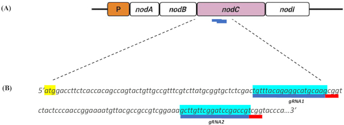
Figure 1. Design of single-guide RNAs (sgRNAs) targeting the nodC locus in Mesorhizobium ciceri. (A) Schematic representation of the M. ciceri nodC locus showing the positions of the two 20-bp sgRNAs designed for CRISPR-Cas9-mediated targeting. sgRNA target sites are shown in blue. (B) Nucleotide sequences of sgRNA1 and sgRNA2 targeting the nodC locus. The protospacer adjacent motif (PAM; 5′-NGG-3′) is indicated in red, and the nodC translation start codon (ATG) is highlighted in yellow. Please click here to view a larger version of this figure.
Two 20 bp target sequences were identified immediately upstream of a 5′-NGG-3′ PAM sequence (Figure 1B). In silico analysis of these candidate sgRNAs using the algorithm by Hsu et al.19 yielded specificity scores of 100, indicating high predicted targeting specificity with no significant off-target complementarity detected within the M. ciceri genome. Minor differences were observed in the predicted on-target efficiency scores of the selected sgRNAs (Table 3), which informed guide selection for downstream experimental evaluation.
| Target position (bp) | Strand | sgRNA sequence (5′ 3′) | PAM | Specificity score | Efficiency score | |
| 77 | + | TGTTTACAGAGGCATGCAAG | CGG | 100 | 43.58 | |
| 139 | + | GCTTGTTCGGATCCGACCGT | TGG | 99.7 | 47.63 | |
Table 3: Candidate single-guide RNAs (sgRNAs) designed for CRISPR-Cas9-mediated editing of the nodC gene in Mesorhizobium ciceri. Candidate sgRNAs were designed using the Benchling CRISPR Guide Design Tool with Bradyrhizobium diazoefficiens USDA 110 as the reference genome. Guides were ranked according to predicted specificity and on-target efficiency scores, and those targeting sequences proximal to the nodC translational start codon and containing the required NGG protospacer adjacent motif (PAM) for Streptococcus pyogenes Cas9 (SpCas9) were selected.
To facilitate HDR and fluorescence-based screening of putative mutants, an HDR cassette was assembled using the pUC18 cloning vector (Figure 2). The gfp reporter cassette was excised from pRJPaph-bjGFP by SacI and KpnI restriction digestion and ligated into the linearized pUC18 backbone (Figure 2A). Following transformation into E. coli, putative positive clones were identified by blue-white screening. Integration of the gfp insert into the pUC18 backbone was further validated by PCR amplification using gene-specific primers, which produced the expected 500 bp amplicon in positive clones (Figure 2A).
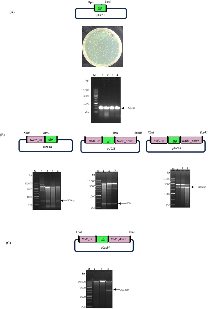
Figure 2. Construction of the CRISPR-Cas9 homology-directed repair (HDR) system for nodC disruption. (A) Construction and validation of the pUC18-gfp intermediate plasmid. The upper panel shows the plasmid map of pUC18-gfp generated by insertion of the green fluorescent protein (gfp) cassette into the pUC18 backbone. The middle panel shows representative colonies following blue-white screening. The lower panel shows colony polymerase chain reaction (PCR) validation using the BjGFP_BP_F1 and BjGFP_BP_R2 primer pair. (B) Sequential assembly of the HDR template in the pUC18 backbone. Plasmid maps and restriction digestion analyses confirm insertion of the upstream homology arm (UHA), downstream homology arm (DHA), and the complete UHA-gfp-DHA HDR cassette. (C) Cloning and validation of the UHA-gfp-DHA HDR cassette into the pCasPP CRISPR-Cas9 vector. The upper panel shows the final pCasPP-HDR plasmid map, and the lower panel shows restriction digestion confirming insertion of the HDR cassette. Please click here to view a larger version of this figure.
The HDR construct was subsequently assembled by introducing nodC homology arms flanking the gfp cassette. A 500 bp UHA and a 400 bp DHA, positioned adjacent to the predicted Cas9 cleavage site, were sequentially cloned into the pUC18-gfp construct through restriction digestion and ligation (Figure 2B). Assembly of the UHA-gfp-DHA cassette was confirmed by restriction digestion analysis, which yielded the expected 2012 bp fragment (Figure 2B). The complete HDR cassette was then subcloned into the broad-host-range CRISPR vector pCasPP (Figure 2C). Restriction digestion analysis confirmed the integrity of the final construct and successful assembly of the genome-editing delivery vector for M. ciceri (Figure 2C).
Biparental mating was performed between the donor E. coli S17-1λpir and the recipient M. ciceri strain to deliver the pCasPP-gRNA-HDR construct into M. ciceri (Figure 3A). Figure 3B illustrates the nod operon organization and the predicted replacement of the nodC locus by the gfp cassette through homologous recombination using the HDR template. Transconjugant colonies were initially identified by GFP fluorescence screening following biparental mating (Figure 3B and 3C). More than 80% of recovered colonies displayed GFP fluorescence with gRNA2, and more than 65% of colonies exhibited fluorescence following introduction of gRNA1 (Figure 3C). Because fluorescence screening alone cannot distinguish plasmid-borne GFP expression from successful genomic integration, downstream molecular validation was performed to confirm locus-specific insertion of the reporter cassette.
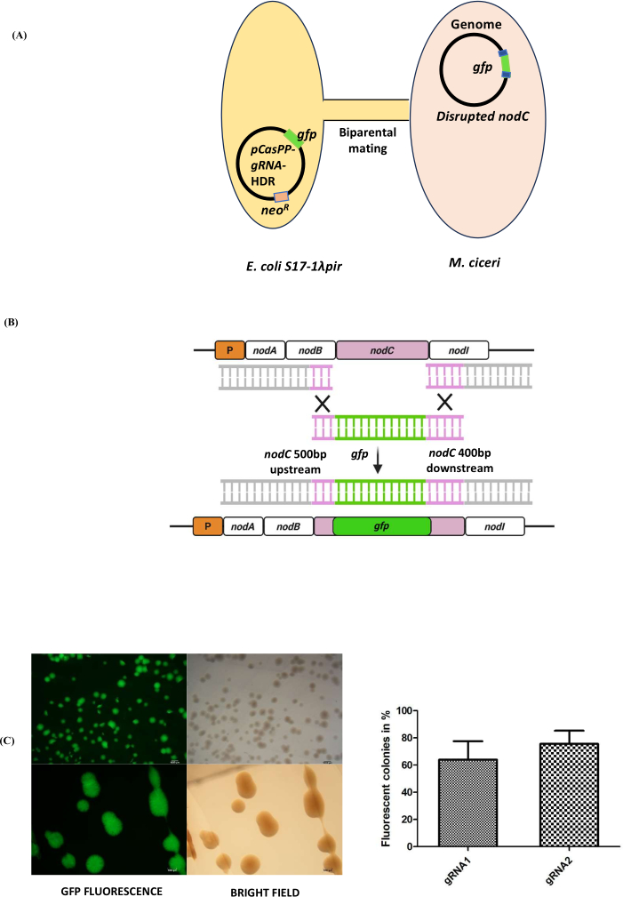
Figure 3. CRISPR-Cas9-mediated disruption of the nodC locus in Mesorhizobium ciceri. (A) Schematic illustration of biparental mating used to transfer the pCasPP-gRNA-HDR construct from Escherichia coli S17-1λpir donor cells into M. ciceri recipient cells. (B) Diagram illustrating homology-directed repair (HDR)-mediated replacement of the nodC locus with the gfp cassette following CRISPR-Cas9 cleavage. The HDR template contains approximately 500 bp upstream and 400 bp downstream homology arms flanking the gfp cassette. (C) Fluorescence-based screening of transconjugant M. ciceri colonies. Representative fluorescence and bright-field stereomicroscopy images of GFP-positive colonies are shown together with the percentage of fluorescent transconjugants recovered following biparental mating using constructs containing sgRNA1 or sgRNA2. Error bars represent standard deviation. Please click here to view a larger version of this figure.
To validate integration of the gfp cassette at the nodC locus, representative GFP-positive colonies were subjected to PCR analysis. Amplification of the genomic region spanning nodB and nodC produced a 1511 bp fragment in the edited clones, consistent with integration of the reporter cassette, whereas the wild-type (WT) strain produced a 430 bp amplicon (Figure 4A). PCR amplification using gfp-specific and nodC-flanking primers generated a distinct 997 bp fragment in the edited clones, while no amplification was detected in the WT control, supporting locus-specific insertion. The orientation of the reporter cassette was confirmed using Sanger sequencing. In addition, GFP expression in the edited strains was evaluated using RT-qPCR, which demonstrated increased GFP transcript accumulation relative to the WT strain (Figure 4B). Expression values were calculated using the 2−ΔΔCt method and normalized to the reference 16s gene, with the WT strain as the control. Data represent the mean ± SD of three biological replicates (n = 3). Statistical significance was determined by one-way ANOVA followed by Dunnett’s test against the WT control.
The study’s current limitation is the absence of a control vector, like an empty-gRNA pCasPP-HDR, to establish the baseline recombination coefficient. Future studies should include such a control.
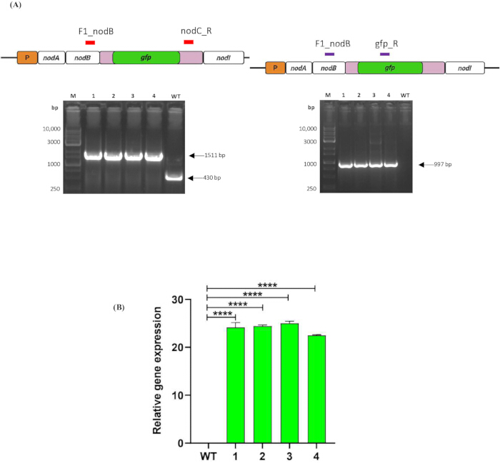
Figure 4. Molecular validation of ΔnodC Mesorhizobium ciceri mutants. (A) PCR-based confirmation of nodC disruption. The left panel shows amplification using the F1_nodB and nodC_R primer pair. Lane M, DNA ladder; lanes 1–4, ΔnodC M. ciceri mutants; lane WT, wild-type M. ciceri. The right panel shows amplification using the F1_nodB and gfp_R primer pair. Lane M, DNA ladder; lanes 1–4, ΔnodC M. ciceri mutants; lane WT, wild-type M. ciceri. Expected amplicon sizes are indicated. (B) Relative green fluorescent protein (GFP) expression in ΔnodC M. ciceri mutants determined by reverse transcription quantitative polymerase chain reaction (RT-qPCR). Gene expression was normalized to the M. ciceri 16S ribosomal RNA (16S rRNA) reference gene and expressed relative to the wild-type (WT) control using the 2^−ΔΔCt method. Error bars represent standard deviation. Statistical significance was determined relative to the WT control. Please click here to view a larger version of this figure.
The biological impact of nodC disruption was evaluated using chickpea infection assays. Plants inoculated with WT M. ciceri formed visible root nodules, whereas plants inoculated with ΔnodC M. ciceri displayed a marked reduction or absence of nodulation (Figure 5A). The uninoculated control plants remained nodule-free, supporting maintenance of sterile experimental conditions (Figure 5A). Quantitative analysis of plant growth parameters further demonstrated differences between WT- and ΔnodC M. ciceri-inoculated plants, including root length, fresh root weight, dry root weight, shoot length, fresh shoot weight, dry shoot weight, and nodule number (Figure 5B). WT-inoculated plants exhibited higher nodule numbers and improved growth-associated parameters compared with ΔnodC M. ciceri-inoculated and uninoculated control plants. These findings are consistent with disruption of nodC-associated nodulation function and support the applicability of this CRISPR-Cas9-based workflow for functional genomics studies in M. ciceri. Quantitative plant phenotype data were evaluated to compare differences among treatment groups. A total of 90 biological replicates were included in the analysis (n = 30 plants per treatment group). Data are presented as mean ± SD. Statistical significance was assessed using one-way analysis of variance (ANOVA) followed by Tukey’s post hoc multiple comparison test. Differences were considered statistically significant at P < 0.05.
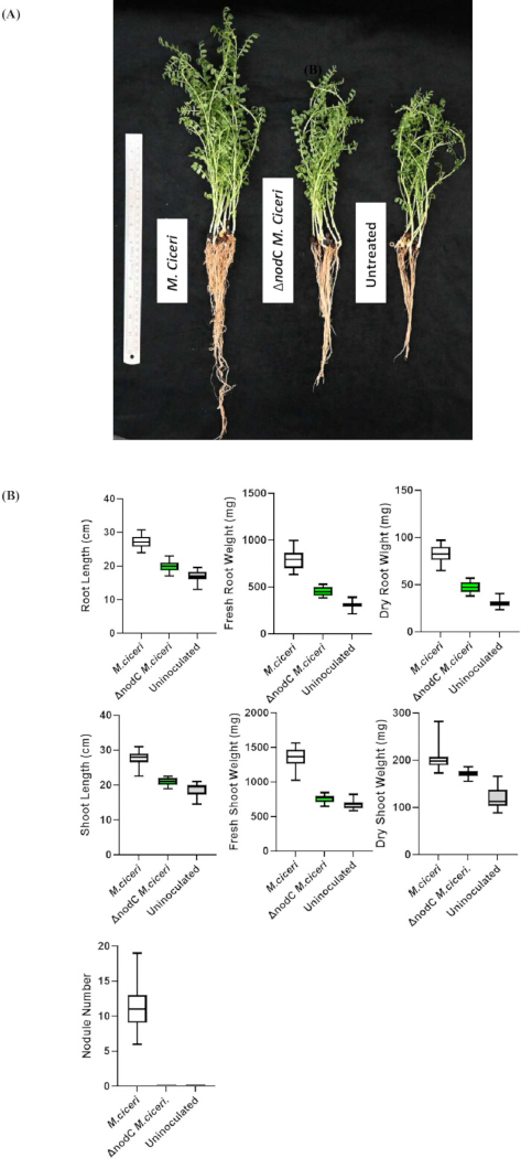
Figure 5. Functional validation of ΔnodC Mesorhizobium ciceri mutants using a chickpea infection assay. (A) Representative chickpea (Cicer arietinum) plants 20 days after inoculation with wild-type M. ciceri, ΔnodC M. ciceri, or uninoculated controls. (B) Quantitative analysis of plant growth and nodulation parameters, including root length, fresh root weight, dry root weight, shoot length, fresh shoot weight, dry shoot weight, and nodule number. Data are presented for plants inoculated with wild-type M. ciceri, ΔnodC M. ciceri, or uninoculated controls. Error bars represent standard deviation. Statistical significance was determined by one-way analysis of variance (ANOVA) followed by Tukey's multiple-comparison test. Please click here to view a larger version of this figure.
Supplementary File 1. Detailed protocol for preparation and validation of chemically competent Escherichia coli cells. This supplementary file provides the complete procedure for preparing and validating chemically competent Escherichia coli DH5α and E. coli S17-1λpir cells, including bacterial culture preparation, competency induction, cell harvesting, washing with CaCl₂–glycerol buffer, aliquoting, cryopreservation, and validation by transformation-efficiency testing. Critical quality-control measures, storage recommendations, and competency acceptance criteria are also included. Please click here to download this file.
Supplementary File 2. Detailed protocol for construction of homology-directed repair (HDR) templates and assembly of CRISPR-Cas9 genome-editing vectors. This supplementary file provides detailed procedures for excision of the green fluorescent protein (GFP) cassette, construction of the pUC18-gfp intermediate plasmid, amplification and cloning of upstream and downstream homology arms, assembly of the complete homology-directed repair (HDR) template, preparation of pCasPP-guide RNA (gRNA) vectors, insertion of HDR templates into CRISPR-Cas9 plasmids, fluorescence-based clone screening, and validation of recombinant plasmids by restriction analysis and Sanger sequencing. Please click here to download this file.
Supplementary File 3. Detailed protocol for biparental mating and generation of GFP-tagged Mesorhizobium ciceri transconjugants. This supplementary file describes donor and recipient strain preparation, membrane-filter biparental mating, recovery and antibiotic selection of transconjugants, plasmid-curing procedures, fluorescence-based screening of recombinant colonies, molecular confirmation of genome-edited strains, and short- and long-term preservation of confirmed GFP-positive M. ciceri transconjugants. Detailed culture conditions, mating parameters, and selection procedures are provided. Please click here to download this file.
Discussion
The establishment of a CRISPR-Cas9 genome-editing system in M. ciceri addresses a major technical limitation in the genetic manipulation of this agriculturally important nitrogen-fixing symbiont. By disrupting the nodC locus using a gfp reporter and HDR, this workflow provides a fluorescence-assisted framework for targeted genomic modification in M. ciceri.
Traditional genetic manipulation approaches in rhizobia have relied primarily on Tn5-based random mutagenesis or sacB-mediated allelic exchange19,26,27. Although these approaches remain valuable, they are labor-intensive, often require multiple rounds of counter-selection, and may leave antibiotic-resistance markers within the genome that complicate subsequent rounds of editing. In contrast, CRISPR-Cas9-mediated genome editing offers a more targeted and streamlined alternative for genomic engineering. Recent studies have reported CRISPR-mediated base editing and Cas12e-based systems in S. meliloti to reduce Cas9-associated toxicity12,13, while dCas9-mediated transcriptional repression has been explored in R. etli11. To our knowledge, this study represents one of the first applications of CRISPR-Cas9-mediated chromosomal disruption in M. ciceri. The protocol incorporates sgRNA-guided targeting, biparental plasmid delivery, and HDR-mediated recombination for locus-specific editing. Integration of the gfp reporter within the HDR cassette also facilitates rapid fluorescence-based screening of putative edited colonies, thereby reducing the number of colonies requiring downstream molecular validation.
Several factors were important for the successful implementation of the workflow. Selection of sgRNAs with high predicted specificity minimized potential off-target effects within the M. ciceri genome, while maintenance of homology arm integrity supported HDR-mediated recombination efficiency. In addition, optimizing donor and recipient growth conditions during biparental mating improved recovery of transconjugants. Fluorescence-based screening enabled rapid identification of putative mutants; however, molecular validation using PCR and Sanger sequencing was still necessary to confirm locus-specific integration of the reporter cassette. Future studies could utilize an empty-gRNA pCasPP-HDR vector control to quantify the baseline recombination coefficient.
Beyond methodological advancement, this platform may support future studies investigating symbiotic signaling and functional genomics in chickpea-associated rhizobia28. Chickpea (Cicer arietinum L.) represents a globally important legume crop and a major contributor to sustainable agricultural systems29,30. The ΔnodC M. ciceri strain generated in this study provides a potentially useful genetic background for investigating host-microbe compatibility and nodulation-associated signaling pathways. Previous studies have suggested that sRNAs function as regulatory effectors during early root nodule symbiosis31. Consequently, the nodulation-deficient ΔnodC M. ciceri strain described here may facilitate future studies focused on the molecular mechanisms underlying rhizosphere communication and nitrogen-fixing symbiosis.
Disclosures
The authors declare no competing financial interests or conflicts of interest.
Acknowledgements
This work is supported by the Department of Biotechnology (DBT)-MK Bhan Grant HRD-16016/15/2023-AFS-DBT for Dr. Nandita Pasari. This work is also supported by a Core Grant provided to Dr. Senjuti Sinharoy from the National Institute of Plant Genome Research, New Delhi, India.
Materials
| Name | Company | Catalog Number | Comments |
|---|---|---|---|
| Agarose | Genetix | PG-40005 | Gel electrophoresis |
| Aluminum foil | Rolia’s | GF83048115 | General laboratory use |
| Bacteriological agar | HiMedia | GRM026 | Media preparation |
| Benchling CRISPR Guide Design Platform | Benchling | N/A | Guide RNA design |
| Boric acid | HiMedia | PCT0102 | McKnight’s solution preparation |
| Borosilicate glass conical flask | Sigma | CLS5100500 | Culture preparation |
| Borosilicate glass tubes | Sigma | DWK73500-1075 | Culture preparation |
| Calcium chloride dihydrate | HiMedia | MB034 | Competent cell preparation |
| Calcium sulfate dihydrate | HiMedia | GRM328 | McKnight’s solution preparation |
| Carbenicillin disodium | Duchefa Biochemie | DUC-C0109.005-18 | Used at 100 µg/mL |
| cDNA synthesis kit | Thermo Fisher Scientific | AB1543A | cDNA synthesis |
| Cling wrap | OA | 10954049 | General laboratory use |
| Cloning kit/T4 DNA ligase | Thermo Fisher Scientific | K1422 | Ligation reactions |
| Congo red | HiMedia | 573-58-0 | Microbial culture work |
| Copper(II) sulfate pentahydrate | HiMedia | PCT0104 | McKnight’s solution preparation |
| Culture tube | Borosil | 9800U08 | Cultivation of primary bacterial culture; 15 cm × 2.5 cm |
| Digital pH meter | Eutech Instruments | EC PH TUTOR DS | pH adjustment |
| Dipotassium phosphate | HiMedia | TC596M | YMB preparation |
| DNA ladder | BIO-HELIX | DM015-R500 | Gel electrophoresis |
| DNase removal kit | Invitrogen | AM1907 | DNA removal from isolated RNA |
| dNTP mix | Thermo Scientific | R0181 | PCR amplification |
| Ethanol | Sigma-Aldrich | 1.00983.0511 | Used at 75% |
| Ethidium bromide | Sigma-Aldrich | E1510 | DNA gel staining |
| FastAP Thermosensitive Alkaline Phosphatase | Thermo Fisher Scientific | EF0651 | Dephosphorylation of vector during cloning |
| FastDigest Buffer | Thermo Fisher Scientific | B64 | Restriction digestions |
| Ferric chloride anhydrous | HiMedia | TC583 | McKnight’s solution preparation |
| Fluorescence stereomicroscope | Nikon | SMZ25 | GFP screening |
| Fungicide, Bavistin | Crystal Crop Protection | Bavistin Carbendazim 50% WP | Seed treatment |
| Gel extraction kit | QIAGEN | 28704 | DNA purification |
| Glycerol | Thermo Fisher Scientific | Q24505 | Competent cells/glycerol stocks |
| Growth incubator with orbital shaker | MRC | LOM-150 | Bacterial culture |
| Hand gloves | Kimtech | 97612 | Personal protective equipment |
| HEPES | HiMedia | MB016 | Oligonucleotide annealing |
| IPTG | G Biosciences | RC1113D | Blue-white screening |
| Laminar airflow cabinet | Biolink | BL-H1300 | Sterile handling |
| LB agar | HiMedia | M1151 | Bacterial culture plates |
| LB broth | HiMedia | G1245 | Bacterial culture |
| Magnesium sulfate | HiMedia | GRM1281-500G | YMB preparation |
| Magnesium sulfate heptahydrate | HiMedia | PCT0008 | McKnight’s solution preparation |
| Manganese(II) sulfate tetrahydrate | Sigma-Aldrich | M3634 | McKnight’s solution preparation |
| Mannitol | HiMedia | MB198 | YMB preparation |
| McKnight’s solution | Laboratory-prepared | N/A | Plant nutrient solution |
| Micro tips, 0.2–10 µL | Tarsons | 521000 | Liquid handling |
| Micro tips, 2–200 µL | Tarsons | 521010 | Liquid handling |
| Micro tips, 200–1000 µL | Tarsons | 521020X | Liquid handling |
| Microcentrifuge tube, 0.5 mL | Tarsons | 500000 | Sample handling |
| Microcentrifuge tube, 1.5 mL | Tarsons | 500010 | Sample handling |
| Microcentrifuge tube, 2.0 mL | Tarsons | 500020 | Sample handling |
| Molybdic acid | Sigma-Aldrich | 232084 | McKnight’s solution preparation |
| NanoDrop spectrophotometer | Thermo Fisher Scientific | 13-400-519 | DNA/RNA quantification |
| Neomycin | Duchefa Biochemie | M0135 | Used at 50 µg/mL |
| Nuclease-free water | HiMedia | ML064-100ML | Molecular biology use |
| PCR master mix | Thermo Fisher Scientific | K0171 | 2× master mix |
| PCR tube strips, 0.2 mL | Tarsons | 610040 | PCR setup |
| Petri plates, 90 mm | PSC | 2041 | Agar plates |
| Pipette, 0.1–2.5 µL | Eppendorf | N10178M | Liquid handling |
| Pipette, 0.5–10 µL | Eppendorf | H40069L | Liquid handling |
| Pipette, 2–20 µL | Eppendorf | H30046M | Liquid handling |
| Pipette, 10–100 µL | Eppendorf | I00395M | Liquid handling |
| Pipette, 20–200 µL | Eppendorf | M38186M | Liquid handling |
| Pipette, 100–1000 µL | Eppendorf | N20610M | Liquid handling |
| Plasmid DNA extraction kit | QIAGEN | 12143 | Plasmid isolation |
| Potassium chloride | HiMedia | TC010 | McKnight’s solution preparation |
| Potassium dihydrogen phosphate anhydrous | HiMedia | TC011M | McKnight’s solution preparation |
| PVDF membrane filter, 0.22 µm, 47 mm | MilliporeSigma | GVWP04700 | Biparental mating |
| Quantitative real-time PCR machine | Bio-Rad | CFX96 | RT-qPCR |
| Refrigerated benchtop centrifuge | Eppendorf | 04-987-373 | Cell harvesting |
| Restriction enzyme, BbsI | Thermo Fisher Scientific | ER1011 | gRNA cloning |
| Restriction enzyme, EcoRI | Thermo Fisher Scientific | FD0274 | Cloning |
| Restriction enzyme, KpnI | Thermo Fisher Scientific | FD0524 | Cloning |
| Restriction enzyme, SacI | Thermo Fisher Scientific | FD1133 | Cloning |
| Restriction enzyme, XbaI | Thermo Fisher Scientific | FD0684 | Cloning |
| RNase-free DNase | QIAGEN | 79254 | RNA cleanup |
| Sand | Ram Surat | Sand | Plant growth substrate |
| Sequencer | Applied Biosystems | ABI3730xl DNA Analyzer | Sanger sequencing |
| SOC medium | Laboratory-prepared | N/A | Transformation recovery |
| Sodium chloride | HiMedia | TC046 | Media preparation |
| Sodium hydroxide | HiMedia | PCT1325 | HEPES pH adjustment |
| Sterile surgical blade | Medicare Surgical | 16020 | Gel excision/tissue handling |
| Sterile syringe | HMD Dispovan | 10ml syringe without needle | Sterile filtration |
| Sterile syringe filter, 0.22 µm | Merck Millex Durapore | 12766842 | Filter sterilization |
| SYBR Green master mix | New England Biolabs | M3003E | RT-qPCR |
| Tetracycline | Duchefa Biochemie | T01050 | Used at 5 µg/mL |
| Thermal cycler | Applied Biosystems | A24811 | PCR amplification |
| Tissue roll | Rolia’s | 07AAECR4885K1ZT | General laboratory use |
| TRIzol reagent | Invitrogen | 15596026 | RNA isolation |
| UV transilluminator | Analytik Jena | TW-43 | Gel visualization |
| UV-Vis spectrophotometer | Eppendorf | D30 | OD600 measurement |
| Vis cuvettes | Eppendorf | 30079345 | OD600 measurement |
| Vortex mixer | Neuation | iSWIX-VT | Sample mixing |
| X-gal | G Biosciences | RC1233 | Blue-white screening |
| Yeast extract | HiMedia | RM668-500G | Media preparation |
| Zinc sulfate heptahydrate | Sigma-Aldrich | Z0251 | McKnight’s solution preparation |
References
- Doudna JA, Charpentier E. The new frontier of genome engineering with CRISPR-Cas9. Science. 2014;346(6213):1258096.
- Jiang W, Bikard D, Cox D, Zhang F, Marraffini LA. RNA-guided editing of bacterial genomes using CRISPR-Cas systems. Nature biotechnology. 2013;31(3):233-9.
- Bertrand C, Thibessard A, Bruand C, Lecointe F, Leblond P. Bacterial NHEJ: a never ending story. Molecular microbiology. 2019;111(5):1139-51.
- Loureiro A, da Silva GJ. Crispr-cas: Converting a bacterial defence mechanism into a state-of-the-art genetic manipulation tool. Antibiotics. 2019;8(1):18.
- Altenbuchner J. Editing of the Bacillus subtilis genome by the CRISPR-Cas9 system. Applied and environmental microbiology. 2016;82(17):5421-7.
- Wang Y, et al. Bacterial genome editing with CRISPR-Cas9: deletion, integration, single nucleotide modification, and desirable “clean” mutant selection in Clostridium beijerinckii as an example. ACS synthetic biology. 2016;5(7):721-32.
- Oh J-H, van Pijkeren J-P. CRISPR–Cas9-assisted recombineering in Lactobacillus reuteri. Nucleic acids research. 2014;42(17):e131-e.
- Cobb RE, Wang Y, Zhao H. High-efficiency multiplex genome editing of Streptomyces species using an engineered CRISPR/Cas system. ACS synthetic biology. 2015;4(6):723-8.
- Wanjofu EI, et al. Nodulation and growth promotion of chickpea by Mesorhizobium isolates from diverse sources. Microorganisms. 2022;10(12):2467.
- Haskett T, et al. Complete genome sequence of Mesorhizobium ciceri strain CC1192, an efficient nitrogen-fixing microsymbiont of Cicer arietinum. Genome Announcements. 2016;4(3):10.1128/genomea. 00516-16.
- Bellahsen O, Díaz-Méndez R, Romero D. Genomic engineering in Rhizobium etli: implementation and evaluation of systems based on dCas9. Frontiers in Microbiology. 2025;16:1604430.
- Liu G, et al. An efficient CRISPR/Cas12e system for genome editing in Sinorhizobium meliloti. ACS Synthetic Biology. 2023;12(3):898-903.
- Wang L, Xiao Y, Wei X, Pan J, Duanmu D. Highly efficient CRISPR-mediated base editing in Sinorhizobium meliloti. Frontiers in Microbiology. 2021;12:686008.
- Schäfer A, et al. Small mobilizable multi-purpose cloning vectors derived from the Escherichia coli plasmids pK18 and pK19: selection of defined deletions in the chromosome of Corynebacterium glutamicum. Gene. 1994;145(1):69-73.
- Rütering M, et al. Tailor-made exopolysaccharides—CRISPR-Cas9 mediated genome editing in Paenibacillus polymyxa. Synthetic Biology. 2017;2(1):ysx007.
- Ledermann R, Bartsch I, Remus-Emsermann MN, Vorholt JA, Fischer H-M. Stable fluorescent and enzymatic tagging of Bradyrhizobium diazoefficiens to analyze host-plant infection and colonization. Molecular Plant-Microbe Interactions. 2015;28(9):959-67.
- Göttfert M. Regulation and function of rhizobial nodulation genes. FEMS microbiology reviews. 1993;10(1-2):39-63.
- Kant C, Pradhan S, Bhatia S. Dissecting the root nodule transcriptome of chickpea (Cicer arietinum L.). PLoS One. 2016;11(6):e0157908.
- Hsu PD, et al. DNA targeting specificity of RNA-guided Cas9 nucleases. Nature biotechnology. 2013;31(9):827-32.
- Doench JG, et al. Optimized sgRNA design to maximize activity and minimize off-target effects of CRISPR-Cas9. Nature biotechnology. 2016;34(2):184-91.
- Norrander J, Kempe T, Messing J. Construction of improved M13 vectors using oligodeoxynucleotide-directed mutagenesis. Gene. 1983;26(1):101-6.
- Simon R, Priefer U, Pühler A. A broad host range mobilization system for in vivo genetic engineering: transposon mutagenesis in gram negative bacteria. Bio/technology. 1983;1(9):784-91.
- Hanahan D, Jessee J, Bloom FR. [4] Plasmid transformation of Escherichia coli and other bacteria. Methods in enzymology. 1991;204:63-113.
- Alexandre A, et al. Natural populations of chickpea rhizobia evaluated by antibiotic resistance profiles and molecular methods. Microbial Ecology. 2006:128-36.
- Weerakkody LR, et al. A rapid, inexpensive and effective method for the efficient isolation of genomic DNA from Gram-negative bacteria. Molecular Genetics and Genomics. 2024;299(1):26.
- Selbitschka W, Niemann S, Pühler A. Construction of gene replacement vectors for Gram− bacteria using a genetically modified sacRB gene as a positve selection marker. Applied microbiology and biotechnology. 1993;38(5):615-8.
- Yadav A, Singh RP, Singh AL, Singh M. Identification of genes involved in phosphate solubilization and drought stress tolerance in chickpea symbiont Mesorhizobium ciceri Ca181. Archives of microbiology. 2021;203(3):1167-74.
- Dicenzo GC, et al. Multidisciplinary approaches for studying rhizobium–legume symbioses. Canadian Journal of Microbiology. 2019;65(1):1-33.
- Calia C, et al. A bibliometric analysis of chickpea agronomic practices in the world during 45 years of scientific research. Legume Science. 2024;6(1):e219.
- Sandhu J, Tripathi S, Chaturvedi S. Chickpea nutritional status and value chain for sustainable development. Sustainable food value chain development: Perspectives from developing and emerging economies: Springer; 2023. p. 175-83.
- Ren B, Wang X, Duan J, Ma J. Rhizobial tRNA-derived small RNAs are signal molecules regulating plant nodulation. Science. 2019;365(6456):919-22.
Reprints and Permissions
This article has been published
Video Coming Soon