This case demonstrates the diagnostic evaluation and multidisciplinary management of primary adrenal high-grade undifferentiated pleomorphic sarcoma presenting initially with brain metastasis, highlighting diagnostic challenges, pathological characterization, treatment decision-making, and disease progression.
A subscription to JoVE is required to view this content. Sign in or start your free trial.
Case Report
Diagnostic Workup of Primary Adrenal High-Grade Undifferentiated Pleomorphic Sarcoma Initially Presenting as Brain Metastasis
In This Article
Summary
Abstract
Primary adrenal high-grade undifferentiated pleomorphic sarcoma (UPS) is an exceptionally rare malignant mesenchymal neoplasm, and presentation with brain metastasis as the initial manifestation is even more uncommon. This report highlights the diagnostic evaluation and multidisciplinary management of a patient presenting with an undifferentiated brain metastasis accompanied by an adrenal mass. A 75-year-old woman presented with speech disturbance, cognitive slowing, memory decline, gait instability, and right-sided weakness. Brain computed tomography and contrast-enhanced magnetic resonance imaging revealed a left frontal lesion with multiple intracerebral nodules. Positron emission tomography/computed tomography further demonstrated a left adrenal lesion and additional metastatic foci. Owing to symptomatic mass effect, the brain lesion was surgically resected. Initial pathology suggested a poorly differentiated malignant tumor, and the tumor origin remained uncertain after the first immunohistochemical assessment. Subsequent multidisciplinary review, serial adrenal imaging, adrenal core needle biopsy, and comparative pathologic evaluation progressively redirected the diagnosis toward a high-grade undifferentiated sarcoma, morphologically consistent with UPS. The patient subsequently received postoperative brain radiotherapy, anthracycline-based chemotherapy, additional systemic therapy, and local treatment for progressive metastatic disease. Despite multimodal management, the tumor rapidly progressed with thigh metastasis, bilateral adrenal involvement, skeletal dissemination, and bilateral pathologic femoral fractures. The patient ultimately died of intracerebral hemorrhage. This case expands the clinical spectrum of primary adrenal UPS and underscores the importance of dynamic diagnostic reassessment, multisite tissue sampling, extended immunohistochemical evaluation, and multidisciplinary collaboration when evaluating undifferentiated brain metastases associated with an adrenal mass.
Introduction
Undifferentiated pleomorphic sarcoma (UPS) is a high-grade malignant mesenchymal neoplasm1,2. In its 2013 classification, the World Health Organization (WHO) standardized the terminology of the former entity malignant fibrous histiocytoma and defined UPS as a group of undifferentiated or dedifferentiated soft tissue tumors lacking a clear line of differentiation3,4. UPS most commonly arises in the deep soft tissues of the extremities, trunk, and retroperitoneum and is characterized by marked aggressiveness, with a substantial risk of local recurrence and distant metastasis despite active treatment5. A long-term study from a large tertiary care center showed that the lung and lymph nodes are among the most common metastatic sites of UPS6. In contrast, adrenal UPS is exceedingly rare, and the currently available evidence for primary adrenal UPS is limited to isolated case reports and a small number of literature reviews7,8,9,10.
Because of its nonspecific clinical presentation and lack of distinctive imaging features, primary adrenal UPS can easily be misdiagnosed preoperatively as pheochromocytoma, adrenocortical carcinoma (ACC), metastatic disease, or other primary adrenal sarcomas11, and malignant adrenal tumors often pose diagnostic challenges12. Accurate classification generally requires integration of imaging findings, endocrine evaluation, histomorphology, and immunohistochemical results and, when necessary, expert consultation or repeat tissue sampling to clarify the tumor origin13,14. UPS accounts for approximately 10%–20% of all soft tissue sarcoma (STS) cases, whereas brain metastasis is an uncommon event in STS15. Previous studies have reported an incidence of only ~0.6% for brain metastasis in STS16, further underscoring the exceptional rarity of primary adrenal UPS presenting initially with brain metastasis. Notably, among the currently available reports of primary adrenal UPS, only two publications described the same 44-year-old woman with a primary lesion arising from the left adrenal gland, and no metastatic disease was observed during 6 months of follow-up after treatment9,10.
To the best of our knowledge, no other case of primary adrenal UPS presenting initially with brain metastasis has been reported. This report describes the diagnostic and clinical decision-making process of a patient with primary adrenal UPS whose first manifestation was brain metastasis and discusses the case in the context of the existing literature. In addition to adding clinical evidence for this exceptionally rare entity, this case illustrates a stepwise diagnostic pathway for patients presenting with an undifferentiated brain metastasis accompanied by an adrenal mass.
Case Presentation:
A 75-year-old woman initially presented with speech disturbance and weakness of the right extremities. No prior malignancy, adrenal disease, or hereditary tumor syndrome was documented in the available medical records. The patient had no recorded symptoms suggestive of adrenal hormone excess, and available endocrine-related and routine laboratory test results, including electrolyte levels, thyroid function tests, myocardial enzyme measurements, adrenocorticotropic hormone levels, and cortisol levels, showed no obvious abnormalities. Family history and smoking history were not documented. Head computed tomography (CT) and contrast-enhanced brain magnetic resonance imaging (MRI) showed a left frontal lobe mass with multiple intracerebral nodules, suggesting malignant disease. Positron emission tomography/CT (PET/CT) further identified a left adrenal lesion, multiple intracerebral masses, and abnormal osseous metabolic lesions. Based on pathology, immunohistochemistry, and repeated expert consultation, the final diagnosis was primary adrenal high-grade UPS with brain and bone metastases. Despite multimodal treatment, including surgery, chemotherapy, radiotherapy, and targeted therapy, the patient ultimately died of intracerebral hemorrhage.
Diagnosis, Assessment, and Plan:
The patient initially presented with speech disturbance, memory decline, cognitive slowing, right-sided weakness, and gait instability, suggesting an intracerebral space-occupying lesion. Contrast-enhanced brain MRI was performed to characterize the intracerebral lesion (Figure 1A). Imaging revealed a left frontal lobe mass with multiple intracerebral nodules, supporting the diagnosis of malignant neoplastic disease. PET/CT was subsequently performed to identify the primary lesion and evaluate systemic metastatic spread (Figure 1B–1E). PET/CT demonstrated a hypermetabolic left adrenal lesion, multiple intracerebral masses, and abnormal metabolic foci in the T12 and L4 vertebrae. These findings favored systemic malignancy with brain metastasis rather than a primary central nervous system neoplasm.
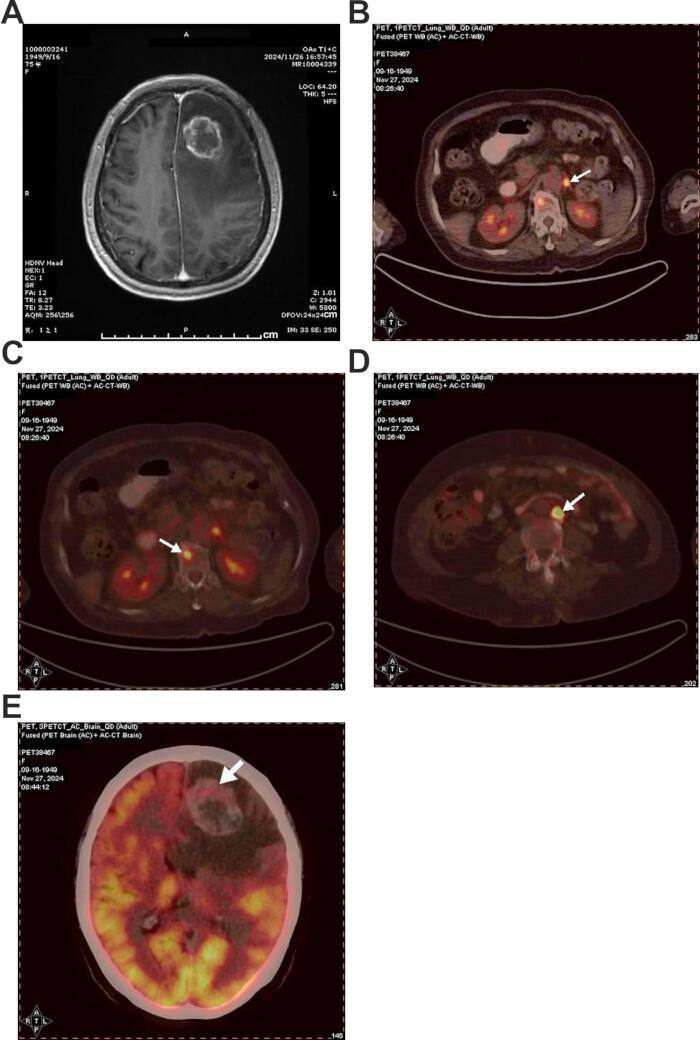
Figure 1. Initial imaging findings demonstrating the intracerebral lesion, adrenal lesion, and metastatic disease at presentation (November 27, 2024). (A) Contrast-enhanced brain magnetic resonance imaging (MRI) showing a ring-enhancing lesion in the left frontal lobe (arrow), associated with surrounding edema and mass effect. (B) Positron emission tomography/computed tomography (PET/CT) fusion image showing a hypermetabolic lesion in the left adrenal region (arrow). (C) PET/CT fusion image showing a hypermetabolic metastatic lesion involving the T12 vertebral body (arrow). (D) PET/CT fusion image showing a hypermetabolic metastatic lesion involving the L4 vertebral body (arrow). (E) PET/CT fusion image of the brain demonstrating increased metabolic activity in the left frontal lobe lesion (arrow). Please click here to view a larger version of this figure.
Because the patient had progressive neurological symptoms and the intracerebral lesion exerted mass effect while representing the most accessible site for obtaining diagnostic tissue, she underwent microscopic supratentorial craniotomy with resection of the intracerebral tumor. Postoperative pathology showed a poorly differentiated malignant tumor with extensive necrosis, initially favoring metastatic ACC. Immunohistochemistry demonstrated a Ki-67 labeling index of approximately 50%, partial cytokeratin (CK) positivity, focal MelanA positivity, and α-inhibin positivity, whereas steroidogenic factor 1 (SF-1) was negative. Glial, melanocytic, and most neuroendocrine markers were negative. Additional immunohistochemical studies showed vimentin positivity and scattered S-100 positivity, whereas synaptophysin (Syn), α-inhibin, calretinin, and chromogranin A (CgA) were negative. Overall, the tumor lacked stable adrenocortical or neuroendocrine differentiation.
During follow-up, a left adrenal nodule was identified, and contrast-enhanced adrenal MRI was performed to assess lesion progression. On January 9, 2025, contrast-enhanced abdominal MRI showed a left adrenal nodule measuring approximately 12.03 mm at its greatest dimension (Figure 2A). Repeat MRI on February 28, 2025 demonstrated enlargement of the lesion to approximately 26.14 mm (Figure 2B), indicating disease progression. Available endocrine-related laboratory test results, including electrolyte levels, adrenocorticotropic hormone levels, and cortisol levels, showed no obvious abnormalities. Specific plasma or urinary metanephrine/catecholamine testing for pheochromocytoma was not documented in the available medical records.
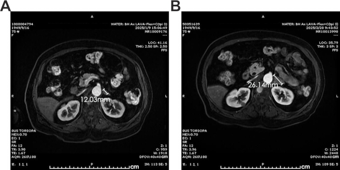
Figure 2. Serial magnetic resonance imaging demonstrating progression of the left adrenal lesion. (A) Axial contrast-enhanced abdominal magnetic resonance imaging obtained on January 9, 2025, showing a left adrenal lesion measuring approximately 12.03 mm in maximum diameter. (B) Follow-up axial contrast-enhanced abdominal magnetic resonance imaging obtained on February 28, 2025, showing enlargement of the left adrenal lesion to approximately 26.14 mm, indicating disease progression. Please click here to view a larger version of this figure.
Immunohistochemistry of the adrenal biopsy specimen showed that the tumor cells were negative for CK, epithelial membrane antigen (EMA), carcinoembryonic antigen (CEA), and CK5/6, providing no evidence of definite epithelial differentiation. Steroidogenic factor 1 (SF-1), MelanA, α-inhibin, and calretinin were negative, arguing against stable adrenocortical differentiation. Synaptophysin (Syn) and chromogranin A (CgA) were also negative, making neuroendocrine differentiation unlikely. Paired box gene 8 (PAX8), S100, desmin, and anaplastic lymphoma kinase (ALK) were negative, whereas murine double minute 2 (MDM2) was negative and cyclin-dependent kinase 4 (CDK4) showed only weak positivity, without supporting a specific line of differentiation. Reticulin staining showed partial destruction of the reticulin framework. Taken together with morphologic features and external expert consultation, these findings favored a high-grade undifferentiated sarcoma. The comparative immunohistochemical findings from the brain lesion, adrenal lesion, and right thigh lesion are summarized in Table 1.
| Specimen | Positive markers | Focally/partially/scattered positive markers | Negative markers | Interpretation |
| Brain lesion | Ki-67 approximately 50%; vimentin positive in additional studies | CK partial positivity; focal MelanA positivity in the initial assessment; scattered S-100 positivity; focal Syn positivity in the initial assessment; α-inhibin positivity in the initial assessment but not reproduced in additional studies | GFAP, Olig-2, SOX10, HMB45, BRAF, TTF-1, CgA, SF-1, calretinin/CR, CD34, ERG, CD21, CD35 | Poorly differentiated malignant tumor without stable evidence of glial, melanocytic, adrenocortical, or neuroendocrine differentiation |
| Adrenal lesion | None supporting a specific line of differentiation | CDK4 weak positivity; CD31 limited to vascular staining; CD68 and CD163 background positivity | CK, EMA, CEA, CK5/6, MelanA, SF-1, α-inhibin, calretinin/CR, Syn, CgA, PAX8, S100, MDM2, desmin, ALK, CD30 | High-grade undifferentiated sarcoma favored; no definite epithelial, adrenocortical, neuroendocrine, melanocytic, or muscular differentiation |
| Right thigh lesion | Vimentin positive | CK focal positivity | MelanA, SF-1, α-inhibin, Syn, CgA, SMA, desmin, MyoD1, CD34, S100, MDM2, CDK4, CD163 | Metastatic poorly differentiated malignant tumor consistent with adrenal sarcoma metastasis |
Table 1: Comparative immunohistochemical profiles of the brain, adrenal, and right thigh lesions. Immunohistochemical findings from the brain lesion, adrenal lesion, and right thigh lesion are summarized according to the available pathology reports and external pathology consultation results. Focal, partial, weak, or scattered staining patterns are presented separately from diffuse positivity because these findings did not provide reproducible evidence of a specific line of differentiation. CD31 staining limited to vascular structures and background staining for CD68 and CD163 were not interpreted as evidence of tumor-cell lineage differentiation. Ki-67 is reported as the proliferative index. Collectively, the immunohistochemical findings did not support definitive glial, epithelial, adrenocortical, neuroendocrine, melanocytic, or myogenic differentiation and, when interpreted together with the morphologic findings and clinical course, favored the diagnosis of high-grade undifferentiated pleomorphic sarcoma. Please click here to download this Table.
Targeted next-generation sequencing (NGS) of the tumor tissue was performed using a panel covering 1,066 tumor-related genes. The assay evaluated single-nucleotide variants, short insertions/deletions, copy number variations, and known fusion genes. The tumor was microsatellite stable (MSS) and had a low tumor mutational burden (TMB-L). Two mutations were identified: TP53 with a variant allele frequency of 20.5% and platelet-derived growth factor receptor alpha (PDGFRA) with a variant allele frequency of 3.5%. These findings provided additional information regarding tumor biology. The TP53 alteration was consistent with genomic instability commonly observed in high-grade sarcomas but was not specific for UPS or adrenal origin. Although the PDGFRA alteration suggested possible involvement of receptor tyrosine kinase signaling, its low variant allele frequency and lack of a documented actionable hotspot mutation limited its clinical significance. Therefore, the NGS findings were considered supplementary information rather than conclusive diagnostic or treatment-directing evidence.
After one cycle of chemotherapy, the patient developed a progressively enlarging painful mass in the right thigh. Abdominal MRI showed further enlargement of the left adrenal lesion, with a maximum diameter of approximately 45.01 mm (Figure 3A). A concurrent axial image demonstrated bilateral adrenal involvement, more pronounced on the left side (Figure 3B). MRI of the right thigh revealed a large soft tissue mass in the right femoral region. In the context of the clinical course and pathology findings, this lesion was considered metastatic (Figure 3C). Core needle biopsy of the newly developed right thigh lesion showed a poorly differentiated malignant tumor with extensive necrosis. Immunohistochemistry demonstrated vimentin positivity and focal CK positivity, whereas adrenocortical, neuroendocrine, myogenic, and other lineage-specific markers were negative. No definite line of differentiation could be established.
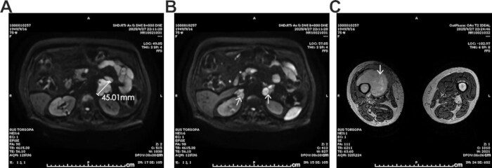
Figure 3. Follow-up magnetic resonance imaging demonstrating progression of adrenal disease and development of a right thigh metastatic lesion (April 27, 2025). (A) Axial abdominal magnetic resonance imaging showing marked enlargement of the left adrenal lesion, measuring approximately 45.01 mm in maximum diameter. (B) Axial abdominal magnetic resonance imaging demonstrating bilateral adrenal involvement (arrows), with more extensive disease on the left side. (C) Axial magnetic resonance imaging of both thighs showing a large soft-tissue metastatic lesion in the right thigh/right femoral region (arrow). Please click here to view a larger version of this figure.
Imaging studies performed on August 9, 2025, indicated further disease progression. Non-contrast head CT showed that the previously seen low-density lesion in the left frontal lobe had decreased in extent, but a new low-density lesion with surrounding edema was identified in the right frontal lobe (Figure 4A). In the context of the patient's history, this finding suggested a new intracerebral metastatic lesion. Non-contrast abdominopelvic CT showed bilateral pathologic femoral fractures, indicating tumor-related osseous destruction (Figure 4C). In addition, bilateral adrenal lesions persisted, with the left side more severely involved, and a large tumorous lesion was identified in the left psoas region. This lesion had indistinct borders with adjacent structures, suggesting local invasion or soft-tissue metastasis (Figure 4B).
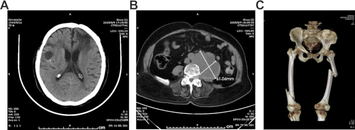
Figure 4. Imaging demonstrating advanced disease progression and skeletal-related complications (August 9, 2025). (A) Non-contrast head computed tomography (CT) showing a new low-density lesion with surrounding edema in the right frontal lobe, consistent with a new intracerebral metastatic lesion. (B) Axial abdominopelvic CT showing a large lesion in the left psoas region measuring approximately 61.06 × 88.38 mm, with indistinct borders relative to adjacent structures, suggesting local invasion or soft-tissue metastatic involvement. (C) Three-dimensional CT reconstruction of the pelvis and bilateral femurs demonstrating bilateral pathological femoral fractures secondary to metastatic disease. Please click here to view a larger version of this figure.
Based on the initial neurological presentation, imaging evidence of an adrenal mass with multisite metastatic disease, converging pathologic findings from the brain and adrenal lesions, and repeated expert consultations supporting a high-grade mesenchymal malignancy, the final diagnosis was primary adrenal high-grade UPS presenting initially with brain metastasis. The principal differential diagnoses included adrenocortical sarcomatoid carcinoma, brain metastasis from ACC, other metastatic sarcomas, other primary high-grade adrenal malignancies, and metastatic undifferentiated malignant tumors of unknown origin involving the central nervous system (Table 2).
| Differential diagnosis | Findings supporting consideration | Findings arguing against this diagnosis / final interpretation |
| Adrenocortical sarcomatoid carcinoma | Adrenal-region mass with widespread metastatic disease; initial brain pathology favored metastatic adrenocortical carcinoma (ACC). | Adrenal biopsy showed no stable adrenocortical or epithelial differentiation. SF-1, MelanA, α-inhibin, calretinin/CR, CK, EMA, CEA, and CK5/6 were negative. Repeated expert pathology review favored high-grade sarcoma/UPS. |
| Brain metastasis from adrenocortical carcinoma | Brain lesion occurred in association with a left adrenal mass, making ACC with brain metastasis an important initial consideration. | SF-1 was negative. MelanA, α-inhibin, and calretinin/CR expression was inconsistent or absent on subsequent testing. Endocrine-related laboratory findings were unremarkable, and integrated pathology favored high-grade sarcoma. |
| Other metastatic sarcomas involving the adrenal gland | High-grade mesenchymal morphology and multisite disease involving the brain, adrenal region, bone, and right thigh. | The adrenal lesion was identified early in the disease course and demonstrated progressive enlargement. No alternative primary sarcoma site was confirmed. Multidisciplinary review favored primary adrenal high-grade UPS with metastatic dissemination. |
| Other primary high-grade adrenal malignancies | Aggressive adrenal-region tumor with rapid systemic progression. | No specific alternative lineage was identified. Syn, CgA, PAX8, S100, desmin, ALK, MDM2, and other lineage-associated markers were negative or non-diagnostic. The final diagnosis remained high-grade undifferentiated sarcoma by exclusion. |
| Metastatic undifferentiated malignant tumor of unknown origin | The initial brain lesion was poorly differentiated and the primary site was uncertain. | Serial adrenal imaging, adrenal biopsy, comparative pathology, right thigh biopsy, molecular testing, and repeated expert review supported primary adrenal high-grade UPS presenting initially with brain metastasis. |
Table 2: Differential diagnostic considerations in primary adrenal high-grade undifferentiated pleomorphic sarcoma presenting initially as brain metastasis. Major differential diagnoses considered during the diagnostic evaluation are summarized together with the principal findings supporting and opposing each diagnosis. Diagnostic distinctions were based on clinical presentation, serial imaging findings, histopathology, immunohistochemistry, molecular testing, and repeated expert pathological consultation. Overall, the absence of stable adrenocortical, epithelial, neuroendocrine, melanocytic, glial, or myogenic differentiation, together with the morphologic findings and clinical course, supported the final diagnosis of primary adrenal high-grade undifferentiated pleomorphic sarcoma with metastatic disease. UPS, undifferentiated pleomorphic sarcoma; ACC, adrenocortical carcinoma; CK, cytokeratin; SF-1, steroidogenic factor 1; IHC, immunohistochemistry. Please click here to download this Table.
A chronological summary of the major diagnostic evaluations, pathology reviews, molecular testing, treatments, disease progression, skeletal complications, and final outcome is provided in Table 3.
| Date | Event | Main finding / decision |
| October 1, 2024 | Initial presentation | Neurological symptoms developed, including speech disturbance, cognitive slowing, gait instability, and right-sided weakness. |
| November 25–27, 2024 | Initial imaging | Brain CT and MRI demonstrated a left frontal lesion with multiple intracerebral nodules. PET/CT demonstrated lesions involving the left adrenal region, brain, T12 vertebra, and L4 vertebra. |
| November 29, 2024 | Brain tumor resection | Surgical resection of the intracerebral lesion was performed. Initial pathology favored metastatic adrenocortical carcinoma. |
| December 2024–January 2025 | Pathology review | External pathology consultation progressively shifted the diagnostic impression toward a high-grade sarcoma. |
| January 3, 2025 | Brain radiotherapy | Postoperative brain radiotherapy was initiated. |
| January 9–February 28, 2025 | Serial adrenal MRI | The left adrenal lesion enlarged from approximately 12 mm to 26 mm, indicating disease progression. |
| March 3–24, 2025 | Adrenal biopsy and multidisciplinary review | Adrenal biopsy findings and multidisciplinary review favored a high-grade sarcoma morphologically consistent with UPS. |
| March 24, 2025 | Molecular testing | NGS demonstrated microsatellite-stable status (MSS), low tumor mutational burden (TMB-L), a TP53 mutation, and a PDGFRA mutation. |
| April 2, 2025 | First-line chemotherapy | Liposomal doxorubicin plus ifosfamide was administered as first-line systemic therapy. |
| April–May 2025 | Disease progression | Progressive adrenal disease and right thigh/femoral metastases were identified. Right thigh biopsy supported metastatic adrenal sarcoma. |
| May 21, 2025 | Second-line therapy | Gemcitabine plus nab-paclitaxel and local tomotherapy were initiated. |
| Late May 2025 | Treatment interruption | Treatment was interrupted because of fatigue, diarrhea, poor oral intake, and grade III myelosuppression. |
| Early June 2025 | Targeted therapy | Oral anlotinib was initiated after hematologic recovery. Improvement in right thigh pain was documented. |
| Aug-25 | Skeletal complications | Bilateral pathologic femoral fractures were managed with femoral artery embolization, internal fixation, curettage, bone cement filling, and radiofrequency ablation. |
| September 5, 2025 | Final outcome | The patient died of intracerebral hemorrhage at a local hospital. |
Table 3: Chronological summary of diagnostic evaluations, treatments, disease progression, skeletal complications, and final outcome. This table summarizes the major clinical events from the patient's initial neurological presentation through the final outcome. Key events include diagnostic imaging, neurosurgical resection, pathology review, adrenal biopsy, molecular testing, systemic therapy, radiotherapy, development of metastatic disease, skeletal complications, orthopedic interventions, and death due to intracerebral hemorrhage. The timeline illustrates the sequential diagnostic reassessment and treatment decision-making process that ultimately led to the diagnosis of primary adrenal high-grade undifferentiated pleomorphic sarcoma presenting initially with brain metastasis. Dates are reported according to the available retrospective medical records; some procedural details and exact assessment time points were not fully retrievable. PET/CT, positron emission tomography/computed tomography; MRI, magnetic resonance imaging; CT, computed tomography; NGS, next-generation sequencing; MSS, microsatellite stable; TMB-L, low tumor mutational burden; UPS, undifferentiated pleomorphic sarcoma. Please click here to download this Table.
After the diagnostic direction became clearer, management was first directed toward the symptomatic brain metastasis to relieve neurological symptoms, reduce mass effect, and obtain adequate tissue for pathological assessment. Local postoperative brain treatment was then administered to enhance intracerebral control. As the diagnosis progressively shifted toward a high-grade sarcoma, treatment was reoriented toward a soft tissue sarcoma-based systemic strategy together with local intervention for progressive lesions. Because of the tumor’s aggressiveness, rapid progression, and early multi-organ dissemination, treatment was palliative, focusing on delaying progression, controlling symptoms, and preserving quality of life. The presence of brain and bone metastases at initial staging indicated a noncurative treatment intent from the outset. Management became predominantly palliative after rapid systemic progression in May 2025, when the patient developed painful right thigh and femoral metastatic disease, progressive adrenal lesions, declining performance status, diarrhea, poor oral intake, and grade III myelosuppression during second-line chemoradiotherapy. No formal palliative care consultation was documented in the available medical records.
Access restricted. Please log in or start a trial to view this content.
Protocol
This retrospective single-patient case report was reviewed and approved by the Ethics Review Committee of Qingdao Central Hospital, University of Health and Rehabilitation Sciences (ethics approval/opinion No. [Y]KY202603601; acceptance No. KY202603601). The committee confirmed that the report complied with institutional ethical requirements and approved its publication. Written informed consent for publication of the clinical information and accompanying images was obtained from the patient’s legal representative. No identifiable personal information was disclosed in this report.
1. Surgical management of the symptomatic brain metastasis
- Initial clinical assessment
- The patient presented with speech disturbance, cognitive slowing, memory decline, and weakness of the right extremities.
- Head CT and contrast-enhanced brain MRI demonstrated a left frontal lobe mass with multiple intracerebral nodules, suggesting malignant disease.
- Surgical candidacy was determined based on progressive neurological symptoms, radiologic evidence of a surgically accessible dominant left frontal lesion with intracerebral mass effect, the need for tissue diagnosis, and the absence of obvious contraindications to surgery.
- Clinical management initially focused on the symptomatic brain metastasis because of the presence of neurological symptoms and intracerebral mass effect.
- Surgical resection of the intracerebral lesion
- Surgical eligibility was assessed based on the patient’s neurological status, the presence of progressive symptoms and intracerebral mass effect, the accessibility of the dominant left frontal lesion, the need to obtain adequate tissue for pathological diagnosis, tolerance of general anesthesia, and routine preoperative evaluations, including complete blood count, coagulation function, liver and renal function, and electrolytes.
- No absolute contraindications to surgery were identified before the procedure.
- Under general anesthesia, a microscopic supratentorial coronal craniotomy was performed for resection of the intracerebral tumor.
- The surgical objective was maximal safe resection of the dominant symptomatic lesion to reduce mass effect, relieve neurological symptoms, and obtain sufficient tissue for pathological diagnosis.
- The resected tumor tissue was submitted for routine histopathological examination and immunohistochemical analysis.
- After induction of general anesthesia, the patient was placed in the supine position with the head slightly elevated by approximately 10°.
- Based on preoperative imaging, a coronal scalp incision and left frontal craniotomy were performed, and an approximately 7 cm × 10 cm left frontal bone flap was removed.
- Tumor resection was performed under a ZEISS K900 surgical microscope. A soft, dark-brown, moderately vascular tumor was identified beneath the left frontal cortex and dissected along its margin under microscopic visualization, while preserving adjacent cortical tissue and draining veins when possible.
- The extent of resection was assessed by intraoperative microscopic inspection and postoperative cranial CT.
- The resected tissue was submitted for routine pathological evaluation, including formalin fixation, paraffin embedding, hematoxylin and eosin staining, and immunohistochemical analysis.
- The available operative record did not document the use of a neuronavigation system.
- The pathological findings from the resected tissue were used to guide subsequent systemic diagnostic workup and treatment planning.
- Postoperative assessment
- Neurological status was assessed during routine postoperative inpatient monitoring by bedside clinical examination, including level of consciousness, language function, cognitive response, and motor function of the extremities.
- The available records documented postoperative improvement in consciousness, language function, and right-sided motor activity.
- Exact postoperative assessment time points and standardized neurological scale scores were not fully retrievable from the retrospective medical records.
- Improvement in consciousness, language function, and right-sided motor activity was observed compared with baseline, indicating short-term symptomatic benefit following resection of the intracerebral lesion.
2. Postoperative local treatment of the brain metastasis
- Postoperative radiotherapy planning
- Postoperative brain radiotherapy was initiated on January 3, 2025, after surgical resection of the intracerebral lesion to improve local control and reduce the risk of intracerebral progression.
- Radiotherapy was delivered using a tomotherapy platform.
- Treatment planning was based on postoperative brain imaging and radiotherapy simulation data.
- Radiotherapy planning was performed using the Accuray Precision Treatment Planning System, version 2.0.1.1.
- Radiotherapy was delivered using a Radixact X5 Treatment Delivery System, a helical tomotherapy platform.
- Detailed information regarding the immobilization method and image-guidance protocol was not fully retrievable from the available retrospective medical records.
- The prescribed dose was 52.5 Gy in 15 fractions of 3.5 Gy each, delivered to 95% of the planning gross tumor volume (PGTV).
- Target volume delineation was based on postoperative brain imaging, preoperative contrast-enhanced MRI findings, the resection cavity, and visible intracerebral metastatic disease.
- The PGTV was generated according to institutional radiotherapy practice to account for setup uncertainty.
- Dose selection was based on the postoperative brain metastasis setting, the need for local intracerebral control, lesion location, expected tolerance of adjacent normal brain tissue, and the patient’s overall metastatic disease status.
- Detailed dose-constraint parameters, target-margin definitions, and target delineation workflow were not fully retrievable from the available retrospective medical records.
- Treatment monitoring
- The patient was monitored throughout radiotherapy for treatment tolerance and neurological complications.
- Monitoring included baseline neurological assessment before radiotherapy and routine on-treatment clinical evaluation during the radiotherapy course.
- Symptom-triggered reassessment was performed when new or worsening neurological symptoms occurred.
- Assessment focused on level of consciousness, language function, motor function of the extremities, headache, vomiting, seizures, and other signs of intracerebral edema or acute neurological deterioration.
- Detailed standardized assessment scales and exact assessment dates were not fully retrievable from the available retrospective medical records.
- No severe acute neurological complications requiring immediate discontinuation of radiotherapy were documented during treatment.
3. First-line systemic therapy
- Treatment selection
- Pathology review and multidisciplinary evaluation progressively shifted the diagnostic impression from a malignant tumor of possible adrenocortical origin to a high-grade sarcoma morphologically consistent with UPS.
- Treatment selection was discussed through multidisciplinary consultation after integration of the brain lesion pathology, adrenal lesion progression, adrenal biopsy findings, and external pathology review.
- Because the tumor was ultimately considered a high-grade soft tissue sarcoma with aggressive metastatic behavior, a sarcoma-based systemic regimen containing an anthracycline and ifosfamide was selected as first-line systemic therapy.
- The treatment goal was disease control and symptom palliation rather than cure.
- CT-guided percutaneous core needle biopsy of the adrenal lesion was performed to obtain tumor tissue for histopathological, immunohistochemical, and molecular analyses.
- Preprocedural CT images were reviewed to select a safe puncture route and avoid adjacent major vessels and organs.
- Under CT guidance, two core tissue samples were obtained from the adrenal lesion using an 18-gauge biopsy gun.
- The obtained adrenal tissue was submitted for formalin fixation, paraffin embedding, hematoxylin and eosin staining, and immunohistochemical analysis.
- Additional tissue was used for next-generation sequencing when sufficient material was available.
- Post-biopsy monitoring was performed according to institutional practice, including observation for puncture-site bleeding, abdominal or flank pain, and other procedure-related complications.
- Tumor tissue underwent targeted next-generation sequencing using a 1,066-gene tumor-related panel by Nanjing Geneseeq Technology Inc., China.
- Detailed information regarding DNA extraction, sequencing instrument model, bioinformatics pipeline, variant-calling criteria, and reporting thresholds was not fully available in the retrospective clinical records.
- Histopathological and immunohistochemical evaluation was performed on tumor specimens obtained from the intracerebral lesion, adrenal lesion, and metastatic sites.
- Tissue processing was performed using an automated enclosed tissue processor, Histo-Tek VP1, followed by paraffin embedding, sectioning, and hematoxylin and eosin staining.
- Immunohistochemical staining was performed using a BenchMark ULTRA PLUS automated immunohistochemistry staining system.
- The immunohistochemical markers used for diagnostic comparison are summarized in Table 1, and the results were interpreted by pathologists in correlation with histomorphology and clinical imaging.
- Chemotherapy administration
- First-line chemotherapy consisted of liposomal doxorubicin administered at 30 mg on day 1 and ifosfamide administered at 1 g on days 2–5 for one treatment cycle.
- The regimen was selected as a soft tissue sarcoma-based systemic treatment after multidisciplinary and pathology review supported a high-grade sarcoma diagnosis.
- Treatment tolerance was assessed using clinical symptoms, performance status, complete blood count, liver and renal function, and electrolyte monitoring.
- Supportive care, including antiemetic treatment, hydration, and laboratory monitoring, was provided according to institutional practice.
- The exact supportive-medication names, planned cycle interval, and formal dose-adjustment criteria were not fully retrievable from the available retrospective medical records.
- The patient was monitored for treatment response and adverse effects during chemotherapy.
- Response assessment was based on clinical symptoms, physical examination, and follow-up imaging when disease progression was suspected or new symptoms developed.
- Safety monitoring included assessment of gastrointestinal symptoms, oral intake, performance status, complete blood count, liver and renal function, and electrolytes.
- A fixed imaging schedule and standardized response-assessment criteria, such as RECIST-based measurements, were not fully documented in the available retrospective medical records.
4. Second-line systemic therapy and targeted therapy
- Assessment of disease progression
- The patient developed a progressively enlarging painful mass in the right thigh after completion of first-line chemotherapy.
- Disease progression was assessed using symptom evaluation, physical examination, and follow-up imaging.
- On April 27, 2025, MRI of the right thigh demonstrated a right femoral-region mass consistent with metastatic disease in the clinical context.
- Additional metastatic involvement was identified in the right femoral medullary cavity, right medial femoral condyle, and multiple lesions in the left femoral medullary cavity.
- Repeat adrenal MRI performed on the same date showed further progression of bilateral adrenal tumors, predominantly on the left side, with the largest lesion measuring approximately 45 mm.
- These findings, together with the new painful soft-tissue mass and multifocal osseous involvement, were interpreted as systemic disease progression and treatment failure after first-line chemotherapy.
- Formal RECIST-based measurements and a prespecified imaging-response schedule were not fully documented in the available retrospective medical records.
- Administration of second-line chemotherapy
- Second-line chemotherapy was initiated using gemcitabine administered at 1.4 g on days 1 and 8 and nanoparticle albumin-bound paclitaxel (nab-paclitaxel) administered at 200 mg on days 2 and 9.
- This regimen was selected after progression following anthracycline- and ifosfamide-containing first-line chemotherapy.
- The treatment goal was to control rapidly progressive metastatic high-grade sarcoma and relieve tumor-related symptoms.
- The treatment was selected according to institutional oncology practice in the setting of progressive metastatic disease.
- Supportive care included clinical monitoring, laboratory monitoring, antiemetic treatment, nutritional support when needed, and hematologic support when cytopenia occurred.
- The planned cycle length, detailed supportive-medication names, and formal dose-modification criteria were not fully retrievable from the available retrospective medical records.
- Treatment response and adverse effects were monitored using clinical symptom assessment, physical examination, performance status evaluation, laboratory testing, and clinically indicated follow-up imaging.
- Safety monitoring included assessment of fatigue, gastrointestinal symptoms, oral intake, complete blood count, liver and renal function, and electrolytes.
- Because the patient experienced rapid clinical deterioration during second-line chemoradiotherapy, response assessment was performed according to the clinical course and symptom-triggered imaging rather than a fixed RECIST-based imaging schedule.
- A prespecified imaging interval and standardized response-assessment schedule were not fully documented in the available retrospective medical records.
- Local radiotherapy for progressive metastatic lesions
- Local tomotherapy (TOMO) was administered to the adrenal lesion and the right thigh metastatic lesion.
- Treatment planning was based on radiotherapy simulation data together with diagnostic imaging of the adrenal and right thigh lesions.
- Gross tumor targets were delineated according to visible disease on imaging and institutional radiotherapy practice.
- Detailed information regarding the tomotherapy platform model, treatment-planning system version, immobilization method, target delineation workflow, organ-at-risk constraints, and image-guidance procedures was not fully retrievable from the available retrospective medical records.
- The prescribed doses were 95% PGTV 1 (PGTV1) 55 Gy in 25 fractions of 2.2 Gy and 95% PGTV 2 (PGTV2) 50 Gy in 25 fractions of 2.0 Gy.
- Target volume delineation was based on gross metastatic disease identified on diagnostic imaging and radiotherapy simulation images.
- PGTVs were generated according to institutional practice to account for setup uncertainty and local disease extent.
- Dose selection was based on the palliative intent of treatment, the need for local control of progressive adrenal and right thigh lesions, expected normal-tissue tolerance, and the patient’s overall metastatic disease status.
- Detailed margin definitions and dose-constraint parameters were not fully retrievable from the available retrospective medical records.
- Administration of targeted therapy
- Treatment was subsequently adjusted to include oral anlotinib administered at 10 mg on days 1–14 of a 3-week treatment cycle.
- Anlotinib was selected after rapid systemic progression, intolerance to continued combined chemoradiotherapy, declining physical condition, and the need for an oral systemic treatment option with potential activity in advanced soft tissue sarcoma.
- Treatment selection was primarily guided by disease progression, clinical condition, and multidisciplinary therapeutic judgment rather than by a definitive actionable molecular alteration.
- The available molecular findings, including TP53 and PDGFRA alterations, were considered supplementary information regarding tumor biology and were not interpreted as conclusive biomarkers directing anlotinib therapy.
- Improvement in pain symptoms was observed following treatment adjustment.
- Improvement in pain symptoms was assessed clinically based on patient-reported right thigh pain and routine symptom evaluation during follow-up.
- The available records documented reduced right thigh pain after initiation of anlotinib.
- A standardized pain score, analgesic-consumption record, or quality-of-life scale was not fully documented in the available retrospective medical records.
5. Treatment-related adverse effects and supportive care
- Assessment of treatment-related adverse effects
- During radiotherapy and chemotherapy, the patient developed marked fatigue, diarrhea, poor oral intake, and grade III myelosuppression manifested by reductions in white blood cell, neutrophil, and platelet counts.
- Adverse events and myelosuppression were classified according to institutional clinical toxicity grading practice using Common Terminology Criteria for Adverse Events (CTCAE)-based terminology.
- The grade III classification was based on the documented severity of hematologic abnormalities, including leukopenia, neutropenia, and thrombocytopenia.
- Hematologic and clinical parameters were monitored during treatment using complete blood count testing, clinical symptom assessment, oral intake evaluation, and performance status observation.
- Treatment-related toxicity was identified based on new or worsening clinical symptoms, including fatigue, diarrhea, poor oral intake, and abnormal laboratory findings, particularly decreases in white blood cell, neutrophil, and platelet counts.
- The available records documented a white blood cell count of 1.48 × 10⁹/L, a neutrophil count of 1.00 × 10⁹/L, and a platelet count of 39 × 109/L during treatment.
- The exact laboratory monitoring schedule and all assessment dates were not fully retrievable from the available retrospective medical records.
- Supportive care
- Supportive management included leukocyte-stimulating therapy, platelet-supportive treatment, nutritional support, and symptomatic treatment.
- These interventions were administered after the development of clinically significant fatigue, diarrhea, poor oral intake, and grade III myelosuppression during combined second-line chemotherapy and local radiotherapy.
- Local radiotherapy was suspended because of declining physical condition and hematologic toxicity.
- Specific supportive-care drug names, dosages, administration duration, transfusion details, and formal treatment thresholds were not fully documented in the available retrospective medical records.
- Clinical status was reassessed following supportive treatment through symptom evaluation, physical condition assessment, oral intake monitoring, and repeat hematologic testing.
- The grade III myelosuppression was considered most likely treatment-related because it occurred during combined chemotherapy and radiotherapy and improved after hematologic supportive care and treatment interruption.
- However, a multifactorial contribution could not be excluded because the patient had aggressive disseminated disease, osseous metastatic involvement, declining nutritional status, and poor general condition.
- Therefore, the myelosuppression was interpreted as predominantly treatment-related with possible disease- and condition-related contributing factors.
6. Management of complications related to bone metastases
- Assessment of skeletal complications
- Bilateral pathologic femoral fractures were identified during disease progression, indicating worsening skeletal metastatic burden and severe bone-related complications.
- Skeletal disease progression was assessed using symptom evaluation, physical examination, and imaging.
- The patient presented after a fall at home with severe bilateral thigh pain and limited mobility.
- Emergency abdominal and pelvic CT demonstrated bilateral femoral pathologic fractures.
- Previous right thigh MRI had shown a right femoral-region metastatic mass, metastatic involvement of the right femoral medullary cavity and medial femoral condyle, and multiple metastatic lesions in the left femoral medullary cavity.
- The diagnosis of pathologic fracture was based on fracture occurrence in bones with known metastatic involvement, severe pain, activity limitation, and imaging evidence of osseous metastatic disease.
- The extent of osseous involvement and fracture-related complications was evaluated before intervention using clinical symptoms, mobility status, and imaging findings.
- Surgical management was considered necessary because the patient had bilateral femoral pathologic fractures, severe pain, marked limitation of movement, and metastatic destruction involving weight-bearing bones.
- The treatment objectives were to stabilize the fractures, reduce pain, preserve or restore limb function, and prevent further skeletal-related complications.
- Formal orthopedic scoring systems for impending or completed pathologic fracture risk were not fully documented in the available retrospective medical records.
- Management of pathologic femoral fractures
- Left lower-limb femoral artery embolization was performed under local anesthesia on August 12, 2025, before left femoral surgery.
- Right lower-limb femoral artery embolization was performed on August 19, 2025, before right femoral surgery.
- The procedural objective was to reduce tumor vascularity and minimize intraoperative bleeding before orthopedic stabilization and tumor debulking.
- Detailed information regarding the selective target vessels, embolization technique, angiographic endpoints, and embolic materials was not fully retrievable from the available retrospective medical records.
- On August 12, 2025, the patient underwent open reduction and plate internal fixation of the left femoral pathologic fracture, curettage of the metastatic lesion, bone cement filling, and radiofrequency ablation.
- On August 20, 2025, the patient underwent open reduction and plate internal fixation of the right femoral pathologic fracture, curettage of the metastatic lesion, soft-tissue tumor resection, bone cement filling, and radiofrequency ablation.
- Lesions were selected for intervention based on completed pathologic fracture, severe pain, impaired mobility, structural instability of the femur, and the need for local tumor control.
- Detailed information regarding the specific fixation hardware model, bone cement type, radiofrequency ablation parameters, and intraoperative margin assessment was not fully documented in the available retrospective medical records.
- Postoperative assessment
- Postoperative clinical status and mobility were assessed during inpatient monitoring and before discharge.
- Assessment included evaluation of pain, limb function, wound recovery, postoperative complications, and tolerance of movement after fixation.
- Rehabilitation and mobilization were provided according to orthopedic postoperative practice and the patient’s general condition.
- Detailed rehabilitation protocols, weight-bearing instructions, standardized mobility scores, and exact postoperative assessment time points were not fully retrievable from the available retrospective medical records.
- Improvement in mobility and general postoperative status was observed after treatment.
- The available records documented that both procedures were completed successfully and that the patient was discharged in improved condition on September 3, 2025.
- A standardized functional outcome scale was not documented in the available retrospective medical records.
7. Follow-up outcome
- Follow-up assessment
- The patient underwent surgical resection of the intracerebral lesion, postoperative radiotherapy, first-line and subsequent systemic therapy, local radiotherapy, and surgical management of complications related to bone metastases.
- Clinical status, disease progression, treatment response, and treatment-related complications were monitored throughout follow-up using symptom assessment, physical examination, laboratory testing, and repeat imaging when clinically indicated.
- Imaging surveillance included brain CT or MRI for intracerebral disease.
- Imaging surveillance also included adrenal MRI or CT for adrenal lesions, thigh MRI for soft-tissue and femoral involvement, and CT imaging for systemic or skeletal complications.
- Disease progression was assessed based on the appearance of new lesions, enlargement of known lesions, worsening metastatic burden, new skeletal-related events, decline in performance status, and emergence of tumor-related symptoms such as pain or impaired mobility.
- A fixed follow-up schedule, prespecified imaging interval, and formal RECIST-based response assessment were not fully documented in the available retrospective medical records.
- Final outcome
- Despite multimodal treatment, the patient ultimately died of intracerebral hemorrhage.
- The available records did not contain sufficient information to determine a single definitive cause of the intracerebral hemorrhage.
- The hemorrhage was considered most likely multifactorial in the context of intracerebral metastatic disease, prior neurosurgical resection, postoperative brain radiotherapy, aggressive systemic progression, advanced age, previous grade III myelosuppression during combined treatment, and prior exposure to antiangiogenic targeted therapy.
- Detailed terminal imaging, laboratory findings, platelet counts, coagulation parameters, and autopsy results were unavailable because the patient died at a local hospital.
- Consequently, the relative contributions of tumor-related bleeding, treatment-related effects, age-related vascular fragility, thrombocytopenia, or other factors could not be definitively established.
- The final clinical outcome was documented following disease progression and treatment failure.
- The patient first developed neurological symptoms in October 2024.
- Diagnostic intracerebral tumor resection was performed on November 29, 2024.
- The diagnosis progressively converged toward primary adrenal high-grade undifferentiated pleomorphic sarcoma after adrenal biopsy and multidisciplinary pathology review in March 2025.
- The patient died on September 5, 2025, approximately 9 months after diagnostic neurosurgical resection and approximately 5.5 months after the final sarcoma-oriented pathological diagnosis.
- The overall treatment intent was non-curative because of brain, bone, and adrenal metastatic disease.
- No formal end-of-life care plan or palliative-care consultation was documented in the available retrospective medical records.
Access restricted. Please log in or start a trial to view this content.
Results
Initial imaging demonstrated a left frontal lobe lesion with multiple intracerebral nodules on brain MRI (Figure 1A). PET/CT identified a hypermetabolic lesion in the left adrenal region (Figure 1B), together with abnormal metabolic foci involving the T12 vertebral body (Figure 1C) and L4 vertebral body (Figure 1D). Increased metabolic activity was also observed in the left frontal lesion on PET/CT brai...
Access restricted. Please log in or start a trial to view this content.
Discussion
A critical aspect of this case was the stepwise diagnostic workflow used to distinguish primary adrenal UPS from other high-grade adrenal malignancies. Previous reports have emphasized that primary adrenal UPS is fundamentally a diagnosis of exclusion and requires integration of imaging findings, histomorphology, and immunohistochemistry9,17. In the present case, the initial pathologic interpretation of the intracerebral lesion favored a malignant tumor of possib...
Access restricted. Please log in or start a trial to view this content.
Disclosures
The authors declare no competing interests.
Acknowledgements
Hui Sun contributed to data collection, literature review, figure preparation, and manuscript drafting. Chao Xin assisted with data collection and manuscript revision. Ruichen Zhang participated in data interpretation and manuscript revision. Zongzhan Wang conceived and supervised the study, critically revised the manuscript, and served as the corresponding author. All authors read and approved the final manuscript. The authors sincerely thank the clinicians, radiologists, pathologists, and nursing staff involved in the diagnosis, multidisciplinary evaluation, and treatment of this patient. Their contributions were essential to the clinical management of this case.
Access restricted. Please log in or start a trial to view this content.
Materials
| Name | Company | Catalog Number | Comments |
|---|---|---|---|
| Anlotinib hydrochloride capsules | Chia Tai Tianqing Pharmaceutical Group Co., Ltd., China | N/A | Targeted therapy |
| Automated immunohistochemistry staining system | Roche Diagnostics / Ventana Medical Systems, Inc., United States | BenchMark ULTRA PLUS | Used for immunohistochemical analysis |
| Brain magnetic resonance imaging system | GE Healthcare, United States | Discovery MR750 3.0T | Used for diagnosis and follow-up imaging |
| Complete blood count analyzer | Mindray Bio-Medical Electronics Co., Ltd., China | CAL 7000 Automated Hematology Analyzer | Used for hematologic monitoring during chemotherapy, radiotherapy, and supportive care |
| Computed tomography system | Philips Healthcare, Netherlands | Brilliance iCT | Used for diagnostic imaging, CT-guided biopsy planning, and follow-up assessment |
| Core needle biopsy gun | Nanjing Canyon Medical Technology Co., Ltd., China | BN-2/181600-1 (18-gauge, 160 mm) | Used for CT-guided percutaneous adrenal biopsy |
| Formalin fixative | Nanchang Yulu Laboratory Equipment Co., Ltd., China | 10% neutral buffered formalin (catalog number not documented) | Used for tissue fixation before histopathological evaluation |
| Gemcitabine | Qilu Pharmaceutical Co., Ltd., China | N/A | Second-line chemotherapy |
| Hematoxylin and eosin staining reagents | Roche Diagnostics / Ventana Medical Systems, Inc., United States | VENTANA HE 600 reagents; Hematoxylin: Roche #07024282001; Eosin: catalog number not documented | Used for routine histopathological staining of tissue sections |
| Histopathology processing platform | Sakura Finetek, Japan | Histo-Tek VP1 automated enclosed tissue processor | Used for routine tissue processing and dehydration before paraffin embedding |
| Ifosfamide | Qilu Pharmaceutical Co., Ltd., China | N/A | First-line chemotherapy |
| Liposomal doxorubicin | Qilu Pharmaceutical Co., Ltd., China | N/A | First-line chemotherapy |
| Nanoparticle albumin-bound paclitaxel | Qilu Pharmaceutical Co., Ltd., China | N/A | Second-line chemotherapy |
| Next-generation sequencing platform | Nanjing Geneseeq Technology Inc., China | Customized 1,066-gene tumor-related panel (catalog number/RRID not applicable) | Used for molecular profiling with a 1,066-gene tumor-related panel |
| Paraffin embedding system/materials | Paishijie Medical, China; Jiangsu Shitai Laboratory Equipment Co., Ltd., China | BM450B paraffin embedding system; tissue-specific paraffin wax (catalog number not documented) | Used for preparation of formalin-fixed paraffin-embedded tissue blocks |
| Positron emission tomography/computed tomography system | Siemens Healthineers, Germany | Biograph Vision PET/CT | Used for systemic staging |
| Radiotherapy treatment-planning system | Accuray Incorporated, United States | Precision Treatment Planning System v2.0.1.1 | Used for tomotherapy treatment planning, dose calculation, and plan review |
| Surgical microscope | Carl Zeiss Meditec, Germany | ZEISS K900 surgical microscope | Used for microscopic resection of the brain metastatic lesion |
| Tomotherapy platform | Accuray Incorporated, United States | Radixact X5 Treatment Delivery System | Used for helical tomotherapy treatment delivery |
References
- Board PDQ Pediatric Treatment Editorial. PDQ cancer information summaries. National Cancer Institute; Bethesda, MD; 2002.
- Okuda M, Kobayashi S, Gabata T, Yamamoto N, Nojima T. Undifferentiated pleomorphic sarcoma arising in a fibrous dysplasia confirmed by GNAS mutation analysis: A case report. JBJS Case Connect. 2020;10:e20.00157.
- Orenstein JM. The so-called malignant fibrous histiocytoma is actually an unusual fibrosarcoma. Ultrastruct Pathol. 2014;38:52-54.
- Jo VY, Doyle LA. Refinements in sarcoma classification in the current 2013 World Health Organization classification of tumours of soft tissue and bone. Surg Oncol Clin N Am. 2016;25:621-643.
- Bandiera G, et al. Long-term follow-up after surgical management of laryngeal malignant pleomorphic sarcoma: A case report. Braz J Otorhinolaryngol. 2023;89:101276.
- Gonzalez MR, et al. Prognostic and predictive factors in undifferentiated pleomorphic sarcoma: A long-term study from a large tertiary care urban center. J Surg Oncol. 2023;128:322-331.
- Yamagishi T, et al. Malignant fibrous histiocytoma of the mediastinum associated with bilateral adrenal gland metastases: A case report. Nihon Kyobu Geka Gakkai Zasshi. 1992;40:1897-1901.
- Kobayashi E, et al. Bilateral adrenal gland metastasis from malignant fibrous histiocytoma: Value of F-18 FDG PET/CT for diagnosis of occult metastases. Ann Nucl Med. 2006;20:695-698.
- Xiaochuan G, et al. Undifferentiated pleomorphic sarcoma of the adrenal gland: A case report and literature review. Front Oncol. 2024;14:1439357.
- Kawasaki T, et al. Commentary: Undifferentiated pleomorphic sarcoma of the adrenal gland: A case report and literature review. Front Oncol. 2026;16:1685761.
- Dobrindt EM, et al. The challenge of differentiating sarcoma from adrenal carcinoma: An observational study. Rare Tumors. 2021;13:20363613211057746.
- Duregon E, et al. Pitfalls in the diagnosis of adrenocortical tumors: A lesson from 300 consultation cases. Hum Pathol. 2015;46:1799-1807.
- Mete O, et al. Overview of the 2022 World Health Organization classification of adrenal cortical tumors. Endocr Pathol. 2022;33:155-196.
- Sangoi AR, et al. Immunohistochemical distinction of primary adrenal cortical lesions from metastatic clear cell renal cell carcinoma: A study of 248 cases. Am J Surg Pathol. 2011;35:678-686.
- Graves L, Jeck WR, Grilley-Olson JE. A league of its own? Established and emerging therapies in undifferentiated pleomorphic sarcoma. Curr Treat Options Oncol. 2023;24:212-228.
- Itoga R, et al. Brain metastasis in soft tissue sarcoma at initial presentation. Anticancer Res. 2021;41:5611-5616.
- Ozaal AMO, Rajendra S. Pleomorphic spindle cell sarcoma of the adrenal gland: An extremely rare cause of a large abdominal mass and a challenging diagnosis. J Surg Case Rep. 2022;2022:rjac049.
- Sabrine D, Zakia B, Kaoutar Z. Adrenocortical sarcomatoid carcinoma: A case report and review of the literature. J Surg Case Rep. 2020;2020:rjaa211.
- Bentaleb S, Bourkadi G, Aynaou H, Salhi H. Adrenocortical sarcomatoid carcinoma revealed by an adrenal incidentaloma: A case report. Cureus. 2024;16:e53720.
- Turla A, et al. Outcome of brain metastases from adrenocortical carcinoma: A pooled analysis. J Endocrinol Invest. 2024;47:223-234.
- Burotto M, et al. Brain metastasis in patients with adrenocortical carcinoma: A clinical series. J Clin Endocrinol Metab. 2015;100:331-336.
- Waack A, Jaggernauth S, Vattipally V. Primary adrenal leiomyosarcoma. Radiol Case Rep. 2023;18:741-744.
- Sakellariou M, et al. Primary adrenal leiomyosarcoma: A case report. Mol Clin Oncol. 2020;12:317-320.
- Naeem Z, et al. Primary adrenal angiosarcoma: A case report and review of the literature. Urol Case Rep. 2023;50:102513.
- Marritt KL, Baniak N, Graham P. Primary adrenal angiosarcoma: A rare case presentation. J Surg Case Rep. 2025;2025:rjaf1015.
- Chen J, et al. Differential diagnosis of rare adrenal cellular schwannomas: A case report. Medicine (Baltimore). 2024;103:e37452.
- Kang W, et al. A case of retroperitoneal liposarcoma mimicking an adrenocortical carcinoma. J ASEAN Fed Endocr Soc. 2019;34:95-98.
- Park C, et al. Case series of soft tissue sarcoma patients with brain metastasis with implications from genomic and transcriptomic analysis. Cancer Res Treat. 2024;56:665-674.
- Zhang E, et al. Brain metastases in sarcomas: A multicenter retrospective cohort study. Cancers (Basel). 2024;16(22):3760.
- Gronchi A, et al. Soft tissue and visceral sarcomas: ESMO-EURACAN-GENTURIS clinical practice guidelines for diagnosis, treatment and follow-up. Ann Oncol. 2021;32:1348-1365.
- Van der Graaf WT, et al. Pazopanib for metastatic soft-tissue sarcoma (PALETTE): A randomised, double-blind, placebo-controlled phase 3 trial. Lancet. 2012;379:1879-1886.
- Dickson MA, et al. Phase II trial of gemcitabine and docetaxel with bevacizumab in soft tissue sarcoma. Sarcoma. 2015;2015:532478.
- De Sanctis R, et al. Efficacy of trabectedin in advanced soft tissue sarcoma: Beyond lipo- and leiomyosarcoma. Drug Des Devel Ther. 2015;9:5785-5791.
- Penel N, et al. Exceptional long-lasting clinical benefit of trabectedin in a patient with metastatic undifferentiated pleomorphic sarcoma: Postscriptum from the T-DIS trial. Am J Clin Oncol. 2019;42:737.
- Tawbi HA, et al. Pembrolizumab in advanced soft-tissue sarcoma and bone sarcoma (SARC028): A multicentre, two-cohort, single-arm, open-label, phase 2 trial. Lancet Oncol. 2017;18:1493-1501.
- D'Angelo SP, et al. Nivolumab with or without ipilimumab treatment for metastatic sarcoma (Alliance A091401): Two open-label, non-comparative, randomised, phase 2 trials. Lancet Oncol. 2018;19:416-426.
- Seligson ND, et al. A multicenter, randomized, non-comparative, phase II study of nivolumab ± ipilimumab for patients with metastatic sarcoma (Alliance A091401): Expansion cohorts and correlative analyses. J Immunother Cancer. 2024;12(9):e009472.
- Nasr LF, et al. High-grade pleomorphic sarcomas treated with immune checkpoint blockade: The MD Anderson Cancer Center experience. Cancers (Basel). 2024;16(9):1763.
Access restricted. Please log in or start a trial to view this content.