Here, we present a protocol for immediate robotic ileal ureter interposition to repair a full-length ureteral avulsion after ureteroscopy, avoiding delayed treatment and prolonged diversion.
Case Report
Robot Assisted Ileal Ureter Replacement For Complete Ureteral Avulsion
In This Article
Summary
Abstract
Complete ureteral avulsion is a rare yet severe iatrogenic complication of ureteroscopic stone treatment, with an incidence of less than 1%. Traditional management typically involves initial percutaneous nephrostomy drainage, followed by a period of delayed open reconstruction—such as ileal interposition, renal autotransplantation, or, in unsalvageable cases, nephrectomy.
This report describes a case of a female patient who experienced an iatrogenic ureteral avulsion during treatment of an 8 mm proximal ureteral stone. After an initial percutaneous nephrostomy, a robot-assisted ileal interposition was successfully carried out the next day. In addition to harvesting and isoperistaltic interposition of a 25 cm ileal segment, the procedure included bilateral inguinal hernia repair. The surgery was technically challenging due to the patient's body habitus, necessitating a change from the initial lateral decubitus position to supine and back to lateral decubitus, resulting in an operative time of approximately 300 min with minimal blood loss. The postoperative course was uneventful; the patient was discharged on postoperative day 7, and the Foley catheter was removed on day 10 following an unremarkable cystogram. The intraluminal drain was removed cystoscopically four weeks postoperatively. The six-month follow-up was uneventful (creatinine 0.8 mg/dL, glomerular filtration rate (GFR) >90 mL/min/1.73 m2). Renal scintigraphy revealed no obstruction. This case demonstrates that robot-assisted repair may be a safe and practical option in selected cases compared to delayed reconstruction, helping patients avoid extended urinary diversion when treated at experienced centers.
Introduction
Ureteral avulsion occurs in <1% of ureteroscopic procedures for stone disease, often necessitating complex reconstruction due to devascularization and length defects1. Traditional management relies on percutaneous nephrostomy for initial drainage, followed by delayed interventions, most commonly ileal interposition or renal autotransplantation2,3,4. These multi-stage approaches carry risks of prolonged diversion, infection, renal deterioration, and reduced quality of life5.
Ileal ureter interposition has emerged as a durable option for long-segment defects, providing reliable antegrade drainage with low complication rates6,7. Robot-assisted ileal ureter replacement has proven feasible and has gained popularity6,8,9.
Recent studies comparing robot-assisted ileal ureter replacement (RAIUR) and robot-assisted renal autotransplantation (RAKAT) for complex long ureteral defects have shown that both techniques yield similar improvements in renal function and have comparable rates of high-grade postoperative complications10. In most cases, both RAIUR and RAKAT offer definitive solutions for complex long ureteral defects. RAIUR, compared with autotransplantation, avoids vascular complications.
This technique is appropriate for stable patients in facilities with robotic expertise, where intraoperative recognition of avulsion allows prompt conversion11. It is suitable for full-length defects with limited contraindications in carefully selected patients. Readers should consider institutional experience, patient comorbidities, and alternative options when evaluating applicability.
Case Presentation
A 63-year-old female patient with arterial hypertension, diverticulosis, and indirect bilateral inguinal hernias presented to our emergency department with acute flank pain and typical symptoms of renal colic. The computed tomography (CT) scan revealed grade I–II hydronephrosis due to an 8 mm impacted stone in the proximal ureter. The patient was slightly overweight, with a body mass index (BMI) of 29, and had normal baseline renal function. Preoperative urinalysis showed no evidence of infection. The patient had no prior urologic history. A double-J stent was placed, and the patient was discharged. After 4 weeks, the patient underwent ureteroscopic lithotripsy (URS). During the initial URS, a semirigid ureteroscope (8 Fr) was used. The 8 mm stone was moderately impacted in the proximal ureter. Stone basketing was attempted with a nitinol stone-retrieval basket. Stone dusting via laser lithotripsy was deemed unnecessary because the stone size was underestimated. After the stone was successfully captured, the basket was initially pulled out with remarkable resistance. The avulsion occurred during basket extraction of the impacted stone in the setting of mild mucosal edema and a pre-existing double-J stent. No significant operative difficulty was noted until the avulsion event, which was recognized immediately by loss of vision and the sudden loss of resistance. A retrograde pyelogram showed loss of ureteral continuity and extensive extravasation (Figure 1A,B). The endoscopic procedure was stopped immediately, and a nephrostomy catheter was placed as a salvage measure to divert urine. The following morning, a CT with a urologic/excretory phase showed the extent of the injury, which involved the entire ureter and a large retroperitoneal hematoma (Figure 2A,B). After extensive consultation with the patient regarding treatment options, including delayed repair in 3 months or RAKAT, she preferred an immediate repair, which was performed the same day.
Diagnosis, Assessment, and Plan
Intraoperative assessment confirmed full-length ureteral loss from renal pelvis to bladder. A percutaneous nephrostomy (PCN) was placed immediately. Contrast-enhanced CT urography was performed the day after the intraoperative avulsion (approximately 18 h after percutaneous nephrostomy placement). The study consisted of a non-contrast phase, an arterial phase, a nephrographic phase (approximately 80–100 s post-contrast), and a delayed excretory phase (5 min) to fully characterize the ureteral injury and assess renal drainage (Figure 2A,B). Findings: 1) Left ureteral injury: Complete discontinuity of the left ureter was confirmed, beginning just distal to the ureteropelvic junction (UPJ) and extending to the ureterovesical junction (UVJ). The proximal ureteral stump was not visualized; the distal stump retracted into the retroperitoneum. 2) Retroperitoneal hematoma: A large, non-enhancing, hyperdense (35–45 HU: Hounsfield Unit) collection measuring approximately 8 cm × 5 cm × 4 cm was present in the left pelvic retroperitoneum, extending from the level of the iliac vessels to the pelvic sidewall. No active contrast extravasation was identified. 3) Minor emphysema: Several small (2–4 mm) gas foci were noted within the left retroperitoneal hematoma and adjacent perirenal space. 4) Renal excretion and urinoma – The left kidney demonstrated preserved cortical enhancement. No discrete urinoma was identified. 5) Vascular assessment showed no pathologies.
Clinical rationale for immediate repair: The choice of immediate RAIUR over delayed reconstruction, renal autotransplantation, or nephrectomy was based on a systematic evaluation of five key domains: patient stability, renal function, injury extent, contamination/inflammation, and institutional expertise. 1) Patient stability: the patient remained hemodynamically stable with no signs of sepsis, ongoing bleeding, or multiorgan dysfunction. 2) Renal function: preoperative serum creatinine was normal, and the PCN provided adequate drainage with preserved cortical perfusion on CT. This created a time window to salvage the kidney rather than commit to nephrectomy. Delayed repair would have exposed the patient to prolonged nephrostomy-related morbidity (infection, dislodgement, renal functional decline). 3) Injury extent: CT confirmed a full-length, Grade V avulsion from the renal pelvis to the bladder. Such extensive loss precludes the use of primary ureteroureterostomy or ureteroneocystostomy alone. Ileal interposition or renal autotransplantation is the established durable solution for long-segment defects. 4) Contamination and inflammation: The injury was acute (<24 h) with a contained retroperitoneal hematoma but no urinoma, abscess, or feculent contamination. Unlike delayed repair (weeks to months), immediate reconstruction was performed before dense fibrosis or chronic infection developed, which would otherwise complicate dissection and anastomotic healing. 5) Team expertise: The surgical team performs approximately 50 robot-assisted radical cystectomies annually, mainly with intracorporeal ileal conduit or neobladder, gaining extensive experience in harvesting ileal segments, mesenteric preservation using indocyanine green (ICG) fluorescence, stapled ileo-ileal anastomosis, and tension-free urinary reconstructions. The department has also successfully performed robotic ileal ureter replacement for benign strictures, facilitating procedural expertise for acute avulsion. Since renal autotransplantation is not routinely performed and the intact renal vessels are visualized on CT, performing it would add unnecessary vascular risks without benefit. Autotransplantation would also have required transfer or delay, both of which were avoided. Nephrectomy was not considered acceptable in this patient with preserved renal function and benign conditions, especially when reconstructive options were available.
Protocol
The patient consented in writing to all treatment steps and anonymous publication. The study complies with the Declaration of Helsinki and was approved by the ethics committees of the Westfalen-Lippe Medical Association and the University of Muenster (2023–500-f-S) for retrospective data management.
1. Perioperative preparation
- Antibiotic prophylaxis: Antibiotic therapy was initiated after the iatrogenic injury. An additional single-dose intravenous cephalosporin was administered 30 min prior to skin incision, and the therapy was continued until discharge. Additionally, a single 500 mg dose of metronidazole was administered intraoperatively before starting bowel manipulation.
- Bowel preparation was not performed according to institutional standards.
- Venous thromboprophylaxis: subcutaneous low-molecular-weight heparin was given the night before the surgery and was continued daily for three weeks after hospital discharge. Intermittent pneumatic compression devices were not applied intraoperatively.
- Baseline laboratories (day of surgery): Renal function, blood gas parameters, hemoglobin level, and coagulation parameters were within the normal range.
- Cross-matched blood was not required according to institutional standards.
- Consent for bowel interposition: Written informed consent was obtained specifically for ileal ureter replacement, including discussion of metabolic acidosis, mucus production, bowel complications (leak, obstruction, ileus), and the rare risk of secondary malignancy.
- Anesthesia risk assessment was performed per institutional standards.
2. Patient positioning in left lateral decubitus
- After the introduction of general anesthesia, the patient was then positioned in lateral decubitus to facilitate optimal exposure of both the upper abdomen and the pelvis.
- The patient was securely fastened to the operating table to prevent shifting.
3. Trocar placement and robot docking
- Pneumoperitoneum was established (12 mm Hg) through a mini-laparotomy paraumbilically.
- A standard transperitoneal renal robotic configuration was used: a supraumbilical camera port and three additional trocars on a pararectal line, one cranial trocar for bipolar forceps, and two caudal trocars. The first one, next to the camera port, was used for the monopolar scissors, and the most caudal one was used for the robotic grasping instrument. Trocars were arranged in a curved arc toward the renal pelvis and the ureter, maintaining 8 cm spacing between them.
- The robotic system was docked from a lateral approach.
- Instruments included monopolar scissors, fenestrated bipolar forceps, robotic surgical forceps, and a needle holder.
4. Robotic-assisted adhesiolysis and total ureter resection
- A systematic abdominal exploration was conducted.
- The renal pelvis was isolated, and the injured proximal ureter was identified following the removal of the retroperitoneal hematoma.
- The mid ureter was then traced to the distal perivesical region. The remaining ureter was fully resected, ensuring clear margins for reconstruction.
5. Transition from lateral decubitus to the Trendelenburg position
- The robotic instruments were removed. The abdomen was covered with sterile gauze, and the patient was placed in the Trendelenburg position.
- The pneumoperitoneum was reestablished using the assistant trocar. The two most cranial trocar incisions were reused, and the ports were inserted through them.
- Two additional trocars were introduced in the right upper abdominal quadrant.
- The existing assistant trocars were reused.
- The two most caudal trocars from the first phase of the surgery were closed temporarily.
6. Ileal segment harvesting using ICG and ileovesical anastomosis with psoas hitch
- A 25 cm ileal segment was selected approximately 20 cm proximal to the ileocecal valve. The segment length was measured with a scale.
- The segment was isolated using an endoscopic stapler, and a side-to-side ileo-ileal anastomosis was performed using a stapled technique.
- ICG fluorescence protocol: ICG powder (25 mg) was reconstituted in 10 mL sterile water for injection (2.5 mg/mL). The first dose (3 mL) was administered to assess perfusion of the bowel segment.
- The enterotomy was closed with a linear stapler. The mesenteric window was closed with a 3-0 monofilament suture. The staple line was not reinforced, and an ileal anastomosis leak test was not performed, as per institutional standards.
- Prophylactic appendectomy was performed, and bilateral large indirect inguinal hernias were repaired to minimize the risk of postoperative bowel-related complications, and mesh repair was performed. The mesh was secured to the abdominal wall using a 3-0 monofilament suture, which took 15 min for both hernias.
- A mesenteric window in the sigmoid colon mesentery was created to facilitate transposition of the ileal graft from right to left, ensuring no tension or torsion.
- The bladder was mobilized while preserving its blood supply, and a psoas hitch was carried out to lessen tension on the planned ileovesical anastomosis using a 2–0 absorbable braided suture. An approximately 20 Ch cystotomy was made on the ventrolateral cranial aspect of the bladder.
- The bowel segment orientation (isoperistaltic), with the proximal side assigned for the pelvic anastomosis and the distal side assigned for the vesical anastomosis, was confirmed.
- An ileovesical anastomosis was performed using 3-0 absorbable barbed sutures. A 15 Ch intraluminal drain was placed within the ileal segment before finishing the anastomosis.
- A watertightness test was performed by instilling 200 mL of saline into the bladder.
- The ileopelvic anastomosis was initiated but was technically challenging in the Trendelenburg position.
7. Transition from Trendelenburg to lateral decubitus position
- The robotic instruments were removed. The abdomen was covered with sterile drape, and the patient was placed in the left lateral decubitus position once again.
- The two trocars that were introduced in the right upper abdominal quadrant for ileal segment harvesting were closed.
- The capnoperitoneum was reestablished using the assistant trocar. The four initially introduced robotic trocars were reused, and the two assistant trocars were reused with the same instrument assignment as in step 3.
- The renal pelvis was spatulated, and perfusion was assessed using ICG fluorescence over approximately 2 cm. A watertight, tension-free anastomosis was achieved using 4-0 barbed suture, starting with the posterior wall.
- The intraluminal drain was placed in the renal pelvis, through the ileal interposition segment to the bladder. The anterior wall of the anastomosis was then completed.
- A watertightness test was performed by instilling saline via the nephrostomy catheter.
- No intra-abdominal drain was inserted.
8. Stent and catheter management
- The nephrostomy tube was removed intraoperatively after confirming adequate anastomotic integrity.
- The Foley catheter was placed in the bladder and inflated with 10 mL of saline.
- The operative field was inspected for hemostasis, graft perfusion, and anastomotic integrity. The robot was undocked, and incisions were closed.
9. Postoperative management
- Postoperative care included monitoring renal function, electrolytes, and urine output, which remained within the normal range throughout hospitalization.
- Following the fast-track protocol established at the institute for radical cystectomy, the patient was allowed to drink on the same day and to start on a gradual diet on the first postoperative day.
- Urine output was monitored closely and was normal throughout the entire hospital stay.
- The Foley catheter was removed after an uneventful cystography on the 10th postoperative day.
- The intraluminal 15 Ch drain was removed 4 weeks postoperatively.
Results
Operative time was 300 min, with an estimated blood loss of 300 mL. The postoperative course was uneventful. Mucus production was managed conservatively with hydration. Histology showed ureteral wall fibrosis, hemorrhage associated with lithiasis, and fibrotic tissue. Ultrasonography and laboratory findings were unremarkable; wounds healed primarily. The patient was discharged on the seventh postoperative day. No complications occurred (Clavien-Dindo grade 0). Anastomotic leak was excluded by intraoperative leak testing, negative cystography on postoperative day 10, and normal creatinine values. Bowel function returned spontaneously (flatus on day 1, bowel movement on day 2); no ileus or obstruction occurred. There were no infectious complications (afebrile, negative urine cultures, healed wounds), metabolic acidosis, or readmissions within 30 days or 6 months. At 4 weeks postoperatively, the intraluminal drain was removed endoscopically after retrograde pyelography showed no pathologies (Figure 3A,B). Late stricture surveillance via renography and renal function (creatinine 0.8 mg/dL; GFR > 90 mL/min/1.73 m2 at 6 months) showed no evidence of obstruction, and mercaptoacetyltriglycine (MAG3) renal scintigraphy also revealed no signs of obstruction.
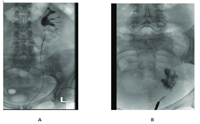
Figure 1: Retrograde imaging before and after ureteral avulsion during ureteroscopy (URS). (A) Pre-avulsion retrograde contrast study showing an intact left ureter.
(B) Post-avulsion retrograde contrast study showing complete ureteral discontinuity with contrast extravasation into the retroperitoneum. Please click here to view a larger version of this figure.
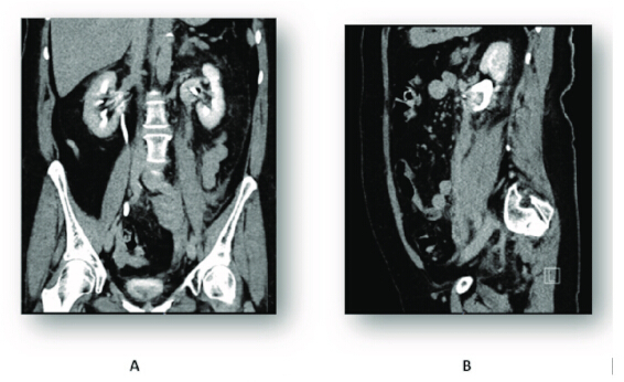
Figure 2: CT urography after the avulsion. Contrast-enhanced computed tomography (CT) urography was performed on the day following ureteral avulsion (pre-reconstruction injury assessment). The study was obtained after urgent placement of a percutaneous nephrostomy and before definitive robotic reconstruction. Imaging demonstrates complete left ureteral injury accompanied by an extensive retroperitoneal hematoma. No urinoma or active contrast extravasation is seen. (A) Coronal view. (B) Sagittal view. Please click here to view a larger version of this figure.
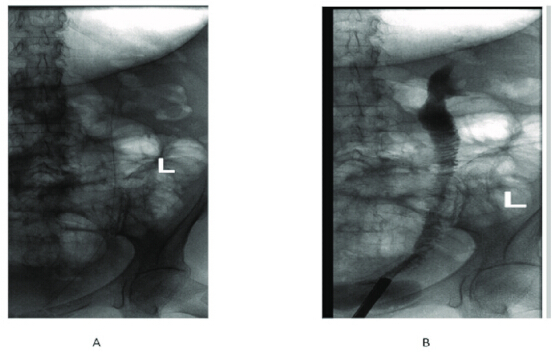
Figure 3: Retrograde imaging after RAIUR. The study was performed 4 weeks after surgery, immediately prior to cystoscopic removal of the indwelling intraluminal drain. The percutaneous nephrostomy had been removed intraoperatively after confirming anastomotic patency. The Foley catheter was removed on postoperative day 10 after negative cystography. (A) The indwelling 15 Ch intraluminal drain is visualized within the interposed ureteral segment. (B) No evidence of contrast extravasation is seen, and contrast passage into the bladder appears unobstructed, confirming anastomotic integrity and graft patency. Please click here to view a larger version of this figure.
Discussion
While immediate RAIUR proved safe and effective in this case, this conclusion must be interpreted within its clinical context. The patient was hemodynamically stable, had preserved renal function, and was treated by an experienced robotic team in urinary diversion. Therefore, immediate RAIUR should be considered a selected-case solution rather than a universally applicable strategy. Ideal candidates are stable patients with complete avulsion, no active infection or uncontrolled bleeding, treated at high-volume robotic centers. Alternatively, delayed reconstruction remains the default.
A primary challenge of immediate repair is the presence of acute inflammation and a retroperitoneal hematoma12,13. The robot-assisted immediate repair addresses these difficulties through high-definition 3D visualization and superior dexterity, which facilitate precise identification of anatomical planes and the execution of watertight anastomoses despite tissue edema5. Furthermore, the use of ICG fluorescence is a critical technical adjunct; it ensures mesenteric vascular integrity of the harvested ileal segment and confirms perfusion of the spatulated renal pelvis, thereby mitigating the risk of ischemic stricture14.
An isoperistaltic ileal segment is commonly used to treat long-segment defects, as it maintains antegrade urinary transport via active peristalsis. To address significant length deficits and ensure a tension-free distal anastomosis, the psoas hitch serves as an essential maneuver. In our patient, metabolic risks—such as hyperchloremic metabolic acidosis—remained negligible, likely due to the use of a relatively short ileal segment. Similarly, mucus production was minimal and effectively managed through conservative hydration strategies.
While immediate RAIUR was appropriate here, other scenarios favor different approaches: Delayed repair (3–6 months) remains preferable when the patient is unstable or when significant contamination or urinoma is present. Delayed repair allows inflammation to subside and facilitates safer dissection. Renal autotransplantation is a better choice when the patient has pre-existing metabolic acidosis (e.g., chronic kidney disease) or when the ileum is unavailable (prior resection, inflammatory bowel disease)3,10. It may also be considered in younger patients to avoid the long-term metabolic and malignant risks associated with an ileal segment. Nephrectomy is justified only when the kidney is non-functional, when the patient is too frail for prolonged reconstruction, or when recurrent infections from a non-draining kidney threaten the contralateral renal unit. In this case, preserved renal function and patient stability made nephrectomy unacceptable.
By opting for immediate robotic rescue, we avoided the complications inherent to chronic urinary diversion, such as recurrent infections, tube dislodgement, and the progressive renal deterioration often seen during the "waiting period" for delayed repair. Additionally, the ability to perform concomitant procedures—such as the bilateral inguinal hernia repair and appendectomy—highlights the efficiency and versatility of the robotic approach without increasing postoperative morbidity.
Despite its advantages, RAIUR is a technically demanding procedure with a significant learning curve. This study has limitations beyond those mentioned. First, it is a single-case report without a comparator group, limiting its generalizability. Second, the follow-up period is only 6 months; late complications such as anastomotic stricture and metabolic acidosis may occur afterward. Third, mucus-related complications—absent here—may occur with longer follow-up and were managed conservatively without objective measurement. Fourth, long-term bowel issues like chronic diarrhea, vitamin B12 deficiency, or adhesive small bowel obstruction remain possible risks of ileal interposition.
Disclosures
The authors have nothing to disclose.
Declaration of AI use: The authors used a language model (Gemini) exclusively for language polishing and grammatical refinement. All scientific content was controlled, verified, and approved by the authors, who assume full responsibility for the final manuscript.
Acknowledgements
The authors received no funding for this work.
Materials
| Name | Company | Catalog Number | Comments |
|---|---|---|---|
| Double-J stent | Boston Scientific | M0061752640; https://www.bostonscientific.com/en-US/products/stents--ureteral/percuflex-plus.html | 6 Fr, 28 cm |
| Endostapler | Endo GIA | EGIAUSTND; https://www.medtronic.com/en-us/healthcare-professionals/products/surgical-stapling/surgical-staplers/laparoscopic-staplers/endo-gia-ultra-universal-stapler.html | endoscopic stapler |
| Guidewire | Terumo | https://www.terumois.com/products/product-type/guidewires/glidewire.html | Hydrophilic |
| Ileal segment | Not applicable | Not applicable | 15-20 cm; autologous patient tissue - not a commercial material |
| Monopolar shears | Intuitive Surgical | 470179; https://www.intuitive.com/en-us/-/media/ISI/Intuitive/Pdf/da-vinci-x-xi-instruments-accessories-catalog.pdf | For the da Vinci X robotic surgical system; standard |
| Nitinol Stone Retreival Basket | Escape | M0063902000 or M0063902010; | stone extractor |
| Prograsp forceps | Prograsp | 471093; https://www.intuitive.com/en-us/-/media/ISI/Intuitive/Pdf/da-vinci-x-xi-instruments-accessories-catalog.pdf | robotic surgical forceps |
| Robotic stapler | Intuitive Surgical | 470430 or 470530; https://www.intuitive.com/en-us/-/media/ISI/Intuitive/Pdf/da-vinci-x-xi-instruments-accessories-catalog.pdf | EndoWrist stapler for the da Vinci X system |
| Robotic surgical system | Intuitive Surgical | https://www.intuitive.com/en-us/products-and-services/da-vinci/systems/x; system-level model/configuration specific | da Vinci X |
| Vicryl sutures (4-0) | Ethicon | https://www.ethicon.com/na/epc/search?keyword=vicryl%204-0 | Absorbable; polyglactin 910 |
References
- Hosseini SR, Gorji A, Fakhr Yasseri AM. Endourological management of mucosal ureteral avulsion: case report. Urol Case Rep. 2021;36:101595. doi:10.1016/j.eucr.2021.101595.
- Chopra S, et al. Initial series of four-arm robotic completely intracorporeal ileal ureter. J Endourol. 2016;30(4):395–399.
- Kaouk J, et al. Single port robotic kidney autotransplantation: initial case series and description of technique. Urology. 2023;176:87–93.
- Yang K, et al. Totally intracorporeal robot-assisted unilateral or bilateral ileal ureter replacement for the treatment of ureteral strictures: technique and outcomes from a single center. Eur Urol. 2023;84(6):561–570.
- Harrison W, Munien K, Desai D. Advancements in minimally invasive bilateral ileal ureter replacement: a promising approach for complex long-segment ureteric strictures. Transl Androl Urol. 2023;12(8):1215–1218.
- Yuan C, et al. Ileal ureteral replacement for the management of ureteral avulsion during ureteroscopic lithotripsy: a case series. BMC Surg. 2022;22(1):262. doi:10.1186/s12893-022-01690-0.
- Wagner JR, Schimpf MO, Cohen JL. Robot-assisted laparoscopic ileal ureter. JSLS. 2008;12(3):306–309.
- Brandao LF, et al. Robotic ileal ureter: a completely intracorporeal technique. Urology. 2014;83(4):951–954.
- Porto BC, et al. Robot-assisted versus laparoscopic ileal ureteral replacement: systematic review and meta-analysis. Cent European J Urol. 2024;77(2):304–309.
- Afferi L, et al. Robotic ileal ureter replacement vs kidney autotransplantation for long ureteric strictures. BJU Int. 2025;135(6):1031–1038.
- Ubrig B, et al. Functional outcome of completely intracorporeal robotic ileal ureteric replacement. Urology. 2018;114:193–197.
- Assimos D, et al. Surgical management of stones: American Urological Association/Endourological Society guideline, PART I. J Urol. 2016;196(4):1153–1160.
- Ragonese M, et al. Immediate ileal ureter replacement for ureteral avulsion during ureteroscopy. IJU Case Rep. 2020;3(6):241–243.
- Pathak RA, Hemal AK. Intraoperative ICG-fluorescence imaging for robotic-assisted urologic surgery: current status and review of literature. Int Urol Nephrol. 2019;51(5):765–771.
Reprints and Permissions
This article has been published
Video Coming Soon