We describe an optimized trans-mitochondrial cybrid generation protocol from mtDNA patient platelets, emphasizing growth parameters and colony isolation. This approach enables the study of mitochondrial DNA variants at variable heteroplasmy levels in a common nuclear background to quantify their functional significance on electron transport chain enzymatic activities and integrated respiratory capacity.
Method Article
Trans-Mitochondrial Cybrid Generation from mtDNA Patient Platelets: An Efficient Protocol Optimizing Colony Selection and Functional Validation
Opens in a new tab
In This Article
Summary
Loading...
$$\rightleftharpoonup{xx}$$
$$\longleftharp{xx}$$,
$$\longrightharp{xx}$$,
Abstract
Loading...
$$\rightleftharpoonup{xx}$$
$$\longleftharp{xx}$$,
$$\longrightharp{xx}$$,
Trans-mitochondrial cybrid cell line generation represents the gold-standard method for determining pathogenicity by enabling biochemical analyses of a specific mitochondrial DNA (mtDNA) variant of interest at high and low percentages (heteroplasmy levels) within an otherwise identical mtDNA and nuclear genome background. Historically, the cybrid generation process has been tedious and poorly efficient. Here, we describe a highly efficient and effective protocol for generating trans-mitochondrial cybrid cell lines by fusing human platelets with a standard osteosarcoma 143B cell line to provide an isogenic nuclear background depleted of mtDNA (Rho0 cells). Cell isolates capture a given mtDNA genome of interest to establish stable cell lines harboring different degrees of heteroplasmy, or to compare divergent effects of distinct mitochondrial haplogroups. Because cybrids from mitochondrial patients may be more difficult to establish with standard protocols, this current methodology focuses on isolating mtDNA variants where the electron transport chain activity is affected. We here demonstrate that colony selection techniques reduce time and improve the yield of generating high-level heteroplasmy mtDNA mutant cybrid lines. A case study is provided of cybrid generation for a variant of unknown significance in MT-ND1, m.3985G>A (p.E227K). We analyze the efficiency of the cybrid generation process using this protocol and run functional studies performed by high-resolution respirometry. High-level heteroplasmy MT-ND1 m.3985G>A cybrid mutants generated by this protocol are shown to have impaired complex I-dependent mitochondrial respiration relative to wild-type control, demonstrating m.3985G>A is likely pathogenic.
Introduction
Loading...
$$\rightleftharpoonup{xx}$$
$$\longleftharp{xx}$$,
$$\longrightharp{xx}$$,
Establishing pathogenicity of mitochondrial DNA (mtDNA) mutations is challenging due to their rarity, heteroplasmy involving variable levels of mutated and wild-type mtDNA genomes in different tissues, diverse pathophysiologic mechanisms, and complex interactions with nuclear genome backgrounds1,2,3,4. For these reasons, trans-mitochondrial cybrid cell lines remain the gold standard method by which to establish pathogenicity of mtDNA variants, where "cybrids" represent the fusion of one cell's nucleus with another cell's mitochondria5,6,7,8,9,10 (Figure 1). Cybrid cell lines contain an isogenic nuclear background and mitochondria with a specified percentage of heteroplasmy for the mtDNA variant under study11,12. In this way, functional assays such as respirometry (by polarography) or electron transport enzyme activity (by spectrophotometry) assays can be performed in cybrid lines ranging between 0% and 100% heteroplasmy levels to determine the potential pathogenic effects of a given mtDNA variant, as well as the relative influence of mitochondrial haplogroups that may differ between cybrid lines1. Other cell types can be used to study pathogenicity and are best used to complement cybrid generation and functional analysis, as every cell type has its own advantages and disadvantages. Patient fibroblasts directly originate from the patient but are limited in the inherited heteroplasmy and lack an isogenic nuclear control to eliminate nuclear gene effects. mtDNA editing is becoming more popular but suffers from off-target editing and limited editable sites in mtDNA13. Induced pluripotent stem cells (iPSCs) can be differentiated to study a mutation's effect on different cell types, such as neurons and retinal ganglion cells, but induction of pluripotency may lead to loss of the mtDNA variant of interest or result in additional de novo variants14. Cybrid study involves the generation of a more artificial system that is based on a cancer cell line (classically 143B osteosarcoma) nuclear background, but classically grows well, making it amenable to use in high-throughput screening studies of candidate interventions. Limitations to high heteroplasmy level cybrid generation include platelet quality, starting heteroplasmy, and the selective disadvantages of the mutant mitochondria, which are discussed at length in this protocol.
Although cybrid generation is the gold standard for functional determination of mtDNA variant pathogenicity, few labs generate cybrids for the study of mitochondrial disease9,10,15,16,17,18,19,20,21,22,23,24,25,26,27. Rather, more cybrids are generated to study cancer and aging28,29. Here, we use a suspected mitochondrial disease patient case study to demonstrate optimization strategies developed to facilitate cybrid generation from mtDNA patient platelets, including selection processes and cell preparations needed to establish cybrid cell lines with high versus low heteroplasmy levels for the target mtDNA variant. Specifically, we demonstrate the methodology to generate cybrid cell lines for a complex I (CI) subunit MT-ND1 variant m.3985G>A, in which we perform biochemical studies that demonstrate the pathogenic nature of this variant. Figure 2 depicts the timeline for cybrid generation, where major methodological steps include fusion, selection, and isolation methods to yield distinct colonies.
Protocol
Loading...
$$\rightleftharpoonup{xx}$$
$$\longleftharp{xx}$$,
$$\longrightharp{xx}$$,
Blood platelets were obtained per the Children's Hospital of Philadelphia's (CHOP) IRB study 08-6177.
1. Preparation of platelets
- Day 1: Draw blood into acid-citrate-dextrose tubes when drawn.
NOTE: Blood should be placed on the rotator immediately. Platelets are ideally harvested a maximum of 4 h after blood draw. - Record the volume of the blood. A volume of 5–6 mL of blood is recommended.
- Centrifuge blood at 200 × g for 20 min at 12 °C.
NOTE: Centrifugation at 12 °C preserves sample integrity and prevents platelet activation. - Transfer the supernatant (plasma) to a new centrifuge tube.
- Record the volume of the plasma.
- Centrifuge the plasma at 1500 × g for 20 min at 12 °C.
- Record the estimated platelet pellet volume.
NOTE: Too many platelets can be cumbersome in this protocol. After fusion, when plating the Rho0 cells and platelets, an excess of platelets can stick to the bottom of the flask and prevent Rho0 cell adherence. It is recommended to use less than 50 µL of the pellet volume of platelets for the fusion. The excess platelets can be frozen by splitting the platelets in the following steps. - Remove the supernatant.
NOTE: If the pellet is large, removal of the pellet can be performed by aspiration. Patient platelets are sometimes drastically reduced in number and are not very sticky. If this is the case, use a pipette to slowly remove the plasma. - If continuing with fresh platelets, resuspend the pellet in 1 mL of Spinner modification of Minimum essential Eagle's medium (SMEM), and transfer to a microcentrifuge tube.
- If freezing, resuspend the pellet (platelets) in 1.05 mL of SMEM and 450 µL of platelet freezing solution (350 µL of fetal bovine serum [FBS] + 100 µL of dimethyl sulfoxide [DMSO]).
NOTE: Some patient platelets are not stable during the freezing procedure, and a high mutant cybrid cannot be obtained after frozen. It is recommended to use the fresh preparation. - If the Rho0 cells are not ready in time, place the platelets at 37 °C with light shaking at 60 rpm.
NOTE: Ultimately, it is best to time the platelets and Rho0 cells to be ready at the same time. It is not suggested to wait for more than 2 h.
2. Preparing Rho0 cells
- Use either freshly thawed 143B osteosarcoma Rho0 cells or Rho0 cells that were growing to near confluence.
NOTE: It is recommended to use Rho0 cells that were growing in the incubator to near confluence. Freshly thawed Rho0 cells can be used, and it is recommended to use a frozen vial of Rho0 cells with a high cell density. Allow the cells to attach for two or more hours prior to fusion. - Wash the T75 flask with 5 mL of PBS.
- Add 1 mL of Trypsin-EDTA 0.25% to cover the Rho0 cells.
- Incubate for 5 min at 37 °C.
- After rocking the flask, wash the bottom of the flask with 5 mL of SMEM.
- Collect Rho0 cells in a 15 mL conical tube.
- Centrifuge at 200 × g for 5 min to concentrate for cell count measurement.
- Remove supernatant and resuspend in 1 mL of SMEM.
- Count the cells.
NOTE: Ideally, there should be 1.5 million cells for each fusion; however, successful fusions have been done with as few as 30,000 cells. - Aliquot 1.5 million cells into 1.5 microcentrifuge tubes and bring the volume to 1 mL.
NOTE: Be sure to have an extra "mock fusion" tube. This tube will only contain Rho0 cells and will be used to help guide the selection process. - Centrifuge the platelets at 1700 × g for 15 min at room temperature.
NOTE: Rho0 cells are sensitive to cold temperature. - Remove the supernatant from the platelets.
- Add 1 mL of Rho0 cells on top of the platelet pellet.
- Centrifuge the Rho0 cells and the platelets at 200 × g for 5 min at room temperature.
3. Fusion
- Remove the supernatant from the Rho0 cells and the platelet pellet.
- Add 150 µL of fusion buffer (50% polyethylene glycol [PEG]/10%DMSO) and triturate for 90 s to resuspend.
NOTE: Some pellets are quite sticky and need to be manually stirred and broken up in addition to pipetting up and down. Record if the pellet was sticky. - Add 12 mL of Cybrid media (DMEM) + 10% FBS + 50 µg/mL uridine to a T75 flask.
- Add the 1 mL of fused platelets and Rho0 cells to the flask.
- Cells are incubated at 37 °C with 5% CO2 in high humidity.
NOTE: All cell growth steps are under these conditions.
4. Growth
- Day 2: Remove Cybrid media and add fresh 12 mL of Cybrid media + 10% FBS + 50 µg/mL uridine.
NOTE: Rho0 cells take about one week to incorporate the mtDNA. - Day 5: Remove Cybrid media and add fresh 12 mL of Cybrid media + 10% FBS + 50 µg/mL uridine.
NOTE: If the Rho0 cells are at 80% confluence, split the cells into two flasks.
5. Selection
- Day 8: Remove media and wash the cells with 5 mL of PBS.
- Remove PBS and add 1 mL of trypsin.
- Cover the bottom of the flask with trypsin and incubate for 5 min at 37 °C.
- Add 5 mL of Cybrid media + 10% FBS + 50 µg/mL uridine.
- Count the cells.
NOTE: Selection should start at 10% confluence, which is 125,000 cells in a T75 or tissue culture-treated petri dish. It is recommended to use petri dishes if the colony picking method is preferred. Use a T75 if using the dilution method. - Add 125,000 cells to the appropriate container and add the rest of the volume up to 12 mL with Selection media (DMEM).
NOTE: Selection media is designed to separate fused 143B cells with mitochondria from the Rho0 cells. Rho0 cells require pyruvate and uridine to survive. Dialyzed FBS does not contain pyruvate and uridine and should only be used for selection. Use two different selection media if possible. One should be 10% dialyzed FBS, and the other should be less restrictive of growth, like 20% dialyzed FBS + 50 µg/mL uridine. A less restrictive selection medium is suggested in parallel for severe mutants. - Freeze remaining cells in two cryovials with 1 mL of freezing solution each.
NOTE: These are preselection cells, and different selections can be tried down the line. - Change the media every other day for about 1 week.
NOTE: The Rho0 cells will float when they die and will wash away after the media change. Use the "mock" fusion cells to determine when selection is complete (no more cells in the flask). This flask will be entirely empty because all the Rho0 cells will have washed away. It is a good idea to wait another day, even when the mock flask is empty, because fused cells can provide the nutrients in the media to help other Rho0 cells persist in the flask. If the cells grow to more than 20% confluence, split and replate. More growth will mask the selection.
6. Colony selection growth method
- Grow the colonies until they are visible by eye with Cybrid media + 20% FBS + 50 µg/mL uridine.
NOTE: Tilt the dish and hold it up to the light. The colonies appear as cloudy circles. Under the microscope, there should not be a lot of background cells in between the colonies. - Circle independent non-overlapping colonies with a marker at the bottom of the flask while holding them up to the light.
- Use a P20 to both scrape (mechanically disturb cells attached to the bottom of the flask) and aspirate 20 µL at the same time within the drawn circle to place cells from the selected colony into the tip of the P20.
NOTE: Scraping manually detaches the cells and pulling up 20 µL is collecting the cells in the pipette tip. - Plate each colony in its own well in a 96-well plate.
NOTE: The advantage of this method is that 143B cells tend to grow well together, so one should not have a problem growing and will get faster results. The disadvantage is that one might have a mixed population.
7. Dilution growth method
- When the cells reach 75% confluence in Cybrid media + 20% FBS + 50 µg/mL uridine, wash the cells with 5 mL of PBS.
- Remove PBS and add 1 mL of trypsin.
- Cover the bottom of the flask with trypsin and incubate for 5 min at 37 °C.
- Add 5 mL of Cybrid media + 20% FBS + 50 µg/mL uridine.
- Count the cells.
- Add 1 × 10⁵ cells in 20 mL of Cybrid media + 20% FBS + 50 µg/mL uridine to 50 mL conical tube 1.
NOTE: This is 5000 cells/mL, which will be 500 cells in 100 µL when plated in a 96-well plate. - Prepare a 50 mL conical tube with a 100-fold dilution (200 µL from Tube 1 in 20 mL of Cybrid media + 20% FBS + 50 µg/mL uridine).
NOTE: This is about 5 cells per well. - Prepare a 50 mL conical tube 3 with a 1:1 dilution (10 mL from Tube 2 in 10 mL of Cybrid media + 20% FBS + 50 µg/mL uridine).
NOTE: This is about 2.5 cells per well. - Prepare a 50 mL conical tube 4 with a 1:1 dilution (10 mL of Tube 3 in 10 mL of Cybrid media + 20% FBS + 50 µg/mL uridine).
NOTE: This is about 1 cell per well. - Plate Tubes 2, 3, and 4 in 96-well plates with 100 µL each.
NOTE: Be sure to mix the cells before plating. It is hard to check the day of to see if one cell is in each well because the cells need to sink to the bottom and attach. The advantage of this method is that a single cell grows into a colony, ensuring the absence of a mixed background. The disadvantage is that severe mutants may not survive if isolated. - The next day, cross out wells that clearly have more than one cell per well.
- After a few days, check the wells again.
NOTE: Now there should be colonies, and it is more apparent if there are single cells near the edges of the wells. If there is more than one colony, remove those wells. If there is no cell, remove the well.
8. Growth
- Start in the 96-well plate and transfer cells to bigger plates step-wise as needed.
NOTE: These cells like to grow best with high cell density, and not transferring enough cells could cause death. Always keep the previous plate with some cells in it and mark which well came from where so that one can always go back if needed. Importantly, 143B cells die and detach if overconfluent, so check these cells every day.
9. Genetic confirmation
- Centrifuge the aliquoted cells at 450 ×g for 5 min.
- Remove media.
- Add 5 µL of DNA Extraction buffer (50 mM NaOH).
NOTE: This number can change if there are a lot of cells. Significant cell pellets can be in 10 µL of buffer. - Incubate at 95 °C for 30 min.
- Add an equivalent volume of DNA Stabilization buffer (0.1 M Tris-HCl; pH 8) to the extraction buffer.
- Perform polymerase chain reaction (PCR) according to the enzyme's guidelines.
NOTE: Some point mutants can use restriction enzymes to distinguish between control and patient cells. Other point mutations cannot be distinguished and require sequencing. In this instance, it is important to use PCR mixes without dyes for high-quality results. Sanger sequencing and next-generation sequencing can be used. 2.5 µL of DNA is sufficient for a 25 µL reaction. - Use PCR amplicons specific to this mutation for Sanger sequencing.
NOTE: The PCR amplicons were sent for Sanger's sequencing. Mutation heteroplasmy level was confirmed both after clone expansion and after functional analysis.
10. High-resolution respirometry using high-resolution respirometry
- Perform air calibration as per the respirometer (e.g., Oroboros Instruments) guidelines.
- Wash cybrids in a T75 flask with PBS.
NOTE: The cells should not be overconfluent; 70%–80% confluent is ideal. - Add 1 mL of trypsin and incubate for 4 min.
- Add 5 mL of Cybrid media and count cells.
- Add 500,000 cells to a tube to centrifuge down at 450 × g for 5 min.
NOTE: The cell number and volumes for the respirometer are with the 0.5 mL chamber. - Wash cells by resuspension with PBS and centrifuge again.
- Resuspend cells in 510 µL of MiR05 buffer and add to an empty chamber.
- After stabilization, add 0.65 µL of digitonin (50 mg/mL), 0.8 µL of malate (2.5 M), and 2 µL of pyruvate (2.5 M).
NOTE: Before the addition of digitonin, this is where basal respiration measurement is taken. Every subsequent step should be done after stabilization. - Add 12 µL of adenosine diphosphate (ADP) (0.25 M).
- Add 4 µL of glutamate (2.5 M).
- Add 12 µL of succinate (1.25 M).
- Add 1 µL of oligomycin (1 mg/mL).
- Add 1 µL of carbonyl cyanide-p-trifluoromethoxyphenylhydrazone [FCCP] (0.2 mM).
NOTE: Titrate with multiple additions until no further increase is observed (typically 3–5). - Add 1 µL of rotenone (2 mM).
- Add 1 µL of antimycin A (1 mg/mL).
- Add 0.5 µL of ascorbate (0.8 M) and 0.5 µL of tetramethylphenylenediamine [TMPD] (0.2 M).
- Open the chamber with a spacer for 20 min.
- Close the chamber and stabilize for 5 min.
- Add 22.5 µL of azide (4 M).
- Collect cells for protein quantification using a bicinchoninic acid (BCA) assay kit.
- Perform calculations after stabilization of the oxygen consumption rate and normalize by protein. Basal Respiration is the stabilized oxygen consumption rate after the addition of cells to the chamber.
NOTE: All other measurements are taken after the noted substrate or inhibitor. - Calculate OXPHOSCI: (Glutamate-Antimycin A)/(FCCP-Antimycin A).
- Calculate OXPHOSCI+CII: (Succinate-Antimycin A)/(FCCP-Antimycin A).
- Calculate ETSCI+CII: (FCCP-Antimycin A).
- Calculate ETSCII: (Rotenone-Antimycin A)/(FCCP-Antimycin A).
- Calculate LEAKCI+CII: (Oligomycin-Antimycin A)/(FCCP-Antimycin A).
- Calculate OXPHOSCIV: (TMPD/ASC-Azide)/(FCCP-Antimycin A).
- Use at least 4–6 biological replicates for respirometry measurements.
Results
Loading...
$$\rightleftharpoonup{xx}$$
$$\longleftharp{xx}$$,
$$\longrightharp{xx}$$,
To demonstrate methodologic differences and the path to protocol optimization, we present the results of cybrid generation from one patient with suspected mitochondrial disease who harbors a variant of unknown significance (VUS) in MT-ND1 of complex I (CI) in the electron transport chain. After clinical mtDNA genome next-generation sequencing in multiple tissues at two different CLIA-approved clinical diagnostic labs, the only variant suspicious of pathogenicity was MT-ND1 m.3985G>A. The heteroplasmy in the patient's blood of MT-ND1 m.3985G>A (p.E227K) mutation was 50%. Multiple protocol optimization insights were demonstrated by generating cybrids for this individual (Table 1). Prior attempts to generate cybrids from frozen platelets were either unsuccessful or only yielded wild-type (0% heteroplasmy) cybrids (Table 1). By contrast, using freshly isolated platelets yielded better results. It became evident that the patient's platelets were not stable after SMEM addition, which was likely the reason for failed heteroplasmy return from the frozen platelets. Importantly, our research group has observed a broader mitochondrial disease patient platelet stability problem in an attempt to generate other complex I disease patient-derived cybrids. High (20%) FBS in the growth media and selection media was found to be essential to the success of generating high-level heteroplasmy cybrids (Table 1). The colony method generated the highest heteroplasmy level at 95%, as compared to the dilution method, where the maximal heteroplasmy level was only 40% (Table 1). Figure 3A indicates the heteroplasmy levels of all sequenced colonies by these two methods from conditions indicated in trials 5–9 in Table 1. Figure 3B,C separates the total sequencing by the dilution method and colony picking method, respectively. Overall, the ideal conditions to generate high-level heteroplasmy cybrid cell lines for this case study included the use of freshly isolated platelets, selected with 20% dialyzed FBS Cybrid media with uridine, grown in Cybrid media + 20% FBS + 50 µg/mL uridine, and using the colony picking method for colony isolation to yield a mutant of 95% heteroplasmy for m.3985G>A.
To demonstrate a pathway towards functional biochemical validation of generated cybrids, we performed high-resolution respirometry to determine if MT-ND1 m.3985G>A (p.E227K) caused a complex I-specific respiratory chain defect. Specifically, we compared a 0% mutant heteroplasmy control cybrid line and a 95% mutant heteroplasmy cybrid line both generated from the same patient who carried m.3985G>A (p.E227K). Basal respiration trended lower (p value > 0.089) in the 95% mutant cybrid line as compared to the 0% mutant control (Figure 4A). Substrate-dependent coupled rate OXPHOSCI was significantly lower in the 95% mutant cybrid as compared to control, ultimately confirming this mtDNA mutation impairs CI activity and is thereby likely pathogenic (Figure 4B). OXPHOSCI+CII was not significantly different (p value > 0.224) between the 0% and 95% mutant cybrid lines (Figure 4C), nor were uncoupled ETSCI+CII (p value > 0.156) and ETSCII respiration rates (p value > 0.196) (Figure 4D,E). LEAKCI+CII and CIV activity were both significantly increased in the 95% mutant cybrid line as compared to control, likely in compensatory response as is commonly seen in complex I disorders30,31,32 (Figure 4F,G). Collectively, these high-resolution respirometry studies in trans-mitochondrial cybrid cell lines generated from the affected proband's platelets support that MT-ND1 m.3985G>A (p.E227K) causes a CI-specific mitochondrial respiratory chain defect and is likely pathogenic.
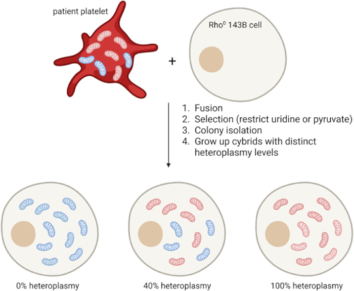
Figure 1: Platelets and Rho0 cells can be fused to yield cybrids with varying degrees of heteroplasmy. Patient platelets harbor mitochondria with different mtDNA mutation levels (heteroplasmy) and do not contain a nucleus. 143B osteosarcoma cells can be treated with ethidium bromide to remove all mitochondria (Rho0 cells). By fusion, selection, colony isolation, and growing the cybrids up, cybrids with distinct heteroplasmy levels can be obtained that have the mitochondria from platelets and an isogenic nucleus from 143B cells. Blue mitochondria indicate mitochondria having no mutation (0% heteroplasmy or homoplasmic wild-type). Increasing amounts of red mitochondria indicate increasing mitochondria that have the mutation at levels up to a potential 100% heteroplasmy (homoplasmic mutant). Please click here to view a larger version of this figure.
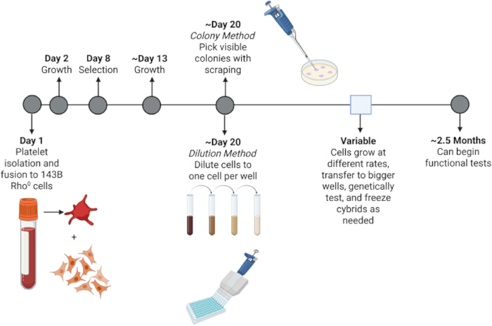
Figure 2: Trans-mitochondrial cybrid generation timeline. Day 1: Platelet isolation from fresh blood and platelet fusion to 143B Rho0 cells. Day 2: Growth in Cybrid media for one week. Day 8: Selection starts, and media is changed every other day until all "mock" cells have died. Day 13: Growth in Cybrid media until cells have grown to colonies or 75% confluence. Day 20: Here two different colony isolation methods can be used. The top shows the colony method, where visible colonies are scraped and simultaneously aspirated with a pipette. Bottom shows the dilution method where cells are serially diluted until about one cell can be plated per well in a 96-well plate. The square indicates a variable time range where each distinct colony will grow at different rates and will need to be transferred to bigger wells, tested genetically using PCR or other methods, and frozen to save the newly generated cybrid line at different times. This whole process should take about two and a half months, and the cybrids can then be used for functional studies. Please click here to view a larger version of this figure.
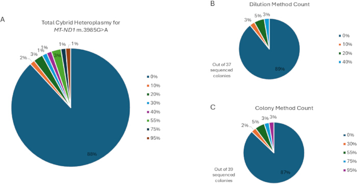
Figure 3: Percentage of heteroplasmy achieved in cybrid cell lines generated using different methods for MT-ND1 m.3985G>A variant. (A) Heteroplasmy of all sequenced cybrids in both dilution and colony isolation methods. (B) Heteroplasmy of all sequenced cybrids from the dilution method. (C) Heteroplasmy of all sequenced cybrids from the colony method. Please click here to view a larger version of this figure.
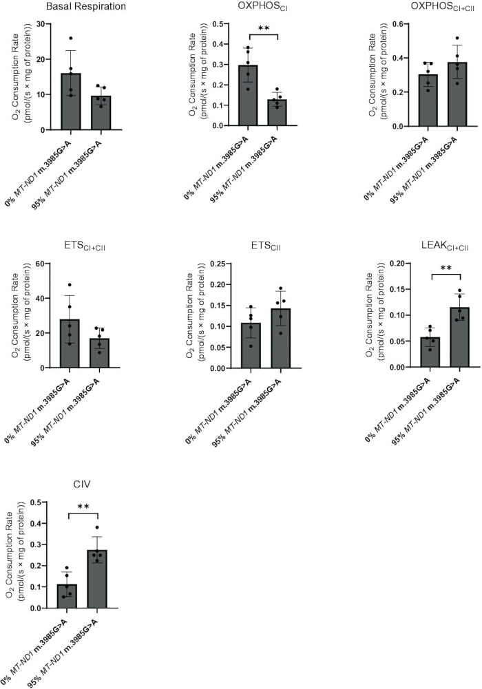
Figure 4: 95% MT-ND1 m.3985G>A (p.E227K) cybrid mutant has a CI-specific respiratory chain defect. Oxygen flux measurements obtained by high-resolution polarography with an Oxygraph-2k (Oroboros) on cybrids grown in Cybrid media. (A) Basal respiration is the measurement before the addition of substrates or inhibitors and is normalized by mg of protein. Each subsequent measurement is taken as the oxygen consumption rate (pmol/s) after the addition of substrate or inhibitor. Removal of non-mitochondrial respiration is performed by subtraction of oxygen consumption after antimycin A addition. All measurements are normalized by mg of protein. (B) OXPHOSCI is the substrate-driven capacity of CI taken after the addition of glutamate. (C) OXPHOSCI+CII is the capacity of both respiratory chain complex I and II (CI+CII) measured following the addition of succinate. (D,E) ETSCI+CII and ETSCII are the maximal respiration and maximal respiration after the addition of rotenone to inhibit CI, respectively. (F) LEAKCI+CII is the non-phosphorylating electron transfer across the mitochondrial inner membrane. (G) Respiratory chain complex IV (CIV) activity is assessed by reducing cytochrome c with TMPD and ascorbate and by the inhibition of CIV using azide. n = 5 biological replicates, ** indicate p-value < 0.01 using Welch’s t-test. Please click here to view a larger version of this figure.
| Trial | Frozen or fresh platelets | Selection Media | Growth media | Colony selection method | Success | Highest heteroplasmy % |
| 1 | frozen | Selection: 5% dialyzed FBS no uridine | Cybrid 10% FBS | Dilution | No | - |
| 2 | frozen | Selection: 10% dialyzed FBS no uridine | Cybrid 10% FBS | Dilution | Yes | 0% |
| 3 | frozen | Selection: 20% dialyzed FBS with uridine | Cybrid 10% FBS | Dilution | Yes | 0% |
| 4 | fresh | Selection: 5% dialyzed FBS no uridine | Cybrid 20% FBS | Dilution | Yes | 0% |
| 5 | fresh | Selection: 5% dialyzed FBS no uridine | Cybrid 20% FBS | Colony | Yes | 0% |
| 6 | fresh | Selection: 10% dialyzed FBS no uridine | Cybrid 20% FBS | Dilution | Yes | 0% |
| 7 | fresh | Selection: 10% dialyzed FBS no uridine | Cybrid 20% FBS | Colony | Yes | 20% |
| 8 | fresh | Selection: 20% dialyzed FBS with uridine | Cybrid 20% FBS | Dilution | Yes | 40% |
| 9 | fresh | Selection: 20% dialyzed FBS with uridine | Cybrid 20% FBS | Colony | Yes | 95% |
Table 1: Conditions tested for MT-ND1 m.3985G>A (p.E227K) cybrid variant, having successful levels of heteroplasmy established.
Discussion
Loading...
$$\rightleftharpoonup{xx}$$
$$\longleftharp{xx}$$,
$$\longrightharp{xx}$$,
Here, we present a highly efficient protocol for the generation of trans-mitochondrial cybrid cell lines from mtDNA patient platelets. We demonstrate the applicability with a case study of an MT-ND1 variant of uncertain significance, the importance of carefully considering growth and selection parameters. Once these major issues are optimized, the successful selection of cybrids harboring mtDNA variants that may impair respiratory chain function. This protocol can be used for the generation of cybrids with mtDNA mutations.
The timeline for our case study demonstrates that cybrid generation takes about 2.5 months from the time of platelet isolation (Figure 2). In terms of active time, day 1 and day 20 are the most time-consuming. The variable time of moving cells from a 96-well plate to bigger wells is challenging, and cells need to be checked daily, given the challenges of working with 143B cells, where overconfluence or too few cells may both cause cells to detach and die. The colony picking method transfers many cells to one well, where they grow quickly and can be quickly transferred. The colony picking timeline is therefore shorter than the dilution method, but has a higher chance of becoming contaminated with another cybrid of a differing level of heteroplasmy. By contrast, the dilution method is prone to many cells being unable to grow because they are isolated in the well.
The method that yielded cybrid cell lines harboring the highest heteroplasmy level was the colony picking method, where we were able to isolate 95% heteroplasmy of the MT-ND1 m.3985G>A variant (Table 1). After ongoing work with this 95% heteroplasmy cybrid line, a phenotype became evident in 10% FBS DMEM media (data not shown), further highlighting the need for consistent use of 20% Cybrid media throughout the entire cybrid generation protocol. Other cybrid protocols give one selection medium that is very restrictive8,9,10,12,15, and as a result, it may be challenging to generate mitochondrial mutants under these conditions. In this case study, it was evident that low dialyzed FBS without supplementation of uridine was too restrictive for mitochondrial mutants, as high heteroplasmy level cybrid lines could not be generated (Table 1). In testing mock Rho0 cells with different selection media, Rho0 cells could still be eliminated with 20% dialyzed FBS and uridine supplementation, which represents a "high nutrient" yet restrictive selection medium. Colony picking is the final change that pushed the mutation towards a high heteroplasmy level and required substantially less screening effort as compared to the dilution method, which is not standard in previously reported cybrid generation protocols. Finally, high heteroplasmy mutants were obtained with this modified selection protocol.
The goal when generating cybrid cell lines is to have a 0% heteroplasmic mutant (homoplasmic wild-type) that may be directly compared to the highest possible achieved mutant heteroplasmy level (ideally, a homoplasmic mutant) to isolate the functional effects of the variant in question. This can be difficult to achieve in a platelet sample having a low starting mutant heteroplasmy level, given the low probability of achieving a resulting clone with a high mutant heteroplasmy level. Conversely, a 100% homoplasmic mutant sample might prevent obtaining a 0% "wild-type" mtDNA genome control unless another platelet sample from a maternal relative, such as a mother or sibling with the same mtDNA haplogroup who does not carry the variant in question, is available. When comparing with a heteroplasmic mutant cybrid line, the preferred 0% control cybrid line would originate from the patient's platelet sample. However, if their 0% mutant cybrids cannot be recovered, using cybrids established from a maternal relative is preferred to control for their otherwise identical mtDNA genome haplogroup background. New advances in mtDNA editing will likely replace cybrids as the gold-standard methodology to isolate and study a mtDNA variant's functional effects. However, cybrid lines will likely remain a useful research tool, given they classically use a common cell line nuclear background (143B) and also enable effects of fixed haplogroup (homoplasmic) variant combinations to be studied that may be difficult to achieve with gene editing techniques. Controlling mutant heteroplasmy levels in cell types such as iPSCs and fibroblasts remains a current limitation of mtDNA editing, where cybrids can be useful, although it is feasible that mtDNA editing in the future might relieve this bottleneck13,14.
Functional biochemical analysis to isolate the impact of a variant (or haplogroup) of interest in the mtDNA remains the overarching goal of cybrid cell line generation. In this regard, we report high-resolution respirometry data confirming that MT-ND1 m.3985G>A (p.E227K) is likely pathogenic. We describe for the first time a decrease in OXPHOSCI respiration and an increase in both OXPHOSCIV and LEAKCI+CII respiration (Figure 4). Indeed, CIV biogenesis is often increased as an adaptive response in CI deficiency (Figure 4), while more work would be needed to elucidate the cause of the increased proton leak. Overall, this work provides functional confirmation that MT-ND1 m.3985G>A (p.E227K) causes a specific and pronounced CI respiratory chain defect and should be considered pathogenic. One limitation in this case study is that only one independent clone was tested by respirometry analysis for the 95% cybrid mutant. As only one stable mutant clonal line of this high heteroplasmy was successfully obtained and maintained, it was not possible to test multiple independently derived clones to conclusively exclude a clone-specific effect on mitochondrial respiration. Broadly, the generation of high heteroplasmy cybrids may vary depending on the precise mtDNA variant, starting heteroplasmy level in the platelet sample, platelet quality, and the selective disadvantage of the mutant mitochondria. Overall, this optimized cybrid generation protocol describes different strategies to enhance the likelihood of successfully generating low and high heteroplasmy lines in which comparative functional analyses may be performed to identify a potentially pathogenic mtDNA variant in an isogenic nuclear background. Cybrids can then be used downstream for respirometry and also cell viability, biochemical analysis, and even high-throughput screening.
Disclosures
Loading...
$$\rightleftharpoonup{xx}$$
$$\longleftharp{xx}$$,
$$\longrightharp{xx}$$,
The authors have no relevant financial conflicts of interest relative to this work.
Acknowledgements
Loading...
$$\rightleftharpoonup{xx}$$
$$\longleftharp{xx}$$,
$$\longrightharp{xx}$$,
We are grateful to the patient and their family, as well as to Rebecca Ganetzky, Sheila Clever, Doug Wallace, and Ryan Morrow for experimental troubleshooting guidance. This work was funded in part by the CHOP Mitochondrial Medicine Mazzullo Family complex I research philanthropic fund and the National Institutes of Health (R35-GM134863 to MJF). The content is solely the responsibility of the authors and does not necessarily represent the official views of the funders, including the NIH.
Materials
| Name | Company | Catalog Number | Comments |
|---|---|---|---|
| ADP | Sigma Aldrich | A5285-1G | |
| Antimycin A | Sigma Aldrich | A8674-25MG | |
| Ascorbate | Sigma Aldrich | 11140-250G | |
| Azide | Sigma Aldrich | S2002-100G | |
| Dialyzed FBS | Fisher Scientific | 35071CV | |
| Digitonin | Sigma Aldrich | D141-100MG | |
| DMEM | Corning | 10-013-CV | Cybrid media |
| DMEM | Corning | 10-017-CM | Selection media |
| FBS | Cytiva | SH30910.03 | |
| FCCP | Apex Bio | B5004 | |
| Glutamate | Sigma Aldrich | G5889-100G | |
| Graphpad Prism | Graphpad | ||
| Hybri-Max | Sigma Aldrich | P7306 | fusion buffer |
| Malate | Sigma Aldrich | M1000-100G | |
| MiR05 buffer | Oroboros Instruments | 60101-01 | |
| O2K respirometor | Oroboros Instruments | C-0050 | |
| Oligomycin | Sigma Aldrich | O4876-5MG | |
| Pyruvate | Sigma Aldrich | P2256-100G | |
| Rotenone | Sigma Aldrich | R8875-1G | |
| SMEM | Sigma | M8167 | |
| Succinate | Sigma Aldrich | S2378-100G | |
| TMPD | Sigma Aldrich | T3134-5G | |
| Trypsin-EDTA 0.25% | Thermo Scientific | 25200056 | |
| Uridine | Sigma Aldrich | U3750-25G |
Reprints and Permissions
Request permission to reuse the text or figures of this JoVE article
Request Permission