Clinically relevant mouse models of anthracycline-induced cardiotoxicity are scarce. Here, a model using immunocompetent mice bearing a tumor receiving anticancer treatment is proposed, mimicking cancer patients’ clinical protocols.
Method Article
A Cardio-Oncological Mice Model of Anthracycline-Induced Cardiotoxicity in Breast Tumor-Bearing Immunocompetent Mice
* These authors contributed equally
In This Article
Summary
Abstract
The recent update from the European Society of Cardiology on preclinical models of cancer therapy-related cardiac dysfunction highlights a critical need for more representative and translational in vivo models. It pinpoints the need to develop models that employ immunocompetent, tumor-bearing animals, incorporating multiple strains and both sexes, to more accurately recapitulate the heterogeneity observed in clinical populations. Therefore, a clinically relevant model of anthracycline-induced cardiotoxicity (AIC) in breast tumor-bearing animals was built. According to mice’ genetic background, either 4T1Luc-cells (200k cells) or PY8119-cells (200k cells) were injected in Balb/c-mice or C57Bl/6J-mice, respectively subcutaneously in the lower mammary gland of males and females. Tumor growth was controlled with bioluminescence and palpation. Once the tumor mass was detectable by palpation, 2 weeks post-tumor cell injection, animals received anticancer Doxorubicin treatment. Mice's tolerance to Doxorubicin was strain-dependent. Cardiac function was evaluated before the start of the protocol (baseline) and weekly after by cardiac ultrasound in ketamine-anesthetized mice. Cardiotoxicity was defined as a decrease in left ventricular fractional shortening of at least 10% from baseline, an absolute value <50%, and a heart rate >300 BPM. The protocol was reproducible with a 99% success of tumor development, and a >80% mice survival rate. Cardiotoxicity was observed in >90% of the treated mice with tumor and Doxorubicin.
Introduction
Cardio-oncology aims to improve cardiovascular outcomes in cancer patients before, during, and after chemotherapy. Preclinical models have played a huge role in better understanding the interplay between these two diseases. Recently, a paradigm-shifting study observed that mice inherently prone to spontaneous intestinal polyps developed more of these tumors after a myocardial infarction and a period with heart failure (HF), demonstrating the direct impact of a cardiovascular event on tumor growth1. Since then, numerous studies have reported consistent findings across diverse tumor types and murine strains, frequently implicating specific molecular pathways or proteins, thereby reinforcing the concept that cancer progression is precipitated by cardiovascular diseases (CVDs)2,3,4,5.
Inversely, the impact of cancer on the risk of developing CVDs has been studied for several decades. Epidemiological data show that cancer survivors are suffering from more CVDs, both due to the shared risk factors of these two diseases and due to the known various toxicities of anti-cancer treatments6,7,8,9,10. Preclinical studies have commonly used cancer-induced cachexia models to identify specific targets that could be modulated in humans to improve long-term cardiovascular outcomes11,12,13,14. However, one could argue that these models hardly reflect a common clinical situation since patients are often treated before reaching this advanced cancer-syndrome, but global cachexia is still suspected to largely contribute to overall cancer death15.
In the first cardio-oncology guidelines of the European Society of Cardiology (ESC), it is concluded that further research is needed to better understand the relationship between cancer and CVDs16. More precisely, in a recent update from the ESC working group on cardio-oncology, it was stated that preclinical research is of utmost importance in addressing this knowledge gap, especially with models using tumor-bearing animals17. Moreover, better correlation between preclinical and clinical studies is needed, since most clinical studies today are conducted on anticancer treatments that are poorly studied in preclinical settings18.
Anthracyclines (ACs), such as Doxorubicin (Dox), are commonly used in chemotherapy for a large number of solid and blood tumors. Even if more modern therapies are available today, their cheapness, well-documented side-effects, and evident effectiveness will lead to further use of these drugs for many more years19,20. A vast amount of evidence suggests that ACs lead to a dose-dependent cardiotoxicity, mainly seen as a left ventricular dysfunction, which is one of the main limiting factors for its clinical use today, and which is often a worrying factor for the cardio-oncologist21,22. This literature mainly comes from breast cancer studies due to its high incidence in humans, but clinical studies on lymphomas, lung cancer, and leukemias provide similar observations23. Therefore, preclinical research has been conducted for several decades to understand the underlying pathophysiological mechanisms of AC-induced cardiotoxicity (AIC), which has demonstrated that oxidative stress, direct and indirect DNA damage, ferroptosis, and mitochondrial damage are commonly implicated23,24.
Nevertheless, there is actually limited evidence on how to prevent or treat AIC. Common HF medications have been tested in clinical trials with mixed results, and the timing of use of the few potential candidates has been difficult to determine21,23,25,26,27,28. Dexrazoxane, an iron-modulating drug, has shown promising results to prevent AIC, but is not systematically used today21. Delivering ACs in forms other than free in the blood, for example, encapsulated in liposomes, has also shown promising results but still has difficulty replacing conventional ACs, partly due to their cost21.
One explanation for this situation is the lack of clinically relevant animal models for studying AIC17. In humans, Dox is administered over time via repeated injections, and most of the AIC is observed during the first year after treatment. However, a large variation in animal protocols is observed across the literature, as shown in a recently published review29. A classic example of an acute cardiotoxicity model is tumor-free mice receiving a single injection of 20 mg/kg of Dox30. In a typical model of chronic cardiotoxicity, many studies describe tumor-free mice receiving one injection per week of 5 mg/kg of Doxorubicin for a total cumulative dose of 20 mg/kg29,31. This chronic cardiotoxicity model can also be observed in tumor-bearing animals at the same Dox dose; however, this remains very uncommon in the literature29,32.
To contribute to reducing this gap of knowledge, a pragmatic and clinically relevant model of breast tumor-bearing immunocompetent mice to study AIC was built, namely in 4T1-bearing Balb/c mice, and its robustness in PY8119-bearing C57Bl/6J mice was confirmed, the latter being, to the authors' knowledge, the first time a cardio-oncological model is built with a triple-negative breast cancer in a C57Bl/6J strain. A good capture of tumor growth via bioluminescence quantification was achieved before treating the animals with Dox, which demonstrated cardiotoxicity, and an organ-collection protocol was implemented to study the tumor microenvironment concomitantly with the cardio-oncological experiment. Moreover, results similar to those described in the literature regarding strain and sex sensitivity were observed. This work is expected to contribute to the standardization and improvement of animal protocols in future cardio-oncological studies, as C57Bl/6J mice are highly valuable for their high survival rate in AIC experiments29.
The advantage of the proposed method is the use of the C57Bl/6J strain, which is easy to genetically engineer, has a well-characterized genome, is cheap to acquire, and is tolerant of high cumulative doses of ACs29,33,34. If knocking out a gene is needed for a specific study with ACs, this model is well-suited for this purpose. Moreover, the use of a triple-negative breast cancer derived from this strain is of translational interest since the women with triple-negative breast cancer are the patients who are very likely to receive ACs and who have the poorest prognosis35. Using this model will likely improve AIC knowledge specifically for these patients.
The strength of this model also lies in the fact that the mice are pre-conditioned with a tumor, which can truly impact cardiovascular remodeling36,37. Also, this model falls between acute and fully chronic models of AIC, with the possibility of extending animal monitoring duration due to their high survival rate, which can be of great value if other AC-cycles are considered.
However, this model is probably not the most well-suited for specific tumor characterization at the end of the protocol, due to its pragmatic approach to tumor inoculation. An adapted version of tumor injection should be performed if this is central to the scientific hypothesis. Also, 4T1 cell injection in Balb/c mice is a well-established model for a more oncological approach, for example, for studying metastasis, which is not described here and should be taken into consideration if metastasis is essential to the scientific question38. This model is performed in adult mice; however, if the aim is to study AIC in a pediatric setting, younger mice should be considered, for which the response to the protocol has not been studied here.
Protocol
All animal experiments were in accordance with the local animal ethics committee (Lariboisière-Villemin) and were approved by the French Ministry of Higher Education and Research (MESRI) under the protocol number APAFIS 46086. The 3R rules were taken into account during the design of the protocol. Balb/c and C57Bl/6J mice were bought from an animal manufacturer such as Janvier Labs or Envigo. For this protocol, mice were aged between 10 weeks and 18 weeks at inclusion to correspond to young adults, similar to the women suffering from triple-negative breast cancer. Animals were kept in groups of 2–5 individuals in a room with 12 h dark/light cycles, room temperature between 20 °C and 24 °C, and humidity between 30% to 70%. Water and food were provided ad libitum. During the protocol, caution was taken to follow humane endpoints predefined before the start of the protocol, as described in Supplementary Table 1. Ensure that an isoflurane filter is used to collect wasted gas for the safety of the isoflurane user. Handle animals with care, wearing gloves and glasses at every step of this protocol. All hazardous materials should follow local institutional regulations; here, all contaminated samples (blood, needles, dox-containing vials, etc.) were discarded in biohazardous healthcare waste bins. The protocol overview is shown in Figure 1A. The reagents and the equipment used are listed in the Table of Materials.
1. Week 0: Cell preparation and injection into the animal
NOTE: The following steps are described for 4T1Luc cells and were identical for the use of PY8119 cells. Nevertheless, this should be adapted for each cell line, depending on the manufacturer's recommendations. To optimize the protocol, one person can handle cell preparation, while the other handles animal preparation.
- When obtaining a confluent cell culture flask (80%–90% confluence), apply 3 mL of trypsin for 5 min at 37 °C and 5% CO2.
- Gently tap the flask to detach all cells, avoid cell clumps, and resuspend in cell culture medium.
- Count the cells using a Malassez chamber (or another technique, depending on what is available).
- Centrifuge the cells at 300 × g for 5 min, then resuspend in an equal volume of PBS to inject 200,000 cells in 100 µL.
NOTE: The number of cells was chosen after a literature review (see Supplementary Table 2). - Aspirate gently using a 26 G syringe 500 µL of the cell suspension. To avoid excessive time spent with the cell suspension in the syringe, filling the syringe with less volume than necessary for 5 animals is recommended.
- Simultaneously (if possible), place the mice in an isoflurane chamber and rapidly induce anesthesia with 3% isoflurane at 1 L/min in ambient air. As soon as the animal is asleep, place the animal in the supine position with the snout in a cone containing isoflurane at 1%.
- Apply soft hair remover cream with a cotton swab on the thoracic and left inferior abdominal wall of the animal for 1 min. Remove the hair and cream with water, then dry the skin with a tissue.
- Ensure sufficient anesthesia with a toe pinch.
- Identify the upper abdominal left mammary nipple. Gently grab it with tweezers and lift it upwards (see Figure 1B).
- Introduce the syringe needle subcutaneously and insert it until the needle is under the mammary gland.
- Inject 100 µL of the cell suspension. A subcutaneous bubble should form under the nipple (see Figure 1C). Control animals should receive PBS. Randomization can be done using randomization software (such as RandoMice, a free-to-use online tool) or by allocating treatment to mice on a predefined 1-to-1 or 2-to-2 ratio (or higher).
- Gently remove the needle, and release the nipple grabbed by the tweezers.
- Weigh the animal to obtain a baseline weight for the protocol.
- Let the animal wake up in ambient air before returning it to its cage.
2. Week 1: Control of tumor growth with bioluminescence
NOTE: This can be done any day after the injection to control growth. Moreover, this could be performed at multiple time points if the tumor growth rate is of interest. However, after 10–14 days, the signal can fade out due to natural tumor cell selection during cancer growth. Also, hair regrowth should not be a problem for proper photon capture if the protocol is performed correctly at the start. See Figure 2A,B for representative results of bioluminescence quantified at days 2, 5, 8, and 11 after tumor injection.
- Put the animals in an isoflurane chamber and rapidly induce anesthesia with 3% isoflurane.
- Once the animals are asleep, inject 10 µL/g body weight of luciferin intraperitoneally (IP) using a 26-G syringe. Avoid injecting luciferin on the left side due to the presence of the tumor, which can sometimes already be palpable after a few days.
- Place the mice in the photon imager in a supine position to optimize photon capture, as the tumor is on the abdominal side. Place the snout into the nose cone and reduce isoflurane to 1%.
NOTE: Correctly placing 5 animals in a photon capturer takes some time; start placing them at least 2 min before the acquisition should start. - Start the acquisition of luminescence activity 10 min after luciferin injection, and run it for 5 min (See Figure 2A). The acquisition mode for the device used here was BLI at 89 ms per frame, with an image size of 220 mm x 165 mm and a BVR Pixel size of 153 µm.
- When the acquisition is complete, let the animal wake up in ambient air before returning it to its cage.
- Use quantification software adapted for the photon imager, following the manufacturer’s recommendations.
3. Week 2: Doxorubicin treatment
NOTE: Echocardiographic assessment should be done before starting the Dox injections. Therefore, use the anesthesia induced after echocardiography to administer the injection while the animal is asleep to minimize stress from the first Dox injection. Also, the cumulative dose should be adapted to each mouse strain. Here, 16 mg/kg for Balb/c mice and 24 mg/kg for C57Bl/6J were used, based on previous in-house data and the literature29 (see Figure 3A–C).
- Resuspend and dilute the doxorubicin powder with saline solution. The dilution should be prepared so that 4 mg/kg of Dox corresponds to a volume of 240–360 µL. Hence, a suggestion is to prepare a solution at 2 mg/mL, then perform a 6-fold dilution with saline solution (final concentration 0.33 mg/mL).
- Two weeks after the tumor injection, ensure that the tumor is palpable in the lower left part of the abdomen, but less than 1 cm.
- Weigh the animal to control weight loss after Dox treatment.
- Using a 26 G syringe, inject the corresponding volume to reach 4 mg/kg IP in the lower right part of the abdomen. Control animals should receive saline solution. Randomization can be done as in step 1.11.
- Repeat this procedure daily for 4 days to reach a cumulative dose of 16 mg/kg (add 2 days to reach 24 mg/kg for C57Bl/6J mice).
4. Week 2–5: Cardiotoxicity monitoring
NOTE: This can be performed on a weekly basis. More frequent assessment will require caution, as the animals are often most vulnerable during weeks 3 and 4 of the protocol, and because some mouse strains are sensitive to general anesthesia, which could lead to increased mortality. Echocardiography is done under ketamine anesthesia to optimize the chances of recording cardiac dysfunction since isoflurane induces myocardial depression, with 40 mg/kg used for Balb/c mice and 80 mg/kg for C57Bl/6J mice39. Doing one ultrasound before the tumor injection starts to quantify the potential effect of tumor growth on cardiac function before chemotherapy is initiated is recommended.
- Weigh the animal. If important weight loss is observed, follow Supplementary Table 1.
- Anesthetize the animals with an IP injection with a 26 G syringe of ketamine at 40 mg/kg (Balb/c mice) or 80 mg/kg (C57Bl/6J mice) to obtain a light anesthesia.
- Put the animal on a heating pad to avoid hypothermia.
- Once the animal is asleep, shave the animal as described in 1.7 to obtain a hair-free thoracic zone.
- Ten minutes after the ketamine injection, perform echocardiography for a maximum of 5 min. Use pre-heated ultrasound gel. Depending on the device used, focus on M-mode and/or do strain-quantification.
- If M-mode is used, record mid-ventricular sections in parasternal long-axis view, beating for at least 4 s, at least 3 times to obtain a representative mean of the mouse's heart function (see Figure 4A,B). Here, the frame rate was set at 100 frames/s, the transmission frequency at 38.4 MHz at 100% power, the receiving frequency at 96 MHz, the speed of sound at 1538 ms, and the sweep speed at 1200 Hz.
- After roughly 5 min, the animal will be too awake to allow echocardiography. Place it back into its cage after cleaning up the gel used for the ultrasound.
- Quantify blindly the cardiac function39,40. Relevant dysfunction in this model is defined as 10% below the baseline value and an absolute value <50% of left ventricular fractional shortening (FS) while heart rate remains >300 BPM. If the pre-treatment FS value is too low, consider excluding the animal from the analysis.
NOTE: At least 3 cardiac cycles are needed for each quantification from the 4 s snippets, but more cycles could be used when possible. If possible, use the same experimenter for all echocardiography and a different person for quantification to blind the analysis. Recommendations regarding quality control of echocardiography are published elsewhere40. - Perform statistical analysis (see step 6). The recommended test to analyze two groups across the entire protocol is a two-way ANOVA, since two factors are being compared: time and treatment.
- Perform this on a weekly basis. The dysfunction appears after only 1week of doxorubicin treatment.
NOTE: If blood sampling is needed at some time point after the Dox, for example, to measure cardiac biomarkers, sample the blood immediately after echocardiography to avoid another anesthesia injection, minimize animal stress, and optimize time.
5. Week 5: Sacrifice procedure
NOTE: Ideally, this organ collection protocol should be performed with two persons.
- After the last echocardiography at week 5, reinject the animals with IP ketamine at 100 mg/kg. Apply local anesthesia with a drop of 1% Tetracaïne to the eye that will be used for bleeding.
- Using a heparin capillary, perform retro-orbital bleeding to collect as much blood as possible in a heparin tube (or EDTA capillary into an EDTA tube).
- When no further blood can be obtained, perform cervical dislocation in accordance with institutionally approved protocols. Place the collected blood immediately onto ice until further handling while proceeding with the sacrifice protocol as seen in step 5.5.
- Centrifuge the blood at 2000 x g for 10 min at 4 °C to separate blood cells and plasma. Snap freeze the plasma tube with liquid nitrogen and store at -80 °C until further analysis.
- Place the animal on a polystyrene foam lid covered with absorbent paper, and secure the 4 legs with pins.
- The tumor should be of an important size (at least 2 cm). Cut with an important margin around the tumor to detach it. Further dissection can be performed after the remaining organ collection described in step 5.15.
- In a separate area, gently dissect the tumor with small, sharp scissors while holding it with tweezers. To increase tension without damaging the tumor, pin the excess skin to the lid. This will make dissection easier.
- Once the tumor is well dissected, gently clean it with PBS to remove blood, hair, and other unattached debris. Weigh the tumor.
- Place a 22 µm nylon mesh in a 5 mL centrifuge tube, gently place the tumor on it, and keep it on ice until centrifugation.
- Centrifuge the tube at 100 x g for 10 min at 4 °C to collect interstitial fluid (see Figure 5A). The obtained volume ranges from 1 to 20 µL, often red-like in color (Figure 5B). Transfer it to a 200 µL microcentrifuge tube and store at -80 °C until further analysis.
NOTE: The rest of the tumor can be cut into smaller pieces and placed in storage tubes, snap-frozen in liquid nitrogen, and stored at –80 °C until further analysis. - To collect bone marrow, cut the right leg with scissors, dissect with a scalpel and tweezers the femur and the tibia so that all muscle, connective tissue, and fat are removed. Cut the epiphysis on both sides of both bones with a scalpel. Put the cleaned bones in PBS in 12-well culture plates while finishing the organ collection of the mice as described in step 5.15.
NOTE: More thorough details of bone-marrow isolation are published elsewhere41. - Pierce the bottom of 500 µL microcentrifuge tubes with an 18 G needle, put the two bones in it, and put the tube in a 1.5 mL centrifuge tube filled with 400 µL of PBS.
- Centrifuge the tubes at 10,000 x g for 1 min at 4 °C. A pellet of cells is seen at the bottom of the 1.5 mL tube; all are bone-marrow cells, while the bone-marrow stroma is mixed in the PBS. Aspirate all the supernatant (bone-marrow stroma) into a separate storage tube, snap-freeze the material in liquid nitrogen, and store it at -80 °C.
- With scissors, make a transverse incision below the thorax at the diaphragmatic level, and cut the sternum along its longitudinal axis to expose the lungs and heart.
NOTE: If needed for further analysis, a whole-blood removal with saline solution can be performed here. - Collect all organs needed for biomolecular and histological analysis. Weigh them and cut them into smaller pieces to make future biomolecular analysis easier. Snap-freeze all the tubes in liquid nitrogen as soon as the organ is cut into smaller pieces, then store them at -80 °C.
- For the heart, separate the apex from the upper part of the heart and place it on plastic molds filled with OCT. Once in OCT place it in cold isopentane before snap-freezing it with liquid nitrogen and storing it at -80 °C.
6. Statistical analysis
NOTE: All statistical analyses and graphs were performed using statistical and graphing software and follow the statistical guide recommendation42.
- Present data are as mean ± SEM.
- Use the same scale for all graphs of fractional shortening.
- Display each individual experimental unit (mouse) on every graph.
- Perform the Shapiro-Wilk test to assess normality of distributions of each experimental group, with a significance level alpha set at p = 0.05.
- For comparison between two groups, use Welch’s or Mann-Whitney’s tests depending on normality of the distribution (Welch’s test for normal distributions in both groups, Mann-Whitney’s test if one or both of the groups have non-normal distributions). Choose the two-tailed options, with a confidence level set at 95%.
- For comparison between more than two groups at one specific time point, again, determine if the distributions of the groups are normal as in step 6.4. Perform one-way analysis of variance (ANOVA) followed by Holm-Sidak’s correction for multiple comparisons if the distribution is normal.
NOTE: If the distribution is not normal Kruskal-Wallis’s test with Dunn’s correction for multiple comparisons should instead be performed. The family-wise alpha threshold is set at 0.05 to determine significance. - For longitudinal data, repeated-measure two-way ANOVA followed by Sidak’s correction for multiple comparisons was used to examine group differences at a specific time point. The family-wise alpha threshold is set at 0.05 to determine significance.
Results
This protocol, as shown in Figure 1A, is a 5-week protocol resulting in the following experimental groups: non-tumor-bearing dox-free mice, non-tumor-bearing dox-treated mice, tumor-bearing dox-free mice, and tumor-bearing dox-treated mice. These groups can be doubled depending on whether both sexes are used. The first 2 weeks of tumor growth, before starting the Dox-treatment, are sufficient for the tumor to grow after 200,000 cells are injected into the 4th left mammary gland of the animals (Figure 1B). Confirmation of subcutaneous injection should be obtained by the appearance of a bubble under the nipple when gently injecting the tumor cell suspension (Figure 1C). Table 1 briefly summarizes key parameters of the protocol for each mouse strain, along with the main outcomes that will be discussed below.
Figure 2A is a representative image of the bioluminescent signal obtained when one mouse without a tumor (left) and one mouse with a tumor (right) are injected with luciferin, and photons are collected for 5 min. This analysis requires tumors expressing the luciferase gene to generate photons upon luciferin injection. In total, 33 Balb/c mice with 4T1Luc cell injections were monitored for 11 days with bioluminescence, while 9 controls were used. Figure 2B confirms that the tumor grows rapidly but that the signal intensity starts to plateau after day 8, and there appears to be an initially more rapid growth of the tumor in females compared to male Balb/c mice, as shown in Figure 2C. Only 1 out of 45 Balb/c mice did not have a tumor after 5 weeks, while all C57Bl/6J mice had a tumor after 5 weeks (Figure 2D).
Figure 3A and Figure 3B demonstrate that cumulative dose reduction of Dox leads to improved survival 3 weeks after the start of Dox: while no tumor-bearing Balb/c mice survived at 24 mg/kg, 50% survived at 16 mg/kg. However, tumor-bearing C57Bl/6J mice are more resistant, even at 24 mg/kg of Dox, since >80% of them survive 3 weeks after the start of chemotherapy (Figure 3D). Sex had an important impact on outcome: females displayed a much better survival than males in Balb/c mice (16 out of 29 females survived compared to 3 out of 16 males). Figure 3C demonstrates the sex differences in Balb/c mice receiving 16 mg/kg of Dox (no statistical test was performed due to the low number of males). In C57Bl/6J mice, there were no statistically significant differences in survival between the two sexes (see Figure 3E).
A representative image of healthy mouse cardiac function, evaluated by echocardiography before Dox treatment, is shown in Figure 4A, and one of a mouse with cardiac dysfunction after Dox is shown in Figure 4B. FS quantified in M-mode in 17 tumor-bearing C57Bl/6J mice treated with 24 mg/kg Dox shows a significant decrease two weeks later (59.3% pre-Dox vs 44.3% post-Dox, p < 0.0001, Figure 4C). Only 1 of the 12 surviving mice (8%) did not meet the predefined criteria for cardiotoxicity. Heart rate slightly but significantly decreases from 623 BPM pre-Dox to 569 BPM post-Dox (p < 0.05, Figure 4D). All parameters quantified by M-mode can be found in Supplementary Table 3.
Confirmation of the cardiotoxic effect of Doxorubicin is obtained through macro- and microscopic analysis. Figure 6A,B demonstrates that absolute heart weight is significantly decreased after Dox treatment in the few surviving tumor-bearing female Balb/c mice, but normalizing heart weight to total body weight only shows a tendency. In an experiment on non-tumor-bearing C57Bl/6 mice, 24 mg/kg of Dox induced expected changes in cardiac mRNA of the following genes one week after the start of Dox: Atrogin and Murf1 increased, in favor of atrophy, NQO1 increased in favor of oxidative stress, and Bax increased in favor of a pro-apoptotic situation (Figure 6C–F).
Figure 5A is a schematic representation of the tumor interstitial fluid protocol. A representative image of the collected fluid is shown in Figure 5B: the volume obtained is small.
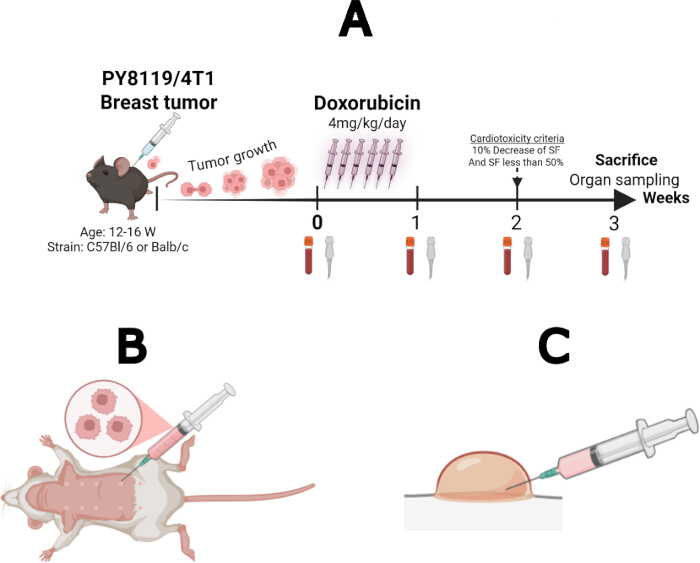
Figure 1: Representative illustrations of the experimental protocol. (A) Schematic overview of the experimental protocol. Created with BioRender (license no. JE29USE7LU). (B) Schematic representation of subcutaneous tumor inoculation. Created with BioRender (license no. SO29JHW7R6). (C) Schematic representation of successful subcutaneous tumor implantation. Created with BioRender (license no. SO29JHW7R6). Please click here to view a larger version of this figure.
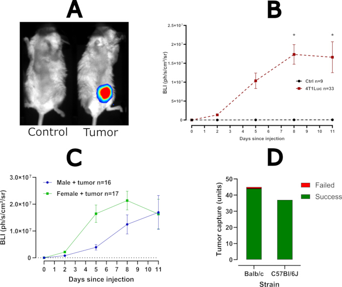
Figure 2: Bioluminescence imaging and tumor implantation rate. (A) Representative bioluminescence images of a control mouse (left) and a tumor-bearing mouse (right). (B) Bioluminescence signal over time (mean ± SEM; repeated-measures two-way analysis of variance [ANOVA]; *P < 0.05). (C) Comparison of bioluminescence signals between male and female Balb/c mice (mean ± SEM). (D) Tumor implantation success rates in both mouse strains. Please click here to view a larger version of this figure.
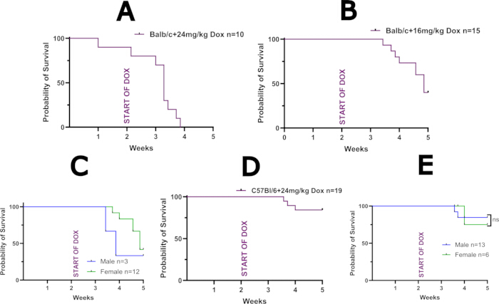
Figure 3: Survival analysis of tumor-bearing mice. (A) Kaplan–Meier survival curve of Balb/c mice treated with 24 mg/kg doxorubicin (Dox). (B) Kaplan–Meier survival curve of Balb/c mice treated with 16 mg/kg Dox. (C) Kaplan–Meier survival curve comparing male and female Balb/c mice treated with 16 mg/kg Dox. No statistical analysis was performed because of the limited number of male mice. (D) Kaplan–Meier survival curve of C57BL/6J mice treated with 24 mg/kg Dox. (E) Kaplan–Meier survival curve comparing male and female C57BL/6J mice treated with 24 mg/kg Dox (log-rank test, P = 0.86). Please click here to view a larger version of this figure.
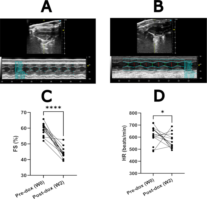
Figure 4: Echocardiographic assessment of doxorubicin-induced cardiotoxicity. (A) Representative M-mode echocardiogram from a healthy mouse. (B) Representative M-mode echocardiogram from a mouse with doxorubicin-induced cardiotoxicity. (C) Fractional shortening (FS) in C57BL/6J mice before and 14 days after doxorubicin treatment. (D) Heart rate (HR) in C57BL/6J mice before and 14 days after doxorubicin treatment. Data are presented for n = 17 mice. Statistical comparisons were performed using Welch's t-test. *P < 0.05; ****P ≤ 0.0001. Abbreviations: FS, fractional shortening; HR, heart rate. Please click here to view a larger version of this figure.
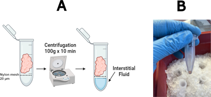
Figure 5: Organ sampling protocol. (A) Schematic representation of the tumor interstitial fluid collection protocol. Created with BioRender (license no. HV29USD91R). (B) Representative image showing collection of tumor interstitial fluid during organ sampling. Please click here to view a larger version of this figure.
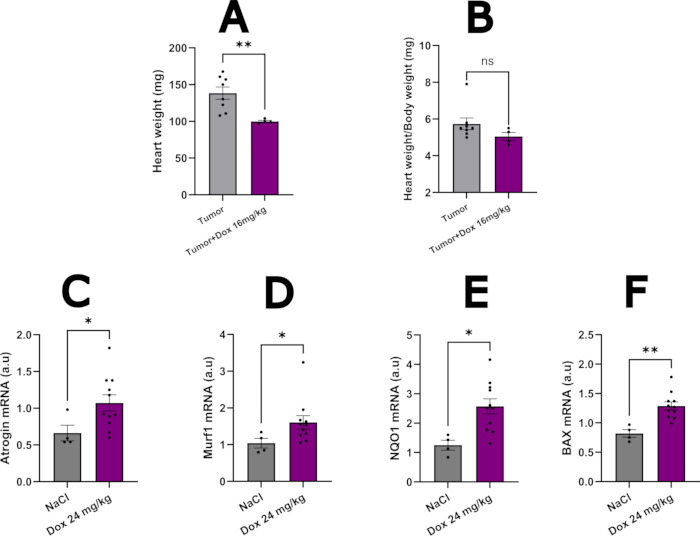
Figure 6: Doxorubicin-induced cardiotoxicity. (A) Heart weight measured 3 weeks after initiation of 16 mg/kg doxorubicin treatment in tumor-bearing Balb/c mice. (B) Heart weight normalized to total body weight measured 3 weeks after initiation of 16 mg/kg doxorubicin treatment in tumor-bearing Balb/c mice. (C) Atrogin mRNA expression in the myocardium of C57BL/6J mice 1 week after initiation of 24 mg/kg doxorubicin treatment. (D) Murf1 mRNA expression in the myocardium of C57BL/6J mice 1 week after initiation of 24 mg/kg doxorubicin treatment. (E) NQO1 mRNA expression in the myocardium of C57BL/6J mice 1 week after initiation of 24 mg/kg doxorubicin treatment. (F) BAX mRNA expression in the myocardium of C57BL/6J mice 1 week after initiation of 24 mg/kg doxorubicin treatment. Statistical analyses were performed using Welch's t-test or the Mann–Whitney U test, as appropriate based on data distribution. ns, P > 0.05; *P < 0.05; **P < 0.01. Please click here to view a larger version of this figure.
| Mice strain | Balb/c | C57Bl/6J |
| Cell line used | 4T1Luc | PY8119 |
| Amount of cell injected | 200 000 | 200 000 |
| Sex used | Male and female | Male and female |
| Number of injection days | 4 | 6 |
| Daily dose | 4 mg/kg | 4 mg/kg |
| Cumulative Doxorubicin dose | 16 mg/kg | 24 mg/kg |
| Ketamine dose | 40 mg/kg | 80 mg/kg |
| Cardiotoxicity incidence | >90% | >90% |
| Survival rate | 19/45 | 16/19 |
Table 1: Key protocol parameters for each mouse strain. Comparison of the principal experimental variables and reagent concentrations used in the Balb/c and C57BL/6J mouse protocols.
Supplementary Table 1: Animal welfare endpoints. Please click here to download this file.
Supplementary Table 2: Literature overview of 4T1 experimental protocols. Please click here to download this file.
Supplementary Table 3: Echocardiographic parameters measured by M-mode in 17 tumor-bearing C57BL/6J mice treated with 24 mg/kg doxorubicin. Please click here to download this file.
Discussion
Here, a model of doxorubicin-induced cardiotoxicity in breast tumor-bearing mice was developed and is believed to be of strong clinical relevance for future studies in cardio-oncology. The aim was to mimic the clinical situation of a patient’s path since their arrival at the hospital with a diagnosed cancer up to treatment with a potentially cardiotoxic chemotherapy. The protocol follows a pragmatic design and uses immunocompetent mice available worldwide, thereby allowing other teams to reproduce it in other labs. Its high tumor-implantation success rate makes it a strong candidate for future cardio-oncological tox studies.
Common pitfalls in this protocol include performing echocardiography on mice with a low heart rate, which can result in an artificially low fractional shortening (FS). Allowing the mice to recover from the initial negative chronotropic effects of the anesthetics before echocardiographic assessment helps ensure accurate FS measurements. In Balb/c mice, further reduction of the anesthetic dose may be necessary if mortality remains high. The same experimenter should perform all echocardiographic assessments throughout the study because inter-observer variability can be significant43. For tumor implantation, withdrawing the syringe during injection can produce a line-shaped tumor, which may grow larger than tumors generated by proper injection. Injection into the correct mammary nipple is also critical, as placement in the upper abdominal nipple can interfere with echocardiographic assessment during later stages of tumor development. Manual counting of cells in the suspension and thorough mixing before injection are recommended to ensure that each mouse receives a comparable number of cells, thereby minimizing variation in tumor size at the end of the protocol. When weekly blood sampling is required, the volume collected should be carefully controlled because mice become more fragile following doxorubicin treatment. A maximum total blood volume of 50 µL at each sampling time point is recommended, as larger volumes can increase mortality during the protocol. The frequency and volume of blood collection should also comply with local animal ethics requirements and institutional animal facility guidelines. Replacing the sampled blood volume with an equivalent volume of saline administered subcutaneously can help compensate for fluid loss after blood collection.
To compare this dox-procedure, a recently published review on animal models of AIC observed that most preclinical models (88%) used repeated injections of Dox and outcome measurements (often via echocardiography) several weeks later29. Most of the articles reported the use of the C57Bl/6J strain (73.8%), probably due to its robustness and low cost29. Unfortunately, a significant number of studies disregarded the impact of a tumor on the biology of the animal before injecting ACs17,29. Only 46 out of 736 studies reported tumor-bearing models, 12 of them used the 4T1 cell line (triple-negative-like breast cancer) in Balb/c mice, and only one was a model of breast cancer in C57Bl/6J mice (more specifically EGFR2-positive-like breast cancer)29. Another study used C57Bl/6J with an EGFR-2-positive-like breast cancer, but did not allow tumor growth to occur before the start of chemotherapy, which limits the translational aspect of this model32.
Xenograft models using immunodeficient mice allow the use of human tumor cell lines but lack a functional immune system, which limits the study of mechanisms of immune-mediated cardiotoxicity and is a major limitation given the growing relevance of immune checkpoint inhibitor-related cardiotoxicity. Large animal models are emerging in the field of cardio-oncology, but will always have the drawbacks of high cost and limited genetic engineering capability44. In vitro and 2D/3D human cell-based systems (e.g., human iPSC-derived cardiomyocytes, cardiac organoids) enable high-throughput mechanistic and toxicological screening but cannot recapitulate the integrated, whole-organism physiology (hemodynamics, neurohormonal regulation, immune crosstalk) captured by in vivo models. Compared with these approaches, this syngeneic, immunocompetent, tumor-bearing model offers a more clinically relevant context to study the interaction between tumor burden and chemotherapy-induced cardiotoxicity, at the cost of greater biological variability, higher mortality in some strains, and lower throughput than simpler in vitro or non-tumor-bearing in vivo models.
Non-tumor-bearing AC cardiotoxicity models, mostly used to date, offer high reproducibility, low cost, and easier echocardiographic and survival assessments but do not capture the systemic effects of tumor burden, cancer-associated inflammation, and cachexia on the heart, which are increasingly recognized as relevant confounders of cardiotoxicity in patients17,45. Moreover, metabolic reprogramming of the heart and, hence, its contractile function can be altered by secreted oncometabolites, such as D-2-hydroxyglutarate or succinate14. Therefore, if a cardio-oncological model is to be built for cardiac biochemical analysis using various omics (proteomics and metabolomics, for example), the presence of the tumor before chemotherapy is essential. There is also some evidence that the tumor by itself can induce overt cardiac dysfunction, although this is not consistent in the literature. While some models demonstrated a detrimental effect of tumor inoculation on FS or electrical conduction parameters, others have found that the presence of the tumor protected mice from the remodeling induced by a transverse aortic constriction11,13,46. Non-tumor-bearing animals are also needed, depending on the preliminary hypothesis. They constitute the only setting in which the direct cardiotoxic effect of the chemotherapeutic agent can be isolated from the confounding influence of the tumor itself, an essential prerequisite for establishing causality and elucidating drug-specific mechanisms of action. These animals will serve as controls for the anticancer therapy effect and its interaction with the tumor on cardiotoxicity. Without this comparator, it would be impossible to disentangle which component of the observed cardiac phenotype is attributable to the drug, to the tumor, or to their interaction. This distinction is critical not only for mechanistic understanding but also for the rational design of cardioprotective strategies. It is therefore important to acknowledge that each of these models addresses distinct and complementary scientific questions and that neither model alone is sufficient to fully recapitulate the complexity of clinical cardio-oncology; a combined or sequential use of both is often required to draw robust mechanistic and translational conclusions.
In this protocol, cardiotoxicity was defined according to the 2022 ESC Guidelines in Cardio-oncology, which define moderate asymptomatic cancer therapy-related cardiac dysfunction as a left ventricular ejection fraction (LVEF) reduction of >10% with an LVEF between 40% and 49%16. However, accurate LVEF quantification requires a 3D module on the ultrasound39. To avoid incorrect systolic cardiac function assessment, and to keep a simple protocol that could be implemented in labs using less recent ultrasounds, it was preferred to use FS as a marker of cardiac dysfunction, which is correctly and easily obtainable through M-mode analysis in mice, with the same criteria as imposed by the ESC. Using this protocol, 90% of surviving mice meet the criteria for cardiotoxicity, making it a strong candidate for future translational studies. The cardiotoxic effect of Dox was confirmed through macroscopic evaluation (decreased heart weight) in Balb/c mice. No tumor-only group in C57Bl/6 mice was included here; hence, no comparison in this strain is available yet. Moreover, in non-tumor-bearing C57Bl/6 mice, the dose of Dox induced changes consistent with previous literature on the mechanisms of anthracycline cardiotoxicity: increased atrophy, oxidative stress, and apoptosis19,20.
However, more modern variables used in patients, such as speckle-tracking global longitudinal strain (GLS), were not quantified here for technical reasons. If this is locally feasible, it would be of added value to quantify GLS, since it is generally considered more sensible to detect early cardiac damage23. Circulating cardiac biomarkers such as troponin can also be assessed to improve the certainty of cardiotoxicity detection; however, the amount of plasma traditionally required for these analyses in mice can be a limiting factor due to the low total blood volume these animals have. Moreover, the frailty of these animals after Dox treatment can increase mortality rates during or after blood sampling. One last limiting factor is the sensitivity of these assays for such biomarkers, which can often hamper interpretation.
Although the same experienced experimenter did all ultrasound acquisitions, a high week-to-week variation of the same Balb/c mice was observed, even without treatment. This can be explained by the strain's sensitivity to ketamine anesthesia47. Indeed, high mortality rates during procedures with anesthesia, leading to low numbers of animals reaching the end of the protocol, were initially noticed, which led to a reduction of the dose of ketamine used from 80 mg/kg to 40 mg/kg for Balb/c mice. Looking at the survival graphs of Balb/c with Dox, it is observed that some mortality occurs at the point of anesthesia (weekly echocardiography), but the majority of deaths occur in the animal facility without any active procedure, emphasizing that Dox is the main contributor to mortality in those mice. Nevertheless, it is important to keep in mind the anesthesia-related death when interpreting survival graphs for this model when comparing two or more strains. To reduce the risk of anesthesia-related fatality, one could also do echocardiography on conscious mice, but this requires training and acclimatization before inclusion of the mice in the protocol. Regarding Dox, even at reduced doses, almost half of the Balb/c mice did not reach the end of the protocol. Induction of cardiotoxicity with even lower doses of Dox could be attempted, with the risk of reducing the number of animals meeting the criteria for cardiotoxicity. Another strategy could be to allow more time for the animal to recover after each Dox injection, which was not performed here but has evidence of improved survival in the literature29.
The observations made while building this model, that Balb/c mice are more sensitive to Dox than C57Bl/6J mice, and that females are more resistant to Dox than males, are in line with previous literature29. Moreover, the similar and reproducible results obtained in both mouse strains further confirm the robustness of this proposed model. By reaching high cumulative doses through moderately high daily doses over a short period of time, high rates of cardiotoxicity with high survival were obtained. Building a breast cancer model (PY8119 is a triple-negative-like breast cancer) in C57Bl/6J will be of great use for further studies, since these mice represent one of the main genetic backgrounds for transgenic models34. The high success rate of tumor inoculations, supporting the effectiveness of the chosen injection method, is also promising for this model. Also, the high Dox tolerance of these mice opens the door to longer studies with Dox re-challenges to mimic the frequent clinical situation of secondary cancer or metastasis. Lastly, the organ sampling protocol leads to the collection of all solid organs, tumors, and their interstitial fluid, and bone marrow and its stroma, which allows for very thorough model analysis.
This model has some limitations. Due to its pragmatic design and to avoid invasive surgery, the precise location of the tumor (i.e., the correct mammary gland rather than subcutaneous fat) cannot be accurately controlled with a subcutaneous injection, which can be of great importance depending on the scientific question being studied48. If precise cancer-site injection is needed, more extensive surgery may be better suited to perform an orthotopic injection, although this seems of true importance mainly if tumor biochemical analysis is planned, since volume, latency, and incidence have similar results regardless of the method of injection48. One could also argue that the use of only ACs is not perfectly correlated with the clinical situation, where patients often receive combination regimens, for example, cyclophosphamide or cisplatin. This could indeed be the next design for this protocol; however, dose adaptation for each chemotherapy used should be carefully performed. Moreover, over 50% mortality in Balb/c mice 3 weeks after the start of Dox treatment remains very high; dose reduction could improve survival, perhaps at the cost of less pronounced cardiotoxicity. Also, regarding the doses used in this model, it should be noted that they are higher than those commonly used in humans today, which may limit the translational potential of this model.
Disclosures
The authors declare no conflict of interest with the manuscript.
Acknowledgements
Thank you to the animal facility team for their help with daily monitoring of animal welfare. Thank you to Léanne Borges, Elias Ait Assa, and Clémence Chabaud for the help with building the animal protocol. For the gift of the PY8119 cells, thank you to Dr Julie Helft from the INSERM Unit 932. Special thanks to Dr Sylvain Provost and his student Mel Gavens for their help with bioluminescence.
Materials
| Name | Company | Catalog Number | Comments |
|---|---|---|---|
| 4T1Luc cells | ATCC | CRL-2539 | |
| Doxorubicin powder | Cliniscience | 23214-92-8 | |
| Echocardiograph | Visual Sonics | VEVO LT-200 | |
| Luciferin | Perkin Elmer | E1605 | |
| Nylon mesh | Merck | NY2002500 | |
| PhotonImager Optima | BioSpace Lab | ||
| PY8119 cells | Gift from Dr Julie Helft |
References
- Meijers WC, et al. Heart failure stimulates tumor growth by circulating factors. Circulation. 2018;138(7):678-691.
- de Wit S, et al. Pressure overload-induced cardiac hypertrophy stimulates tumor growth in tumor-prone Apc(Min) mice. Circ Heart Fail. 2023;16(9):e010740.
- Avraham S, et al. Early cardiac remodeling promotes tumor growth and metastasis. Circulation. 2020;142(7):670-683.
- Langier Goncalves I, et al. Heart failure promotes cancer progression in an integrin β1-dependent manner. Int J Mol Sci. 2023;24(24):17367.
- Koelwyn GJ, et al. Myocardial infarction accelerates breast cancer via innate immune reprogramming. Nat Med. 2020;26(9):1452-1458.
- Wong J, Soh CH, Wang B, Marwick T. Long-term risk of heart failure in adult cancer survivors: a systematic review and meta-analysis. Heart. 2024;110(19):1188-1195.
- Kuiper JG, et al. Risk of heart failure among colon and rectal cancer survivors: a population-based case-control study. ESC Heart Fail. 2022;9(4):2139-2146.
- Mamidanna R, Nachiappan S, Bottle A, Aylin P, Faiz O. Defining the timing and causes of death amongst patients undergoing colorectal resection in England. Colorectal Dis. 2016;18(6):586-593.
- Armstrong GT, et al. Aging and risk of severe, disabling, life-threatening, and fatal events in the Childhood Cancer Survivor Study. J Clin Oncol. 2014;32(12):1218-1227.
- Herrmann J. Adverse cardiac effects of cancer therapies: cardiotoxicity and arrhythmia. Nat Rev Cardiol. 2020;17(8):474-502.
- Kelm NQ, Straughn AR, Kakar SS. Withaferin A attenuates ovarian cancer-induced cardiac cachexia. PLoS One. 2020;15(7):e0236680.
- Graca FA, et al. Progressive development of melanoma-induced cachexia differentially impacts organ systems in mice. Cell Rep. 2023;42(1):111934.
- Xiong S, et al. Development of an efficient mice model of cancer-associated cardiac cachexia. Anim Models Exp Med. 2025; 8(11):1960-1970.
- Karlstaedt A, Moslehi J, de Boer RA. Cardio-onco-metabolism: metabolic remodelling in cardiovascular disease and cancer. Nat Rev Cardiol. 2022;19(6):414-425.
- Murphy KT. The pathogenesis and treatment of cardiac atrophy in cancer cachexia. Am J Physiol Heart Circ Physiol. 2016;310(4):H466-H477.
- Lyon AR, et al. 2022 ESC Guidelines on cardio-oncology developed in collaboration with the European Hematology Association (EHA), the European Society for Therapeutic Radiology and Oncology (ESTRO) and the International Cardio-Oncology Society (IC-OS). Eur Heart J. 2022;43(41):4229-4361.
- Ghigo A, et al. Update on preclinical models of cancer therapy-related cardiac dysfunction: challenges and perspectives. A scientific statement of the Heart Failure Association (HFA) of the ESC, the ESC Council of Cardio-Oncology, and the European Association of Preventive Cardiology. Eur J Heart Fail. 2025;27(6):1028-1046.
- Cagnazzo R, et al. Mismatch between preclinical cardio-oncology and clinical oncology research. JACC CardioOncol. 2025;7(6):770-773.
- Mattioli R, et al. Doxorubicin and other anthracyclines in cancers: activity, chemoresistance and its overcoming. Mol Aspects Med. 2023;93:101205.
- Martins-Teixeira MB, Carvalho I. Antitumour anthracyclines: progress and perspectives. ChemMedChem. 2020;15(11):933-948.
- Fabiani I, Chianca M, Cipolla CM, Cardinale DM. Anthracycline-induced cardiomyopathy: risk prediction, prevention and treatment. Nat Rev Cardiol. 2025;22(8):551-563.
- Rivero-Santana B, et al. Anthracycline-induced cardiovascular toxicity: validation of the Heart Failure Association and International Cardio-Oncology Society risk score. Eur Heart J. 2025;46(3):273-284.
- Camilli M, Cipolla CM, Dent S, Minotti G, Cardinale DM. Anthracycline cardiotoxicity in adult cancer patients: JACC: CardioOncology state-of-the-art review. JACC CardioOncol. 2024;6(5):655-677.
- Díaz-Guerra A, et al. Anthracycline cardiotoxicity induces progressive changes in myocardial metabolism and mitochondrial quality control. JACC CardioOncol. 2024;6(2):217-232.
- Omland T, et al. Sacubitril-valsartan and prevention of cardiac dysfunction during adjuvant breast cancer therapy: the PRADA II randomized clinical trial. Circulation. 2025: 152(16):1136-1145.
- Henriksen PA, et al. Multicenter, prospective, randomized controlled trial of high-sensitivity cardiac troponin I-guided combination angiotensin receptor blockade and beta-blocker therapy to prevent anthracycline cardiotoxicity: the Cardiac CARE trial. Circulation. 2023;148(21):1680-1690.
- Sobiborowicz-Sadowska AM, Kamińska K, Cudnoch-Jędrzejewska A. Neprilysin inhibition in the prevention of anthracycline-induced cardiotoxicity. Cancers (Basel). 2023;15(1):312.
- Stansfeld A, et al. Pharmacological strategies to reduce anthracycline-associated cardiotoxicity in cancer patients. Expert Opin Pharmacother. 2022;23(14):1641-1650.
- Li M, et al. Optimizing dose selection for doxorubicin-induced cardiotoxicity in mice: a comprehensive analysis of single and multiple-dose regimens. Eur J Pharmacol. 2025;1003:177883.
- Gambardella J, et al. Infiltrating macrophages amplify doxorubicin-induced cardiac damage: role of catecholamines. Cell Mol Life Sci. 2023;80(11):323.
- Chen Y, et al. Cysteine imaging reveals early redox dysregulation and identifies gnetol as a ferroptosis-modulating agent in doxorubicin cardiotoxicity. Redox Biol. 2026;92:104131.
- Luo W, et al. Critical role of the cGAS-STING pathway in doxorubicin-induced cardiotoxicity. Circ Res. 2023;132(11):321587.
- Mouse Genome Sequencing Consortium. Initial sequencing and comparative analysis of the mouse genome. Nature. 2002;420(6915):520-562.
- Mekada K, Yoshiki A. Substrains matter in phenotyping of C57BL/6 mice. Exp Anim. 2021;70(2):145-160.
- Yin L, Duan JJ, Bian XW, Yu SC. Triple-negative breast cancer molecular subtyping and treatment progress. Breast Cancer Res. 2020;22(1): doi.org/10.1186/s13058-020-01296-5.
- Akbay EA, et al. D-2-hydroxyglutarate produced by mutant IDH2 causes cardiomyopathy and neurodegeneration in mice. Genes Dev. 2014;28(5):479-490.
- Springer J, et al. Prevention of liver cancer cachexia-induced cardiac wasting and heart failure. Eur Heart J. 2014;35(14):932-941.
- Herndon ME, et al. The highly metastatic 4T1 breast carcinoma model possesses features of a hybrid epithelial/mesenchymal phenotype. Dis Model Mech. 2024;17(9):dmm050771.
- Lindsey ML, Kassiri Z, Virag JAI, De Castro Brás LE, Scherrer-Crosbie M. Guidelines for measuring cardiac physiology in mice. Am J Physiol Heart Circ Physiol. 2018;314(4):H733-H752.
- Zacchigna S, et al. Towards standardization of echocardiography for the evaluation of left ventricular function in adult rodents: a position paper of the ESC Working Group on Myocardial Function. Cardiovasc Res. 2021;117(1):43-59.
- Assad N, et al. Crosstalk between circulating DPP3 and immune cells in the context of cardio-systemic stress. iScience. 2026;29(3):115114.
- Motulsky HJ. GraphPad Statistics Guide. 2016. http://www.graphpad.com/guides/prism/10/statistics/index.htm.
- Garcia-Elcano L, et al. Accurate estimation of interobserver variability in echocardiography segmentation measured from 35 experts and impact on derived clinical metrics. Eur Heart J Cardiovasc Imaging. 2025;26(Suppl 1): jeae333.044.
- Medina-Hernandez D, et al. SGLT2i therapy prevents anthracycline-induced cardiotoxicity in a large animal model by preserving myocardial energetics. JACC CardioOncol. 2025;7(2):171-184.
- Asnani A, et al. Preclinical models of cancer therapy-associated cardiovascular toxicity: a scientific statement from the American Heart Association. Circ Res. 2021;129(1):e21–e34.
- Awwad L, Shofti R, Haas T, Aronheim A. Tumor growth ameliorates cardiac dysfunction. Cells. 2023;12(14):1853.
- Levin-Arama M, et al. Subcutaneous compared with intraperitoneal ketamine-xylazine for anesthesia of mice. J Am Assoc Lab Anim Sci. 2016;55(6):794-800.
- Fleming JM, Miller TC, Meyer MJ, Ginsburg E, Vonderhaar BK. Local regulation of human breast xenograft models. J Cell Physiol. 2010;224(3):795-806.
Reprints and Permissions
This article has been published
Video Coming Soon