This protocol presents a standardized targeted biopsy workflow for upper gastrointestinal precancerous lesions using magnifying endoscopy with narrow-band imaging (ME-NBI). The goal is to systematically identify high-risk focal mucosal abnormalities based on predefined microscopic criteria, thereby enhancing diagnostic accuracy, improving biopsy efficiency, and minimizing unnecessary sampling.
A subscription to JoVE is required to view this content. Sign in or start your free trial.
Method Article
Standardized Magnifying Endoscopy with Narrow-Band Imaging-Guided Targeted Biopsy Workflow For Upper Gastrointestinal Precancerous Lesions
In This Article
Summary
Abstract
Although early detection of upper gastrointestinal precancerous lesions is vital, subtle mucosal variations often limit conventional endoscopic screening. To address this limitation, we present a standardized targeted biopsy protocol using magnifying endoscopy with narrow-band imaging (ME-NBI). This systematic workflow integrates key microstructural and microvascular criteria—specifically demarcation lines, irregular microsurfaces, atypical microvasculature, and marked glandular distortion—to identify high-risk focal areas for mucosal sampling. In a prospective clinical evaluation involving 161 patients, this standardized ME-NBI workflow was compared with a conventional white-light endoscopic assessment. The application of this protocol significantly enhanced the first-pass positive rate, overall biopsy positivity, and the diagnostic yield of high-risk lesions per biopsy sample, while simultaneously reducing the total number of biopsy samples required per patient. Furthermore, the standardized workflow demonstrated high diagnostic performance, achieving an area under the curve (AUC) of 0.928, and yielded strong interobserver agreement. By establishing objective criteria for target selection, this reproducible workflow reduces operator subjectivity, supports early neoplasia detection, and provides a practical framework for clinical implementation.
Introduction
Esophageal and gastric cancers remain leading causes of gastrointestinal cancer-related mortality worldwide1,2. Despite recent advancements in diagnostic and therapeutic modalities, early detection remains the most critical factor in improving patient survival and quality of life3. The accurate identification and timely management of upper gastrointestinal precancerous lesions—spanning mild, moderate, and severe dysplasia—are therefore essential4. However, under conventional endoscopy, these precursor lesions frequently present with only subtle mucosal changes, poorly defined margins, and heterogeneous distributions. Consequently, accurate diagnosis relies heavily on the endoscopist’s ability to visually identify and sample the most representative high-risk areas against a complex background mucosa5.
In standard clinical practice, biopsies guided by conventional white-light endoscopy (WLE) remain highly subjective. Endoscopists typically select targets based on macroscopic features such as surface erythema, depression, hypopigmentation, or nodularity, and may resort to random multi-quadrant sampling for extensive lesions. This experience-based paradigm has significant limitations. Given the patchy distribution and cellular heterogeneity of dysplasia, the highest-grade neoplastic cells often occupy only a restricted fraction of the total lesion area6,7. Because conventional WLE cannot readily resolve these subtle histological variations, empirical biopsy protocols frequently result in sampling error, pathological under-grading, and missed diagnoses. Furthermore, the necessity for multiple or repeat biopsies increases procedural time, exacerbates patient discomfort, and induces mucosal scarring that complicates subsequent endoscopic surveillance or resection8.
The rationale behind using magnifying endoscopy combined with narrow-band imaging (ME-NBI) is its ability to address these optical limitations. Recent assessments of narrow-band imaging algorithms support the use of advanced optical techniques for resolving subtle microvascular and mucosal variations beyond conventional white-light imaging9. Furthermore, comparative evidence underscores that hyperspectral reconstruction of standard white-light endoscopy substantially improves the precise segmentation of early precancerous boundaries10. By filtering broadband white light and leveraging the specific absorption peaks of hemoglobin, ME-NBI significantly enhances the optical contrast of the superficial mucosal microstructure and microvascular architecture. This optical magnification enables gastroenterologists to distinctly visualize morphological aberrations in epithelial gland openings and capillary loops. However, despite its diagnostic utility, a universally standardized workflow for its application in targeted biopsies remains undefined. Decisions regarding when to initiate magnified observation, how to weight specific morphological criteria, and precisely where to biopsy rely predominantly on subjective physician experience. This lack of standardization leads to notable discrepancies in diagnostic accuracy and interobserver agreement, particularly among endoscopists with varying levels of expertise11.
The overall goal of this method is to establish and systematically evaluate a reproducible, evidence-guided workflow for targeted biopsies of upper gastrointestinal precancerous lesions using ME-NBI. This protocol clearly defines the sequential screening steps, the indications for magnified observation, the criteria for recognizing high-risk focal areas, and the rules for optimizing biopsy allocation. Operationally, high-risk focal regions are delineated by a distinct demarcation line, which encapsulates localized structural disruptions. Within these demarcated borders, the workflow directs investigators to identify irregular microsurface patterns, microvascular abnormalities, and severe glandular distortion as actionable indicators for targeted sampling. The primary advantage of this technique over conventional empirical biopsy is its ability to reduce subjective variation, thereby increasing the detection rate of high-risk histopathology and improving the diagnostic yield per biopsy sample while reducing unnecessary sampling. Ultimately, this workflow provides clinicians with a standardized framework to improve diagnostic consensus across clinical teams, offering an objective methodology for endoscopy facilities aiming to improve the early detection of gastrointestinal neoplasia12.
Access restricted. Please log in or start a trial to view this content.
Protocol
This prospective investigation received formal approval from the Institutional Review Board and Ethics Committee of the Central Hospital Affiliated to Shandong First Medical University (Approval No. XAU7781902). Written informed consent was obtained from all individual participants or their legal guardians prior to initiating any endoscopic examinations or mucosal biopsy procedures.
1. Patient enrollment and case selection
- Recruit patients aged over 18 years who are referred for a diagnostic upper gastrointestinal endoscopy.
- Ensure the enrolled patients present with upper gastrointestinal mucosal abnormalities, possess a high-risk background for precancerous lesion development, or have suspicious lesions requiring further histopathological evaluation.
- Exclude patients with a previous diagnosis of upper gastrointestinal malignancy, active gastrointestinal bleeding, or severe thrombocytopenia that makes them unsuitable for biopsy.
- Allocate eligible patients to either the conventional white-light assessment group or the standardized ME-NBI workflow group using a computer-generated randomization sequence to ensure balanced cohorts and minimize selection bias.
NOTE: Figure 1 illustrates the overall workflow of patient selection, grouping, and the distinct observational phases between conventional white-light endoscopy and ME-NBI targeted biopsy13,14.
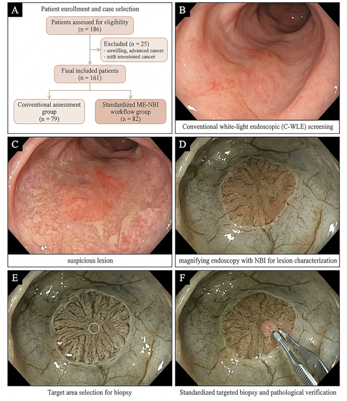
Figure 1: Standardized endoscopic workflow for targeted biopsy of upper gastrointestinal precancerous lesions using magnifying endoscopy with narrow-band imaging. (A) Patient enrollment and study grouping; (B) Conventional white-light endoscopic (C-WLE) screening view; (C) Suspicious precancerous lesion identified under white-light endoscopy; (D) Magnifying endoscopy with narrow-band imaging showing lesion microsurface and microvascular patterns; (E) Targeted biopsy site selection based on ME-NBI findings; (F) Standardized targeted biopsy workflow with pathological verification. Please click here to view a larger version of this figure.
2. Standardized endoscopic examination
- Initiate the upper gastrointestinal examination using a routine conventional white-light endoscopic (C-WLE) system.
- Observe all anatomical regions of the upper gastrointestinal tract sequentially and systematically.
- Record the precise anatomical position, estimated size, color changes, surface roughness, minor protrusions or depressions, and margin clarity of any identified suspicious lesions.
- Switch the endoscopic system to the magnifying endoscopy with narrow-band imaging (ME-NBI) mode immediately after recognizing potentially precancerous lesions or high-risk background mucosa under white light.
- Examine the focal areas in detail, specifically focusing on lesion borders, microsurface structures, microvascular morphologies, and glandular opening architectures.
- Assess the background mucosal features using ME-NBI to establish an internal baseline when evaluating cases with unclear focal lesions or widespread atrophic changes.
3. Target area selection and biopsy execution
- Select the targeted biopsy site strictly based on the following integrated ME-NBI criteria. Classify a focal mucosal region as a high-risk optical impression when a clear demarcation line encapsulates a localized area presenting with a synchronous combination of irregular microsurface patterns, asymmetric or tortuous microvascular arrangements, and marked glandular structural distortion.
- Identify and target the focal area that visually exhibits the most severe or atypical structural abnormality when examining lesions with high internal heterogeneity.
- Obtain mucosal samples directly from these identified high-risk targeted regions using biopsy forceps.
NOTE: The standardized definitions, variables, and operational coding criteria for all endoscopic features are detailed in Table 1. Representative macroscopic and microscopic appearances used to guide target selection are provided in Figure 2 and Figure 315.
Table 1: Standardized endoscopy features definitions and coding criteria for targeted biopsy of upper gastrointestinal precancerous lesions. Please click here to download this Table.
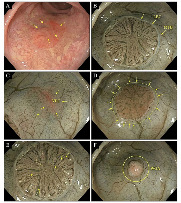
Figure 2: Representative endoscopic features of upper gastrointestinal precancerous lesions under conventional white-light endoscopy and magnifying endoscopy with narrow-band imaging. (A) Conventional white-light endoscopic appearance of a suspicious upper gastrointestinal precancerous lesion; (B) Magnifying NBI image showing intestinal metaplasia-related microface features with light blue crest and marginal turbid band; (C) Magnifying NBI view of focal vascular or epithelial change within a high-risk mucosal area; (D) Magnifying NBI image demonstrating a demarcation line and heterogeneous microsurface architecture; (E) Magnifying NBI view highlighting irregular microsurface and microvascular patterns optimal for targeted biopsy; (F) Magnifying NBI appearance of a localized abnormal glandular structure associated with high-risk pathology. Please click here to view a larger version of this figure.
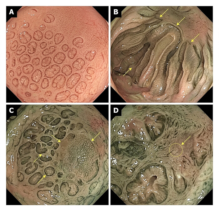
Figure 3: Representative glandular and microsurface patterns of upper gastrointestinal precancerous lesions under magnifying endoscopy with narrow-band imaging. (A) Regular glandular and microsurface pattern in low-risk background mucosa; (B) Intestinal metaplasia-associated glandular pattern with mildly altered but preserved microsurface architecture; (C) Heterogeneous microstructural pattern with focal glandular distortion in a suspicious precancerous lesion; (D) High-risk focal glandular abnormality selected as the prioritized targeted biopsy site. Please click here to view a larger version of this figure.
4. Histopathological evaluation and data analysis
- Grade all obtained biopsy specimens independently in accordance with the revised Vienna classification system for gastrointestinal epithelial neoplasia.
- Engage a third independent pathologist to re-evaluate the specimen and reach a definitive consensus if a grading disagreement arises between the initial two raters. Ensure this adjudicating pathologist remains strictly blinded to all clinical data, prior endoscopic impressions, and assigned procedural groupings by evaluating de-identified slides coded solely with randomized alphanumeric identifiers.
- Calculate the targeted biopsy efficiency indicators, including the average number of biopsy blocks per patient, the average number per lesion, and the diagnostic positivity rate per biopsy block.
- Extract the key endoscopic features detected via C-WLE and ME-NBI, and perform univariate and multivariate logistic regression analyses to determine independent predictors of high-risk pathology.
- Construct Receiver Operating Characteristic (ROC) curves and calculate the Area Under the Curve (AUC) to assess the diagnostic discrimination power of the workflow.
- Evaluate interobserver agreement for both endoscopic interpretation and the initial histopathological grading using the Kappa statistic, and apply Spearman's rank correlation to assess the relationship between specific image features and histopathological severity.
- Perform all statistical comparisons using appropriate tests, including Student's t-tests, Pearson chi-squared tests, or Fisher's exact tests. Define the threshold for statistical significance a priori at P < 0.05.
Access restricted. Please log in or start a trial to view this content.
Results
Baseline clinicopathological and lesion characteristics
Prior to formal enrollment, 25 candidates were specifically excluded from the initial eligibility pool of 186 patients due to preexisting conditions precluding safe and meaningful targeted biopsies, namely a previous diagnosis of upper gastrointestinal tract malignancy, active gastrointestinal bleeding, or severe thrombocytopenia. A total of 161 patients were included in the study. Of these, 79 underwent conventional endoscopic evaluation, and 8...
Access restricted. Please log in or start a trial to view this content.
Discussion
Accurate assessment of upper gastrointestinal precancerous lesions is vital for early diagnosis and improved patient outcomes. Historically, the lack of uniform criteria for targeted sampling and unstandardized biopsy procedures resulted in highly variable diagnostic yields and operator dependencies16. The standardized ME-NBI protocol detailed here addresses this deficiency by defining a clear, reproducible reference point for biopsy selection. A critical step in this method involves the systemati...
Access restricted. Please log in or start a trial to view this content.
Disclosures
The authors have no conflicts of interest to disclose.
Acknowledgements
We would like to express our sincere gratitude to the clinical and nursing staff at the endoscopy center of the Central Hospital Affiliated to Shandong First Medical University for their invaluable assistance during the patient recruitment and endoscopic procedures. We also sincerely thank all the patients who participated in this study. This research received no specific grant from any funding agency in the public, commercial, or not-for-profit sectors.
Access restricted. Please log in or start a trial to view this content.
Materials
| Name | Company | Catalog Number | Comments |
|---|---|---|---|
| Disposable Biopsy Forceps | Olympus Medical Systems | EndoJaw FB-230U | Collection of targeted mucosal biopsy specimens from high-risk areas identified by ME-NBI |
| Excel | Microsoft Corporation | Microsoft 365 (https://www.microsoft.com) | Data entry, organization, and management of endoscopic, pathological, and clinical variables |
| Hematoxylin and Eosin Stain Kit | Vector Laboratories | Catalog Number: H-3502 | Histopathological staining and morphological evaluation of tissue sections |
| Magnifying Gastroscope | Olympus Medical Systems | GIF-H290Z | Detailed assessment of lesion borders, microsurface patterns, microvascular patterns, and glandular architecture for targeted biopsy |
| Materials / Software | Provider | Version / Identifier | Purpose |
| Rotary Microtome | Leica Biosystems | RM2235 | Preparation of serial tissue sections for histological examination |
| SPSS Statistics Software | IBM Corporation | Version 26.0 (https://www.ibm.com/spss) | Statistical comparisons, logistic regression, ROC curve analysis, inter-observer agreement analysis, and correlation analysis |
| Tissue Embedding Paraffin | Leica Biosystems | Catalog Number: 39601006 | Tissue dehydration, embedding, and preparation for sectioning |
| Video System Center | Olympus Medical Systems | EVIS LUCERA ELITE CV-290 | Routine upper gastrointestinal examination and initial identification of suspicious mucosal lesions |
References
- Zhu Y, Wu K, Wang FY. Efficacy of magnifying endoscopy with narrow-band imaging in the diagnosis of early gastric cancer and gastric intraepithelial neoplasia. Turk J Gastroenterol. 2024;35(4):299-306.
- Tao J, et al. Risk prediction model for precancerous gastric lesions based on magnifying endoscopy combined with narrow-band imaging features. Front Oncol. 2025;15:1554523.
- Niu W, Liu L, Dong Z. A deep learning model based on magnifying endoscopy with narrow-band imaging to evaluate intestinal metaplasia grading and OLGIM staging: a multicenter study. Dig Liver Dis. 2024;56(9):1565-71.
- Kanemitsu T, et al. Magnifying endoscopy with narrow-band imaging for diagnosis of subtype of gastric intestinal metaplasia. J Gastroenterol Hepatol. 2023;38(1):94-102.
- Lyu D, Zhao J, Jin HF, Lyu B. The role of endoscopic grading of gastric intestinal metaplasia (EGGIM) in assessing the extent and degree of gastric intestinal metaplasia. J Dig Dis. 2025;26(3-4):129-34.
- Dinis-Ribeiro M, et al. Management of epithelial precancerous conditions and early neoplasia of the stomach (MAPS III): European Society of Gastrointestinal Endoscopy (ESGE), European Helicobacter and Microbiota Study Group (EHMSG) and European Society of Pathology (ESP) guideline update 2025. Endoscopy. 2025;57(5):504-54.
- Morgan DR, et al. ACG clinical guideline: diagnosis and management of gastric premalignant conditions. Am J Gastroenterol. 2025;120(4):709-37.
- Leung WK, et al. Asian Pacific Association of Gastroenterology task force recommendations on surveillance for Helicobacter pylori-associated gastric premalignant conditions. Gut. 2026;75(4):685-704.
- Yang KY, et al. Assessment of narrow band imaging algorithm for video capsule endoscopy based on decorrelated color space for esophageal cancer. Cancers (Basel). 2023;15(19):4715.
- Wang YK. Hyperspectral reconstruction of white light endoscopy images for enhanced segmentation of early esophageal lesions in patients. iScience. 2026;29(5):115460.
- Moss SF, Shah SC, Tan MC, El-Serag HB. Evolving concepts in Helicobacter pylori management. Gastroenterology. 2024;166(2):267-83.
- Aoun J, et al. Optimizing endoscopic detection of precancerous gastric conditions: a single-center prospective study. Endosc Int Open. 2025;13:a25576356.
- Young E, Wan N, Philpott H, Singh R. The West catching up with the East: high-magnification NBI is accurate for the diagnosis of gastric neoplasia in a Western population. J Gastroenterol Hepatol. 2025;40(5):1128-34.
- Manti M, et al. Narrow-band imaging for the detection of early gastric cancer among high-risk patients: a systematic review and meta-analysis. Medicina (Kaunas). 2025;61(9):1613.
- Horiuchi Y, Hirasawa T, Fujisaki J. Application of artificial intelligence for diagnosis of early gastric cancer based on magnifying endoscopy with narrow-band imaging. Clin Endosc. 2024;57(1):11-7.
- Nabi Z, et al. Image-enhanced endoscopy in upper GI tract: state-of-the-art review. Indian J Gastroenterol. 2025.
- Waddingham W, Graham DG, Banks MR. Latest advances in endoscopic detection of oesophageal and gastric neoplasia. Diagnostics (Basel). 2024;14(3):301.
- Parlar K, Cakir M, Ozer O, Sharma P. Future of image enhanced endoscopy of esophageal adenocarcinoma. Clin Endosc. 2025;58(4):503-13.
- Yuan XL, et al. Effect of an artificial intelligence-assisted system on endoscopic diagnosis of superficial oesophageal squamous cell carcinoma and precancerous lesions: a multicentre, tandem, double-blind, randomised controlled trial. Lancet Gastroenterol Hepatol. 2024;9(1):34-44.
- Park JY. Image-enhanced endoscopy in upper gastrointestinal disease: focusing on texture and color enhancement imaging and red dichromatic imaging. Clin Endosc. 2025;58(2):163-80.
- Wang YK. Computer-aided endoscopic diagnostic system modified with hyperspectral imaging for the classification of esophageal neoplasms. Front Oncol. 2024;14:1423405.
- Fujishiro M. Advanced diagnostic and therapeutic endoscopy for early gastric cancer. Cancers (Basel). 2024;16(5):1039.
- Futakuchi T, et al. Texture and color enhancement imaging improves the visibility of gastric neoplasms: clinical trial with image catalogue assessment using conventional and newly developed endoscopes. BMC Gastroenterol. 2023;23(1):389.
Access restricted. Please log in or start a trial to view this content.