This protocol describes methods for fabricating verteporfin-integrated lipid nanoparticles that are activated by visible light or X-ray radiation for spatiotemporally controlled delivery of CRISPR-Cas9 ribonucleoproteins and chemotherapeutic agents, with potential translational relevance to ophthalmic and oncological applications.
Method Article
Light- and X-ray-Activated Liposomes for Controlled Gene Editing and Drug Delivery
In This Article
Summary
Abstract
Photodynamic therapy (PDT) exploits photosensitizer activation to generate reactive oxygen species (ROS), principally singlet oxygen. Beyond direct cytotoxicity, this photochemistry can be repurposed for on-demand cargo release from lipid nanocarrier systems, enabling spatiotemporal control over therapeutic delivery that is not achievable with conventional lipid nanoparticles. This protocol presents methods for fabricating and characterising two distinct verteporfin (VP)-integrated lipid nanoparticle formulations: (1) light-triggered liposomes composed of DOTAP, DOPE, cholesterol, and VP for delivery of CRISPR-Cas9 ribonucleoprotein (RNP) complexes; and (2) X-ray-triggered liposomes composed of DOTAP, DOPC, VP, and gold nanoparticles for controlled chemotherapy drug release. Upon activation at 690 nm (visible light) or by clinical X-ray radiation (6 MeV), VP generates singlet oxygen that oxidises unsaturated lipid components, destabilising the nanoparticle membrane and releasing encapsulated cargos. Protocols are provided for liposome formulation by thin-film hydration and membrane extrusion, physicochemical characterisation, light- and X-ray-triggered cargo release assessment, in vitro gene knockout in human cells, and in vivo validation using a quantitative zebrafish visual reporter system and a mouse xenograft tumour model. Representative results demonstrate knockout of up to approximately 326 slow-muscle fibres per zebrafish embryo via light activation and significant tumour growth suppression via X-ray-triggered doxorubicin release.The clinical precedent for 689–690 nm verteporfin activation in the eye motivates evaluation of this platform for ophthalmic delivery, although retinal biodistribution, pharmacokinetics, and large-animal safety remain to be established. These methods provide an experimental approach with translational potential for externally controlled therapeutic cargo release.
Introduction
Lipid nanoparticles (LNPs) have emerged as the most clinically validated non-viral delivery platform for nucleic acid therapeutics, as demonstrated by their deployment in COVID-19 mRNA vaccines1. However, conventional LNPs release their cargo upon cellular uptake through endosomal escape, offering no external control over the timing, location, or extent of payload delivery2. This absence of spatiotemporal control limits their utility in applications where precise, on-demand release is therapeutically advantageous, including site-specific gene editing, targeted chemotherapy in heterogeneous tumours, and titrated gene silencing in anatomically accessible organs such as the eye3,4.
Photodynamic therapy (PDT) provides a well-established mechanism for externally triggered biological effects. Upon activation at specific wavelengths, photosensitizers generate singlet oxygen (1O2) and other ROS that can oxidise nearby biomolecules5. Verteporfin (VP), a benzoporphyrin derivative clinically approved as Visudyne for the photodynamic treatment of subfoveal choroidal neovascularisation in age-related macular degeneration (AMD), is particularly well suited as a nanoparticle photosensitizer component6. VP is activated clinically at 689 nm for ophthalmic photodynamic therapy, providing an established ocular light-delivery precedent6,7,8. VP is approximately four-fold more photodynamically active than first-generation hematoporphyrin derivatives at tissue-penetrating wavelengths6.
This dual functionality, whereby VP serves simultaneously as a clinically approved photosensitizer and as a light-responsive trigger for nanoparticle cargo release, forms the mechanistic basis of the protocols described herein. When integrated into the lipid bilayer at low molar ratios, VP generates 1O2 upon illumination that, consistent with prior literature, is proposed to oxidise unsaturated phospholipid acyl chains, destabilising the membrane and releasing encapsulated cargos9. The short diffusion radius of 1O2 (~20 nm in biological environments) confines membrane destabilisation to the immediate vicinity of the photosensitizer, minimising damage to cargo while efficiently disrupting the bilayer9,10. Furthermore, the X-ray activation modality overcomes the tissue penetration depth limitation of visible light. When gold nanoparticles (3–5 nm) are co-embedded in the lipid bilayer, clinical X-ray irradiation (6 MeV) enhances VP activation through Auger electron-mediated energy transfer, extending applicability to deep-seated tumours11,12. Direct singlet-oxygen measurements using Singlet Oxygen Sensor Green (SOSG) confirm that 1O2 generation increases with X-ray dose and is enhanced approximately 1.4-fold by gold-nanoparticle co-loading relative to VP alone11.
This platform has particular translational relevance for ophthalmic gene therapy. Current anti-VEGF treatments for neovascular AMD require frequent intravitreal injections, with real-world outcomes declining under chronic undertreatment13,14, while emerging AAV gene therapies reduce this burden but express anti-VEGF constitutively15,16 and therefore cannot be titrated or withdrawn, a concern given the link between sustained VEGF suppression and geographic atrophy, retinal ischaemia, and fibrosis17,18. A light-triggered, CRISPR-based approach using VP-liposomes could instead enable clinician-controlled, spatially delimited VEGF-A knockout, providing clinician-controlled spatial and temporal gating of the initial editing event while retaining the durability of gene editing. The same triggered-release principle extends to large-gene retinal dystrophies such as Stargardt disease (ABCA4)19,20 and to uveal melanoma, where VP’s convergent mechanisms are well matched to the tumour’s biology21,22.
The overall goal of this protocol is to provide comprehensive, reproducible methods for fabricating VP-integrated lipid nanoparticles using two distinct formulations, one activated by visible light (690 nm) and one by X-ray radiation, and for assessing delivery efficacy in both in vitro and in vivo model systems. Because these formulations differ in their lipid composition, cargo-loading strategy, and activation modality, they are presented as separate protocols. This protocol is intended for researchers investigating triggered-release nanoparticle platforms, PDT-based delivery strategies, spatiotemporally controlled gene editing, or light-activated ophthalmic and oncological drug delivery.
Protocol
All animal experiments involving zebrafish were performed under standard conditions according to protocols approved by the Animal Research Ethics Committee at Macquarie University (ARA: 2015-034). Mouse xenograft experiments were performed in compliance with the Macquarie University Animal Ethics Committee (approval No. 2017/001). All procedures were conducted in compliance with the Animal Research Act 1985 and the Animal Research Regulation 2010.
Part A: Light-triggered VP-liposomes for CRISPR-Cas9 delivery (based on Aksoy et al.9)
1. Preparation of light-triggered VP-liposomes encapsulating CRISPR-Cas9 RNP
- Dissolve DOTAP, DOPE, cholesterol (Chol), and VP in 500 µL of chloroform at a molar ratio of 1:0.94:1:0.06 in a glass round-bottom flask9.
CAUTION: Chloroform is toxic and volatile. Perform all steps involving chloroform in a chemical fume hood with appropriate personal protective equipment, including nitrile gloves and safety glasses. - Evaporate the chloroform under a stream of argon gas until a thin lipid film forms on the flask wall.
- Place the flask under vacuum (e.g., in a freeze-dryer or desiccator connected to a vacuum pump) overnight (≥12 h) to remove residual solvent completely.
- Prepare the hydration solution by combining sgRNA (0.01 µM final concentration) and Cas9 protein (0.1 mg/mL final concentration) in 500 µL of nuclease-free water9. Pre-incubate the sgRNA and Cas9 at room temperature for 5 min to allow ribonucleoprotein (RNP) complex formation before hydration.
NOTE: The RNP complex is loaded during the hydration step, not added post-formation. This ensures encapsulation within the aqueous core of the liposome. - Hydrate the dried lipid film by adding the 500 µL RNP solution directly onto the film and mix until the suspension is homogenised. Leave the hydrated lipid suspension for 2 h at room temperature to allow complete hydration of the lipids.
- Extrude the resulting multilamellar suspension 11 times through a 200 nm polycarbonate membrane using a mini-extruder to produce unilamellar liposomes of uniform size9.
NOTE: Extrusion, not sonication, is the size-reduction method for this formulation. Pass the suspension through an odd number of times (11) to ensure that the final product is on the opposite side of the membrane from the starting material, reducing contamination with unextruded vesicles. - Store VP-liposomes at 4°C, protected from light. Use within 48 h of preparation for optimal activity. This 48 h working limit is conservative: VP release remains below 3% over 96 h in 10% FBS9.
NOTE: For in vitro and in vivo applications, prepare and handle liposomes using standard aseptic technique, and sterilize the final suspension by passage through a 0.22 µm filter prior to use.
2. Physicochemical characterization of VP-liposomes
- Dilute the liposome suspension 1:100 in nuclease-free water. Measure the hydrodynamic diameter, polydispersity index (PDI), and zeta potential by dynamic light scattering (DLS). Perform all DLS measurements in triplicate (n = 3 technical replicates) per sample and report values as mean ± standard deviation.
NOTE: Expected characteristics of a successful Part A formulation are a hydrodynamic diameter of 167.5 ± 1.9 nm, PDI < 0.3, zeta potential of +28 ± 1.1 mV, and Cas9 encapsulation efficiency of approximately 75.09%. Representative physicochemical characteristics of successful formulations are summarised in Table 1.9 - For transmission electron microscopy (TEM), deposit 10 µL of diluted liposome suspension onto a carbon-coated copper grid. Stain with 2% uranyl acetate for 20 s. Image at 100 kV9, acquiring at least 10 representative images per batch.
| Parameter | Part A: VP-liposome (light-triggered) | Part B: VP/AuNP-liposome (X-ray-triggered) | |||
| Hydrodynamic diameter | 167.5 ± 1.9 nm | ~100–150 nm | |||
| Polydispersity index (PDI) | <0.3 | < 0.3 (typical) | |||
| Zeta potential | +28 ± 1.1 mV | Positive (DOTAP-based) | |||
| Encapsulation efficiency | Cas9 RNP: 75.09% | Doxorubicin: ammonium sulphate remote loading; quantified following 0.1% Triton X-100 lysis | |||
| Gold-nanoparticle loading | Not applicable | 156 ± 24 AuNPs per liposome (ICP-MS) | |||
| Cargo-release stability | <3% VP release over 96 h in 10% FBS | Stable over 48 h across 0%, 10%, 25%, and 50% FBS and at pH 7.4, 6.0, and 5.0 | |||
| Activation condition | 690 nm, 0.15 mW/cm² LED irradiation | 6 MeV clinical X-ray irradiation | |||
| Primary cargo | CRISPR-Cas9 RNP complex | Doxorubicin | |||
| Activation modality | Visible light | X-ray radiation | |||
| Intended application | Triggered gene editing | Triggered chemotherapy drug release | |||
Table 1: Physicochemical characterization benchmarks and stability profiles of the two verteporfin (VP)-liposome formulations. Representative formulation characteristics, loading parameters, stability profiles, and activation conditions for the light-triggered VP-liposome formulation (Part A) and the X-ray-triggered VP/AuNP-liposome formulation (Part B). These values provide expected quality-control benchmarks for successful formulation preparation and characterization and may be used as reference criteria when reproducing the protocols described herein.
3. Light-triggered cargo release and singlet oxygen measurement
- For light-triggered release assays, prepare VP-liposomes using the same lipid formulation and hydration/extrusion protocol. Dispense 100 µL of the liposome suspension in PBS into a dialysis device suspended in 12 mL of PBS stirred at 80 rpm.
- Irradiate samples with a 690 nm LED light source at 0.15 mW/cm2 for defined intervals (2, 4, and 6 min)9.
- Sample the external dialysate at 0, 1, 3, 6, and 24 h, and quantify released VP by fluorescence; obtain total VP by lysing an equivalent aliquot with 0.1% Triton X-100.
- Calculate VP release as Rvp (%) = (Ft − F0)/(Fmax − F0) × 100, where Ft, F0, and Fmax are the fluorescence values at time t, baseline, and after Triton lysis, respectively (n = 3).
- Confirm membrane destabilization by measuring the change in hydrodynamic diameter by DLS before and after irradiation.
NOTE: Protect all VP-containing samples from ambient light throughout preparation and storage. Use amber tubes or aluminum foil wrapping.
4. In vitro transfection and gene knockout assay
- Seed HEK293-GFP cells (or an equivalent reporter line) in 96-well plates at a density appropriate for ~70% confluency at the time of treatment. Culture the cells in complete medium (DMEM + 10% FBS + 1% penicillin–streptomycin) at 37°C and 5% CO2.
- Add VP-liposomes encapsulating anti-GFP sgRNA/Cas9 RNP to the wells at 50 ug/mL. For fluorescence microscopy analysis, incubate for 2 h; for flow cytometry analysis, incubate for 1 h.
- Replace the medium with fresh complete medium. Irradiate treated wells with 690 nm LED light at 0.15 mW/cm2. For fluorescence microscopy analysis, irradiate for 2, 4, or 6 min; for flow cytometry analysis, irradiate for 4 min. Include dark controls (liposomes without irradiation) and light-only controls (irradiation without liposomes).
- Assess GFP knockout at 48 h post-treatment by fluorescence microscopy or flow cytometry.
NOTE: Guide RNAs should be validated before use. In the source study, candidate eGFP guides were screened for cutting efficiency by restriction fragment length polymorphism (RFLP) analysis, and the selected guide (sg_eGFP_06) achieved 88.24% cutting. Off-target risk was minimised in silico by confirming guide uniqueness (BLASTN against the reference genome using Bowtie/Bowtie2, with no mismatch tolerated in the first ten 3’ bases). Genome-wide off-target sequencing was not performed and is a recommended step for therapeutic translation9. - To measure encapsulation efficiency, centrifuge a sample of liposomes at 15,000 rpm for 1 h at 4 °C, followed by filtration of the supernatant through a 20 nm Whatman Anotop 10 syringe filter.
- Quantify free (unencapsulated) Cas9 in the filtrate by BCA protein assay.
- Calculate encapsulation efficiency as EE (%) = [(Ctot − Csup) / Ctot] × 100, where Ctot is the total Cas9 added and Csup is the unencapsulated Cas9 recovered in the supernatant. See Table 1 for representative formulation performance metrics and expected encapsulation efficiencies.
5. In vivo validation: zebrafish visual reporter assay
- Prepare the injection MasterMix by combining VP-liposomes encapsulating eGFP-targeting sgRNA and Cas9 protein in the ratios specified in the original protocol9. The final 8 uL MasterMix contains 3.5 uL sgRNA (75 ng/uL), 1.0 uL Cas9 protein (500 ng/uL), 2.5 uL VP-liposomes, and 1.0 uL phenol red. For the control MasterMix, substitute 2.5 uL nuclease-free water for the liposomes.
- Microinject 2 nL of MasterMix into the cell (not the yolk) of one-cell-stage zebrafish embryos using a calibrated microinjection system9.
CAUTION: Perform all zebrafish work under approved animal ethics protocols. Handle embryos gently to minimise mechanical damage during injection. - At 2 hours post-fertilisation (hpf), irradiate injected embryos with 690 nm LED light at 0.15 mW/cm2 for defined durations (e.g., 0, 1, 2, and 5 min) to activate cargo release9.
- At 72 hpf, anaesthetise embryos in Tricaine and mount them in 1% low-melting-point agarose. Using the smyhc1:eGFP transgenic reporter line, quantify slow-muscle fibre knockout as the loss of eGFP fluorescence using an inverted fluorescence microscope and a confocal microscope.
- Acquire focus-stacked images and count eGFP-negative (knocked-out) slow-muscle fibres per embryo, analysing typically 16 embryos per group (e.g., in Imaris)9.
NOTE: This assay provides a quantitative, visual readout of gene knockout efficiency. In the smyhc1:eGFP reporter line, slow-muscle fibres are normally eGFP-positive; successful knockout is scored as the loss of eGFP fluorescence in these fibres9.
Part B: X-ray-triggered VP-liposomes for chemotherapy drug delivery (based on Deng et al.11)
6. Preparation of X-ray-triggered VP/AuNP-liposomes loaded with doxorubicin
- Combine 350 µL of DOTAP (100 mg/mL in chloroform) and 370 µL of DOPC (100 mg/mL in chloroform) in a glass round-bottom flask, and make up to a total volume of 1.0 mL with chloroform11.
NOTE: Part B uses DOPC (1,2-dioleoyl-sn-glycero-3-phosphocholine), not DOPE. The two formulations differ in their helper lipid, and this distinction is critical for reproducibility. - Add 50 µL of VP (2.3 mg/mL in DMSO) and 40 µL of a gold nanoparticle suspension (3–5 nm diameter) to the lipid mixture. This yields approximately 156 ± 24 gold nanoparticles per liposome, as quantified by ICP-MS11.
NOTE: The gold nanoparticles must be 3–5 nm in diameter to embed within the lipid bilayer. Larger particles (e.g., 50 nm) will not incorporate into the membrane and will not mediate the Auger electron energy transfer required for X-ray activation11.
NOTE: The base formulation described here is non-targeted. In the source study, an optional active-targeting modification was introduced by post-insertion of DSPE-PEG2000-Folate to direct the liposomes to folate-receptor-expressing tumour cells; this step can be added without altering the core VP/AuNP release mechanism11. - Evaporate the chloroform under argon and dry under vacuum overnight as described in Steps 1.2–1.3.
- Hydrate the lipid film with 1.0 mL of 250 mM ammonium sulphate buffer (pH 5.5). Vortex, leave the hydrated suspension overnight, then extrude 11 times through a 200 nm polycarbonate membrane11.
- Remove free ammonium sulphate by dialysis against PBS (pH 7.4) at 4 C with four buffer exchanges, to establish a transmembrane ammonium sulphate gradient.
- Load doxorubicin (Dox) by incubating the dialysed liposomes at a drug-to-lipid weight ratio of 1:10 at 60°C for 1 h11. The ammonium sulphate gradient drives Dox accumulation into the liposome interior.
- Remove unencapsulated Dox by dialysis against PBS at 4 C with four buffer exchanges.
- Characterize the loaded liposomes by DLS (expected size: ~100–150 nm) and measure encapsulation efficiency lysing the liposomes with 0.1% Triton X-100 and quantifying Dox fluorescence (excitation 485 nm, emission 590 nm).
NOTE: Successful Part B formulations typically exhibit a hydrodynamic diameter of approximately 100–150 nm, PDI < 0.3, and incorporation of approximately 156 ± 24 gold nanoparticles per liposome. Representative physicochemical characteristics are summarised in Table 1.11
7. X-ray-triggered drug release and in vitro cytotoxicity
- Irradiate calcein-loaded VP/AuNP-liposomes using a clinical linear accelerator (6 MeV photon beam) at defined doses (1, 2, and 4 Gy)11. Quantify cargo release by measuring calcein fluorescence (excitation 485 nm, emission 510 nm), and obtain total release following disruption with 0.1% Triton X-100."
- For cytotoxicity assays, seed HCT116 human colorectal cancer cells in 96-well plates (e.g., 2 × 10⁴ cells/mL in 10% FBS).
- Treat the cells with Dox-loaded VP/AuNP-liposomes and irradiate with X-rays as described above, including LipoDox-only, X-ray-only, and untreated controls. "Assess cell viability by MTS assay at 0, 2, 4, and 24 h after 4 Gy irradiation11.
8. In vivo validation: mouse xenograft tumour model
- Inoculate 6–7-week-old female BALB/c nude mice subcutaneously in the flank with 5 × 10⁶ HCT116 cells in 100 µL of serum-free McCoy’s 5A medium11.
- When tumours reach ~100 mm3, inject Dox-loaded VP/AuNP-liposomes (10 mg/kg Dox equivalent) intratumourally in a volume of 20 µL11.
- At 24 h post-injection, irradiate the tumours with 4 Gy X-ray radiation (6 MeV) using a clinical linear accelerator with appropriate shielding of non-target tissue.
- Monitor tumour volume every 2 days using digital callipers. Calculate tumour volume as V = (π/6) × L × W2, where L is the longest dimension and W is the perpendicular width11.
- At the experimental endpoint, excise the tumours, fix them in 10% neutral-buffered formalin, and prepare 6 µm cryosections for histological analysis (H&E staining)11.
9. Statistical analysis
- Quantitative data are presented as mean ± standard deviation (SD) unless otherwise stated.
- Statistical analyses for the zebrafish gene-knockout studies were performed using one-way ANOVA, whereas comparisons between two groups were analysed using a two-tailed Student's t-test, as reported in the source studies. Statistical significance was defined as p < 0.05.
- For the X-ray-triggered liposome studies, tumour-growth, body-weight, cargo-release, and histological datasets were analysed using Student's t-tests as described in the original publication.
- The definition of n for each experiment is provided in the corresponding figure legends.
- Statistical analyses were performed using GraphPad Prism version 7.0 (GraphPad Software).
NOTE: This protocol reproduces experimental workflows and representative datasets from previously published studies. Readers should refer to the original publications for complete statistical-analysis details where applicable9,11.
Results
Light-triggered gene editing (Part A):
Following the Part A protocol, VP-liposomes encapsulating CRISPR-Cas9 RNP exhibited light-dependent cargo release. Minimal release was observed under dark conditions, whereas irradiation at 690 nm (0.15 mW/cm2) produced dose-dependent release that increased with illumination time (Figure 1A)9. The zebrafish eGFP reporter assay provided a quantitative visual readout of gene knockout efficiency. Light-activated embryos showed knockout of 308.37 ± 40.21 eGFP-positive slow-muscle fibres per embryo, compared with 53.15 ± 35.38 fibres in dark controls (n = 80), increasing to 326.12 ± 36.55 fibres after 5 min of irradiation (n = 60) (Figure 1B,C)9. In vitro, light-activated VP-liposomes achieved GFP knockdown comparable to that of Lipofectamine 2000 (approximately 53% versus 50% reduction), while uniquely providing light-gated spatiotemporal control (Figure 1C)9. These findings demonstrate spatiotemporal control, as the same nanoparticle formulation produced markedly different knockout efficiencies depending on light exposure (Figure 1A–C).
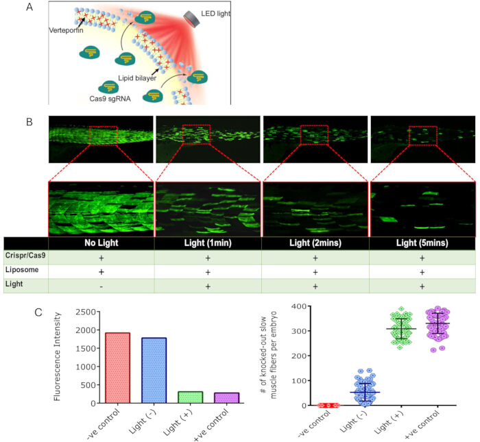
Figure 1. Light-triggered CRISPR-Cas9 delivery and spatiotemporal gene knockout in zebrafish embryos. (A) Schematic illustration of verteporfin (VP)-liposomes (DOTAP:DOPE:Chol:VP = 1:0.94:1:0.06) encapsulating Cas9/sgRNA ribonucleoprotein (RNP) complexes. Upon irradiation with 690 nm light, VP generates reactive oxygen species that destabilise the liposomal membrane and trigger intracellular release of the CRISPR-Cas9 cargo. (B) Representative fluorescence images of smyhc1:eGFP zebrafish embryos at 72 h post-fertilisation (hpf) following treatment with VP-liposomes and increasing light-exposure durations (0, 1, 2, and 5 min; 690 nm, 0.15 mW/cm2). Loss of eGFP fluorescence indicates knockout of slow-muscle fibres. Insets show magnified views of the boxed regions. (C) Quantification of fluorescence intensity and the number of knocked-out slow-muscle fibres per embryo, demonstrating light-dependent CRISPR-Cas9 activation and gene knockout. Data are presented as mean ± SD. Zebrafish experiments were analysed by one-way ANOVA (n = 80 embryos for fluorescence-intensity measurements and n = 60 embryos for fibre-count analysis; p < 0.0001). HEK293-GFP validation experiments were analysed using a two-tailed t-test (n = 4). Scale bars = 500 µm (overview images) and 100 µm (magnified images). Adapted with permission from Aksoy et al.9 Copyright © 2020 American Chemical Society. Please click here to view a larger version of this figure.
Suboptimal results in the zebrafish assay may include: (a) fewer than 50 disrupted fibres per embryo, indicating poor liposome quality, insufficient Cas9 activity, or a suboptimal light dose; (b) embryo mortality exceeding 20%, suggesting mechanical damage during microinjection or excessive VP phototoxicity; or (c) disrupted fibres in unirradiated controls, indicating premature cargo leakage from the liposome formulation.
X-ray-triggered drug delivery (Part B):
VP/AuNP-liposomes prepared according to the Part B protocol showed dose-dependent calcein release over 1–4 Gy (Figure 2A,B)11. In the HCT116 xenograft model, mice receiving Dox-loaded VP/AuNP-liposomes followed by 4 Gy X-ray irradiation showed significant tumour-growth suppression compared with free Dox, X-ray alone, and non-irradiated liposome controls (Figure 2C)11. Histological analysis of tumour sections revealed larger necrotic areas in the combined-treatment group, as assessed by H&E staining (Figure 2D).
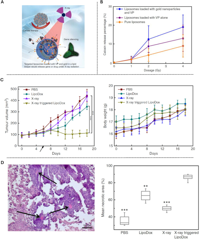
Figure 2. X-ray-triggered doxorubicin release and tumour growth suppression. (A) Schematic illustration of verteporfin (VP)/gold nanoparticle (AuNP)-integrated liposomes containing doxorubicin (Dox). Exposure to X-ray radiation activates VP through AuNP-mediated energy transfer, generating singlet oxygen that destabilises the lipid bilayer and triggers cargo release. (B) Calcein-release profiles from liposomes exposed to increasing X-ray doses (0–4 Gy), demonstrating dose-dependent cargo release from VP/AuNP-liposomes. Error bars represent standard deviation from four measurements. (C) Tumour-volume (left) and body-weight (right) measurements in HCT116 xenograft-bearing mice treated with PBS, LipoDox, X-ray alone, or X-ray-triggered LipoDox. Data are presented as mean ± SD (n = 4 mice per group). Statistical significance was evaluated using the t-test (P < 0.05, P < 0.01, P < 0.001). (D) Representative haematoxylin and eosin (H&E)-stained tumour sections and morphometric analysis of mean tumour necrotic area. Arrows indicate representative necrotic regions. Box plots show the median and interquartile range, with whiskers extending to 1.5 × IQR. Data were analysed using the t-test (n = 5). Scale bar = 50 µm. Adapted from Deng et al.11 under the terms of the Creative Commons Attribution 4.0 International License. Please click here to view a larger version of this figure.
Suboptimal results in the xenograft model may include: (a) no difference in tumour growth between irradiated and non-irradiated liposome groups, indicating insufficient VP-mediated release or inadequate gold-nanoparticle incorporation; or (b) systemic toxicity (weight loss > 15%), suggesting Dox leakage from liposomes prior to X-ray activation.
Discussion
Several steps in these protocols are critical for success. In Part A, the lipid molar ratio (DOTAP:DOPE:Chol:VP = 1:0.94:1:0.06) must be maintained precisely, as VP concentration directly determines the magnitude of 1O2 generation and, consequently, cargo-release efficiency9. Loading the Cas9/sgRNA RNP during the hydration step, rather than by post-formation complexation, is essential for aqueous-core encapsulation. Extrusion through a 200 nm polycarbonate membrane (11 passes) produces uniform unilamellar vesicles; sonication should not be substituted, as it may denature the Cas9 protein. For Part B, the gold-nanoparticle size (3–5 nm) is critical, as the particles must be small enough to embed within the lipid bilayer for effective Auger electron-mediated energy transfer to VP under X-ray irradiation11. The ammonium sulphate–gradient method used for Dox loading requires complete dialysis of external ammonium sulphate before drug addition; incomplete dialysis will reduce loading efficiency.
The lipid composition can be adapted for different applications while maintaining the VP-mediated release mechanism. For example, substituting a fraction of DOTAP with a PEGylated lipid (e.g., DSPE-PEG2000 at 5 mol%) may improve circulation time for systemic administration, although this will reduce the positive surface charge and may decrease cellular uptake in vitro2. If DLS measurements show PDI > 0.3, increase the number of extrusion passes or verify membrane integrity. If singlet oxygen generation is low despite correct VP incorporation, verify the LED light-source wavelength and power density with a calibrated power meter, as VP activation is wavelength-specific (peak excitation at 425 nm for the Soret band and therapeutic activation at 689–690 nm for the Q-band)9. For the zebrafish assay, if embryo mortality exceeds 20% in treated groups, reduce the injection volume or liposome concentration to minimise VP-associated phototoxicity. A structured troubleshooting framework, keyed to the benchmarks in Table 1, is as follows. PDI > 0.3: increase the number of extrusion passes and verify membrane integrity. Low or absent triggered release despite correct VP incorporation: confirm the 690 nm output with a calibrated power meter and verify the VP molar ratio. Fewer than ~50 knocked-out slow-muscle fibres per embryo: remake the liposomes, confirm RNP activity, or increase the irradiation time. Loss of eGFP signal in unirradiated controls: check storage conditions by using liposomes within 48 h of preparation and storing them at 4°C in the dark, and confirm membrane stability. In the xenograft model, no difference between irradiated and non-irradiated liposome groups indicates the need to confirm 3–5 nm gold-nanoparticle embedding and verify the delivered X-ray dose.
The primary limitation of the light-triggered modality (Part A) is the penetration depth of 690 nm light, which restricts direct activation to superficially accessible tissues or those amenable to fibre-optic light delivery5. The X-ray-triggered modality (Part B) overcomes this depth limitation but requires access to a clinical linear accelerator and exposes surrounding tissue to ionising radiation, albeit at therapeutic doses11,12. Both formulations currently rely on DOTAP, a cationic lipid associated with dose-dependent cytotoxicity that may limit the therapeutic window for systemic administration2. The transient nature of RNP-mediated gene editing is advantageous for safety but means that incomplete knockout in a target cell population cannot be augmented by repeat dosing at the same site without re-administration of nanoparticles. Additionally, these protocols have been validated in cell lines and small-animal models; translation to larger animal models and, ultimately, to human clinical application will require formulation optimisation, pharmacokinetic studies, and formal toxicology assessment under GLP conditions. In particular, this protocol does not include pharmacokinetic, biodistribution, or retinal-penetration data. Although light penetration through ocular and other tissues is well characterised in the literature as wavelength-, pigmentation-, and tissue-dependent (and can be augmented with optical-clearing agents), quantitative pharmacokinetic, biodistribution, and retinal-delivery studies remain important next steps before translational claims can be substantiated.
Conventional LNP systems, including those used in mRNA vaccines, release cargo passively upon cellular uptake through endosomal escape and offer no temporal or spatial control over delivery1,2. Viral vectors such as AAV provide efficient gene delivery but are limited by packaging capacity (~4.7 kb), immunogenicity upon re-dosing, and constitutive transgene expression that cannot be modulated post-administration3. The VP-liposome platform addresses these limitations by providing an externally controllable release mechanism that is independent of cargo type, compatible with large payloads (proteins, RNP complexes, and small molecules), and non-viral and potentially compatible with repeat administration, subject to repeat-dose immunogenicity and toxicology studies. Unlike optogenetic or thermally responsive systems, the VP-based approach leverages a clinically approved photosensitizer with established safety data and existing clinical laser infrastructure, particularly in ophthalmology, where the 689 nm Visudyne laser is already standard equipment6. Sang et al. further demonstrated that VP-containing lipid–polymer hybrid nanoparticles enhance radiodynamic therapy efficacy in colorectal cancer models, confirming the adaptability of VP-mediated ROS generation across different nanoparticle architectures12.
The methods described herein have particular relevance to three ophthalmic conditions. In neovascular AMD, the burden of anti-VEGF injections remains a critical barrier to optimal outcomes. Real-world registry data show marked undertreatment relative to clinical trial protocols (a mean of 7.3 versus 12 injections in year one, with 38.8% of patients discontinuing treatment and minimal visual-acuity gain)14, as well as long-term visual decline despite frequent dosing23. AAV gene therapies being developed to reduce this burden—ixoberogene soroparvovec15, ABBV-RGX-31416, and the RMAT-designated 4D-150 (83% injection reduction)24—nonetheless express anti-VEGF transgenes constitutively and cannot be titrated or withdrawn. This is a potential concern given evidence linking continuous VEGF suppression to geographic atrophy (CATT17; a 4,609-eye meta-analysis18; and atrophy prevalence rising to 78.3% over eight years25). A light-triggered VP-liposome system carrying CRISPR-Cas9 targeting VEGF-A could instead enable spatially delimited gene knockout, providing clinician-controlled spatial and temporal gating of the initial editing event while retaining the durability of gene editing. Preclinical studies support the feasibility of this approach. LNP co-delivery of Cas9 mRNA and sgRNA targeting VEGFA reduced laser-induced choroidal neovascularisation (CNV) in mice26, PEG-variant LNPs achieved genome editing in retinal pigment epithelium (RPE)27, and peptide-guided LNPs delivered mRNA to photoreceptors in non-human primates28. However, no LNP-based retinal gene therapy has entered clinical trials as of early 202629, underscoring the translational opportunity. In Stargardt disease, the approximately 6.8 kb ABCA4 coding sequence exceeds AAV packaging capacity, necessitating dual-vector strategies with reduced transduction efficiency19. Clinical candidates include the dual-AAV platforms SB-007 and VG80130, alongside the gene-agnostic modifier therapy OCU410ST31. Non-viral nucleic-acid delivery can bypass AAV packaging constraints. PEG-ECO nanoparticles carrying an ABCA4 plasmid reduced A2E accumulation in Abca4-/- mice20, while separate studies have demonstrated delivery of large retinal DNA constructs32 and LNP-mediated mRNA expression in ocular tissues33. Because localised treatment of the macula and fovea is sufficient to preserve central vision, VP-liposomes are well suited to delivering high concentrations of gene-therapy cargo precisely to these regions. For uveal melanoma, verteporfin acts through three complementary mechanisms: PDT-mediated cytotoxicity, YAP/TAZ pathway inhibition (with YAP1 expression correlating with the strongest metastatic markers, monosomy 3 and BAP1 loss)21, and immunogenic cell death through cGAS-STING activation. Combining VP-mediated YAP inhibition and PDT with anti-PD-L1 therapy converted immunologically cold tumours into immunologically active tumours, markedly suppressing tumour growth and extending survival in orthotopic models22. These findings are consistent with the reported 80% response rate for verteporfin-based PDT as a primary treatment for choroidal melanoma34. The eye’s optical accessibility for 689 nm activation, together with the well-defined location of most uveal melanomas, positions VP-liposomes as a promising platform for delivering localised treatment while potentially reducing the morbidity associated with current brachytherapy and proton-beam approaches.
Beyond the ophthalmic and oncological applications discussed above, the VP-liposome platform is applicable to any therapeutic context requiring externally controlled, on-demand payload release. Potential applications include light-triggered gene editing in dermatological conditions accessible to transcutaneous illumination, X-ray-triggered chemotherapy release synchronised with clinical radiotherapy fractionation schedules, and spatially patterned gene knockout for studies in developmental biology. Several photoactivatable gene-editing systems have been demonstrated in non-retinal tissues, including the Mag-ABE photoactivatable base editor for spatiotemporally controlled genome editing in vivo35 and an NIR-activated upconversion nanoparticle system for spatiotemporally controlled CRISPR-Cas9 editing and photodynamic therapy36. In ophthalmology, increasing interest in durable VEGF suppression strategies further highlights the need for delivery systems that combine long-term therapeutic efficacy with external control over the timing and location of biological activity37. The integration of these fabrication methods with emerging retinal LNP-delivery technologies and photoactivatable gene-editing systems represents a convergence of individually validated components whose combined application in the eye remains an open and potentially high-impact research frontier. Ongoing work in our group is directed towards optimising formulations for subretinal and intravitreal delivery routes, evaluating safety profiles in large-animal ocular models, and adapting the X-ray-triggered modality for concurrent use with standard radiotherapy protocols in clinical oncology.
Disclosures
Y.A.A. is a director and co-founder of EosGene Therapeutics Pty Ltd, which holds patents related to the light-activated lipid nanoparticle technology described herein. E.M.G. and W.D. are co-founders of EosGene Therapeutics.
Acknowledgements
The authors acknowledge the facilities and technical assistance provided by the Macquarie University Zebrafish Facility (zebrafish work) and the Macquarie University Animal Ethics Committee. This work was supported in part by the ARC Centre of Excellence for Nanoscale BioPhotonics (CE140100003). The authors also acknowledge support from EosGene Therapeutics Pty Ltd.
Materials
| Name | Company | Catalog Number | Comments |
|---|---|---|---|
| 20 nm filtration membrane (Anotop 10 or equivalent) | Whatman (Cytiva) | N/A | Used for separation of free Cas9 during encapsulation-efficiency measurements |
| 690 nm LED light source | Thorlabs | M690L4 | Light source for triggering VP-mediated liposome cargo release; adjustable power density |
| Antibiotic–Antimycotic (100X) | Thermo Fisher Scientific | 15240062 | Cell culture supplement; 1% final concentration |
| Argon gas cylinder and regulator | N/A | N/A | Used for solvent evaporation during lipid-film preparation |
| Avanti Mini Extruder | Avanti Polar Lipids | 610000 | Manual extrusion system used with 200 nm polycarbonate membranes for liposome size standardization |
| BALB/c nude mice (female, 5–6 weeks) | Animal Resources Centre | N/A | Immunodeficient mice used for HCT116 xenograft tumour model |
| BCA Protein Assay Kit | Thermo Fisher Scientific | N/A | Used for quantification of free Cas9 and encapsulation-efficiency measurements |
| Bio-Rad ChemiDoc MP Imaging System | Bio-Rad Laboratories | 12003154 | Gel and western blot imaging system |
| Calcein | Sigma-Aldrich | C0875 | Fluorescent dye for release assays; self-quenching at high concentrations |
| Cary 5000 UV-Vis-NIR Spectrophotometer | Agilent Technologies | N/A | UV-Vis absorption spectroscopy of liposomes and verteporfin |
| CCD841 CoN cells (human normal colon epithelial) | ATCC | CRL-1790 | Normal colon epithelial cell line; used as non-cancer control |
| Chloroform | Sigma-Aldrich | 288306 | Organic solvent for lipid-film preparation |
| Cholesterol | Sigma-Aldrich | C8667 | Liposome membrane component used in light-triggered formulation |
| Clinical linear accelerator (6 MeV photon beam) | Varian Medical Systems | N/A | Clinical X-ray source for irradiation studies |
| Digital callipers | N/A | N/A | Used for xenograft tumour-volume measurements |
| Dimethyl sulfoxide (DMSO) | Sigma-Aldrich | 472301 | Solvent for verteporfin stock preparation |
| DOPC (1,2-dioleoyl-sn-glycero-3-phosphocholine) | Avanti Polar Lipids | 850375P | Lipid component used in X-ray-triggered formulation |
| DOPE (1,2-dioleoyl-sn-glycero-3-phosphoethanolamine) | Sigma-Aldrich | P1223 | Helper lipid used in light-triggered formulation |
| DOTAP (1,2-dioleoyl-3-trimethylammonium-propane) | Avanti Polar Lipids | 890890P | Cationic lipid used in both formulations |
| Doxorubicin hydrochloride | Sigma-Aldrich | D1515 | Chemotherapeutic drug loaded into X-ray-triggered liposomes |
| DSPE-PEG2000 | Avanti Polar Lipids | 880120P | PEGylated lipid used in X-ray-triggered formulation |
| Dulbecco's Modified Eagle Medium (DMEM) | Thermo Fisher Scientific | 11965092 | Cell culture medium for HEK293 and PC12 cells |
| Dulbecco's Phosphate-Buffered Saline (DPBS) | Thermo Fisher Scientific | 14190144 | Wash buffer |
| Fetal Bovine Serum (FBS) | Thermo Fisher Scientific | 10099141 | Cell culture supplement; 10% final concentration |
| Fluorolog-Tau3 Spectrofluorometer | HORIBA Scientific | N/A | Fluorescence spectroscopy for verteporfin characterization |
| Focus-stacking software (e.g., Zerene Stacker) | N/A | N/A | Used for focus-stacked image generation in zebrafish imaging |
| Freeze dryer (Alpha 1-4 LDplus) | Martin Christ / John Morris Scientific | N/A | Used for vacuum drying and solvent removal |
| GFP sgRNA | Thermo Fisher Scientific | A35943 | Single-guide RNA targeting eGFP; used for knockout validation |
| Gold nanoparticles (3–5 nm, citrate-stabilized) | NanoComposix | N/A | Radiosensitizer used in X-ray-triggered formulation |
| GraphPad Prism | GraphPad Software | 7.0 | Statistical analysis and data visualization |
| HCT116 cells (human colorectal carcinoma) | ATCC | CCL-247 | Colorectal cancer cell line used for in vitro and in vivo studies |
| HEK293/GFP cells | GenTarget | SC001 | Stable GFP-expressing cell line used for CRISPR knockout validation |
| Hoechst 33342 | Thermo Fisher Scientific | H3570 | Nuclear stain; 5 µg/mL working concentration |
| Image-analysis software (e.g., Imaris) | Bitplane (Oxford Instruments) | N/A | Used for quantification of knocked-out slow-muscle fibres |
| ImageJ/Fiji | National Institutes of Health (NIH) | Open source | Image-analysis software |
| Inverted fluorescence microscope | Leica Microsystems | DMi3000 | Used for zebrafish reporter imaging and GFP fluorescence assessment |
| IVIS SpectrumCT In Vivo Imaging System | PerkinElmer | 128201 | In vivo fluorescence imaging system |
| JEOL JEM-1400 Transmission Electron Microscope | JEOL | JEM-1400 | TEM imaging of liposomes |
| Leica SP2 Confocal Microscope | Leica Microsystems | N/A | Confocal imaging of cellular uptake |
| Low-melting-point agarose | N/A | N/A | Used for mounting zebrafish embryos during imaging |
| Lipofectamine 2000 | Thermo Fisher Scientific | 11668019 | Transfection reagent used as positive control |
| Living Image Software | PerkinElmer | N/A | Image acquisition and analysis software |
| McCoy's 5A Medium | Thermo Fisher Scientific | 16600082 | Cell culture medium for HCT116 cells |
| Microinjection system | N/A | N/A | Used for one-cell-stage zebrafish embryo microinjection |
| MTS Cell Proliferation Assay | Promega | G3582 | Cell viability assay |
| NanoDrop Spectrophotometer | Thermo Fisher Scientific | ND-2000 | RNA purity and concentration measurements |
| Nuclease-free water | Thermo Fisher Scientific | R0582 | Used for CRISPR-Cas9 RNP preparation |
| Opti-MEM Reduced Serum Medium | Thermo Fisher Scientific | 31985070 | Transfection medium |
| Paraformaldehyde (4%) | Sigma-Aldrich | P6148 | Fixative for histological analysis |
| PC12 cells (rat pheochromocytoma) | ATCC | CRL-1721 | Neuronal-like cell line used for uptake studies |
| Polycarbonate membrane filters (200 nm) | Avanti Polar Lipids | 610006 | Extrusion membranes for liposome sizing |
| RNeasy Plus Mini Kit | QIAGEN | 74134 | RNA extraction kit |
| Singlet Oxygen Sensor Green (SOSG) | Thermo Fisher Scientific | S36002 | Fluorescent probe for singlet oxygen detection |
| Slide-A-Lyzer MINI Dialysis Devices (10 kDa MWCO) | Thermo Fisher Scientific | 69570 | Dialysis devices for liposome preparation and drug loading |
| SYBR Gold Nucleic Acid Gel Stain | Thermo Fisher Scientific | S11494 | Nucleic-acid gel stain |
| Tg(ubi:EGFP) zebrafish | Zebrafish International Resource Center | ZDB-GENO-080606-2 | Transgenic zebrafish used for in vivo CRISPR validation |
| TNF-α (human recombinant) | Life Technologies | RTNFAI | Cytokine used in TNFAIP3 induction studies |
| TNFAIP3 sgRNA | Thermo Fisher Scientific | N/A | sgRNA targeting TNFAIP3 |
| Tricaine methanesulfonate (MS-222) | N/A | N/A | Zebrafish anaesthetic used prior to imaging |
| Triton X-100 | Sigma-Aldrich | T8787 | Detergent used for liposome lysis and release assays |
| TrueCut Cas9 Protein v2 | Thermo Fisher Scientific | A36498 | Cas9 nuclease used for CRISPR-Cas9 RNP preparation |
| Trypsin-EDTA (0.25%) | Thermo Fisher Scientific | 25200056 | Cell dissociation reagent |
| Uranyl acetate (2%) | Electron Microscopy Sciences | 22400 | Negative stain for TEM imaging |
| Verteporfin | Sigma-Aldrich | SML0534-5MG | Photosensitizer incorporated into liposome formulations |
| Zetasizer Nano ZS | Malvern Panalytical | ZEN3600 | Dynamic light scattering and zeta-potential measurements |
References
- Polack FP et al. Safety and efficacy of the BNT162b2 mRNA Covid-19 vaccine. N Engl J Med. 2020;383(27):2603-15.
- Cullis PR, Hope MJ. Lipid nanoparticle systems for enabling gene therapies. Mol Ther. 2017;25(7):1467-75.
- Wang D, Tai PWL, Gao G. Adeno-associated virus vector as a platform for gene therapy delivery. Nat Rev Drug Discov. 2019;18(5):358-78.
- Kim S et al. Highly efficient RNA-guided genome editing in human cells via delivery of purified Cas9 ribonucleoproteins. Genome Res. 2014;24(6):1012-19.
- Wilson BC, Patterson MS. The physics, biophysics and technology of photodynamic therapy. Phys Med Biol. 2008;53(9):R61-R109.
- Scott LJ, Goa KL. Verteporfin. Drugs Aging. 2000;16(2):139-46.
- Treatment of Age-related Macular Degeneration With Photodynamic Therapy (TAP) Study Group. Photodynamic therapy of subfoveal choroidal neovascularization in age-related macular degeneration with verteporfin: one-year results of 2 randomized clinical trials—TAP Report 1. Arch Ophthalmol. 1999;117(10):1329-45. doi:10.1001/archopht.117.10.1329.
- Boettner EA, Wolter JR. Transmission of the ocular media. Invest Ophthalmol. 1962;1(6):776–83.
- Aksoy YA et al. Spatial and temporal control of CRISPR-Cas9-mediated gene editing delivered via a light-triggered liposome system. ACS Appl Mater Interfaces. 2020;12(47):52433-44.
- Ogilby PR. Singlet oxygen: there is indeed something new under the sun. Chem Soc Rev. 2010;39(8):3181-209.
- Deng W et al. Controlled gene and drug release from a liposomal delivery platform triggered by X-ray radiation. Nat Commun. 2018;9(1):2713.
- Sang R et al. Unlocking the in vivo therapeutic potential of radiation-activated photodynamic therapy for locally advanced rectal cancer with lymph node involvement. EBioMedicine. 2025;116:105724.
- Fleckenstein M, Schmitz-Valckenberg S, Chakravarthy U. Age-related macular degeneration: a review. JAMA. 2024;331(2):147-57.
- Wykoff CC et al. Impact of anti-VEGF treatment and patient characteristics on vision outcomes in neovascular age-related macular degeneration: up to 6-year analysis of the AAO IRIS Registry. Ophthalmol Sci. 2024;4(2):100421.
- Khanani AM et al. Safety and efficacy of ixoberogene soroparvovec in neovascular age-related macular degeneration in the United States (OPTIC): a prospective, two-year, multicentre phase 1 study. EClinicalMedicine. 2024;67:102394.
- Campochiaro PA et al. Gene therapy for neovascular age-related macular degeneration by subretinal delivery of RGX-314: a phase 1/2a dose-escalation study. Lancet. 2024;403(10436):1563-73.
- Maguire MG et al. Five-year outcomes with anti-vascular endothelial growth factor treatment of neovascular age-related macular degeneration: the Comparison of Age-related Macular Degeneration Treatments Trials. Ophthalmology. 2016;123(8):1751-61.
- Eshtiaghi A et al. Geographic atrophy incidence and progression after intravitreal injections of anti-vascular endothelial growth factor agents for age-related macular degeneration: a meta-analysis. Retina. 2021;41(12):2424-35.
- Li R et al. Split AAV8 mediated ABCA4 expression for gene therapy of mouse Stargardt disease (STGD1). Hum Gene Ther. 2023;34(13-14):616-28.
- Sun D et al. Effective gene therapy of Stargardt disease with PEG-ECO/pGRK1-ABCA4-S/MAR nanoparticles. Mol Ther Nucleic Acids. 2022;29:823-35.
- Brouwer NJ et al. Targeting the YAP/TAZ pathway in uveal and conjunctival melanoma with verteporfin. Invest Ophthalmol Vis Sci. 2021;62(4):3.
- Zhang S et al. Combination of YAP inhibition and photodynamic therapy induces dual DNA damage and activates STING pathway to enhance immunotherapy in uveal melanoma. Redox Biol. 2026;89:103965. doi:10.1016/j.redox.2025.103965.
- Cheema MR, DaCosta J, Talks J. Ten-year real-world outcomes of anti-vascular endothelial growth factor therapy in neovascular age-related macular degeneration. Clin Ophthalmol. 2021;15:279-87.
- Kay C et al. Interim results from the phase 2b population extension cohort in the PRISM clinical trial evaluating 4D-150 in adults with neovascular (wet) age-related macular degeneration. Invest Ophthalmol Vis Sci. 2025;66(8):1861.
- Vofo BN et al. Long-term macular atrophy growth in neovascular age-related macular degeneration: influential factors and role of genetic variants. Eye (Lond). 2025;39(9):1717-23.
- Cao D et al. Dynamically covalent lipid nanoparticles mediate CRISPR-Cas9 genome editing against choroidal neovascularization in mice. Sci Adv. 2025;11(28):eadj0006.
- Gautam M et al. Lipid nanoparticles with PEG-variant surface modifications mediate genome editing in the mouse retina. Nat Commun. 2023;14(1):6468.
- Herrera-Barrera M et al. Peptide-guided lipid nanoparticles deliver mRNA to the neural retina of rodents and nonhuman primates. Sci Adv. 2023;9(2):eadd4623.
- Wu H et al. Innovative gene delivery systems for retinal disease therapy. Neural Regen Res. 2026;21(2):542-52.
- Riedmayr LM et al. mRNA trans-splicing dual AAV vectors for (epi)genome editing and gene therapy. Nat Commun. 2023;14(1):6578.
- Khanani AM et al. A novel modifier gene therapy to treat Stargardt disease: Phase 1 GARDian1 Trial Insights. Eye (Lond). 2026;40:429–30.
- Goddaer S et al. Overcoming size barriers in retinal gene therapy via lipid nanoparticle-mediated delivery of full-length EYS DNA. Drug Deliv Transl Res. 2026;16(8):2921–36. doi:10.1007/s13346-025-02036-y.
- Chambers CZ et al. Lipid nanoparticle-mediated delivery of mRNA into the mouse and human retina and other ocular tissues. Transl Vis Sci Technol. 2024;13(7):7.
- Yordi S, Soto H, Bowen RC, Singh AD. Photodynamic therapy for choroidal melanoma: What is the response rate? Surv Ophthalmol. 2021;66(4):552-59.
- Zou Q et al. Photoactivatable base editors for spatiotemporally controlled genome editing in vivo. Biomaterials. 2023;302:122328.
- Zeng J et al. Near-infrared optogenetic nanosystem for spatiotemporal control of CRISPR-Cas9 gene editing and synergistic photodynamic therapy. ACS Appl Mater Interfaces. 2025;17(1):701-10.
- Campochiaro PA. Sustained suppression of VEGF for treatment of retinal and choroidal vascular diseases. Am J Ophthalmol. 2025;277:1-6.
Reprints and Permissions
This article has been published
Video Coming Soon