Atlas and Leo Baby stents demonstrated similar angiographic and clinical efficacy in intracranial aneurysm coiling. Complication rates showed no statistical difference, though Atlas had a numerically higher trend. Further multicenter prospective studies are warranted to guide device selection.
Research Article
A Retrospective Comparative Study of Leo Baby and Atlas Stents for the Treatment of Intracranial Aneurysms: Embolization Outcomes and Complications
* These authors contributed equally
In This Article
Summary
Abstract
Intracranial aneurysms (IAs) are increasingly managed with endovascular techniques, with stent-assisted coiling offering superior outcomes for complex morphologies. The Neuroform Atlas (laser‑cut, open‑cell) and Leo Baby (braided, closed‑cell) are low‑profile stents deployable through small microcatheters, improving navigability in tortuous vasculature. However, direct comparative data on their relative efficacy and safety remains scarce. This retrospective study compares embolization outcomes and procedure‑related complications between the two devices. A total of 96 patients with IAs treated with stent-assisted coiling between January 2022 and December 2023 were enrolled: 46 received Atlas and 50 received Leo Baby. Baseline demographic and aneurysm characteristics were comparable between groups. Immediate post‑procedural angiography demonstrated similar complete occlusion rates (Atlas 89.1% vs. Leo Baby 88.0%; P = 0.3281), and no significant difference was found in Raymond grade distribution at 6‑month follow‑up (P = 0.5067). Favorable outcomes (modified Rankin Scale score 0–2) at 1 year were achieved in >90% of patients in both cohorts (P = 0.1528). Peri-procedural complications showed no statistically significant difference between the two groups (28.2% vs. 12.0%; P = 0.0857); however, post-hoc power analysis revealed that the current sample size provided only 42% power to detect this observed difference, indicating a high risk of Type II error. In conclusion, these findings suggested that both Atlas and Leo Baby stents afford similar angiographic and clinical efficacy for IA embolization; however, the numerically higher complication rate observed with the Atlas stent should be interpreted with caution due to insufficient statistical power, and our results should be considered hypothesis-generating rather than confirmatory. Further largescale, prospective multicenter studies are warranted to confirm these observations and refine device selection in clinical practice.
Introduction
Intracranial aneurysms (IAs) are increasingly managed with intravascular interventional therapy, a shift driven by minimally invasive and safe characteristics of the approach. For complex cases, such as wide-necked aneurysms, stent-assisted coiling provides superior outcomes compared to simple embolization1. This technique ensures dense packing at the aneurysm neck and alters local blood flow dynamics, thereby promoting healing and mitigating postoperative recurrence. Despite these benefits, technical obstacles persist. The deployment of traditional stents (such as Solitaire AB, Enterprise, Neuroform EZ) can be difficult in the presence of tortuous parent arteries or steep angulation at the aneurysm neck2. These challenges are frequently attributable to the limited compliance of the devices and the requisite use of larger-profile microcatheters (0.021 or 0.027 inches)3,4,5.
The Neuroform Atlas and Leo Baby represent new-generation microcatheter-deliverable stents for IA embolization. Their low-profile design allows deployment through smaller microcatheters (0.0165 or 0.017 inches). This enhances their suitability for IAs located in deep or tortuous vasculature due to improved compliance and conformability2,6. A fundamental distinction lies in their architecture: Atlas is laser-cut, whereas Leo Baby is braided. This structural difference translates into divergent performance, particularly in neck coverage and flow diversion efficacy2,7. Braided stents typically offer greater metal coverage and pore density, which may accelerate intra-aneurysmal flow remodeling and endothelialization8. However, this same characteristic is associated with a potentially higher risk of thromboembolic complications9.
Despite evidence supporting the individual efficacy of both the Leo Baby and Atlas stents, a lack of direct, systematic comparative studies persists. It remains unclear whether their distinct design philosophies translate into differences in critical outcomes such as aneurysm occlusion rates, device-related complications, and prognosis. Therefore, this study aimed to perform a comparative analysis to directly evaluate the effectiveness and complication profiles of the Leo Baby and Atlas stents in the treatment of IAs, thereby providing empirical evidence to inform clinical decision-making.
Protocol
The study was approved by the Ethics Committee of the First Affiliated Hospital of USTC (Approval number: 2026-RE-015). Written informed consent was obtained from all participants prior to their inclusion, in accordance with the ethical principles of the Declaration of Helsinki (1964).
Patients
This retrospective study included patients with IAs who underwent stent-assisted treatment with either the Atlas or Leo Baby stent at our institution between January 2022 and December 2023. The inclusion criteria were as follows: IA confirmed by computed tomography angiography (CTA) or digital subtraction angiography (DSA) from our hospital or a referring center; aneurysm located distal to the circle of Willis; a modified Rankin Scale (mRS) score of <5 at admission; and availability of complete clinical and angiographic follow-up data within two years after the procedure. The exclusion criteria included aneurysms situated in proximal cerebral vessels; treatment with other stent types or simple coiling without stent assistance; known coagulation dysfunction; or any contraindications to endovascular intervention. For all enrolled patients, demographic information, aneurysm characteristics (including location, size, and neck width), and details of the treatment procedure were collected and analyzed. All patients were scheduled for regular clinical and imaging follow-up. This was a retrospective analysis of patients treated between 2022 and 2023.
The choice between the Atlas and Leo Baby stents was based on predefined institutional criteria: Atlas was preferred for emergency coiling of ruptured saccular aneurysms without contraindications; Leo Baby was preferred for dissecting aneurysms, regardless of rupture status, due to its higher mesh density and flow-diversion effect; Leo Baby was also preferred when the aneurysm neck involved branching vessels or bifurcation sites, where the “tulip” or “lampion” effect of the braided stent could preserve branch patency; Atlas was preferred for saccular aneurysms with tortuous parent arteries or mild parent artery stenosis, given its superior navigability and lower profile; and Atlas was the preferred choice for Y-stent configurations. These criteria were applied consistently during the study period. The distribution of stent diameter and length specifications for Atlas and Leo Baby groups were shown in Supplementary Table 1. Because five patients in the Atlas group and three patients in the Leo Baby group received two stents, a total of 51 Atlas stents and 53 Leo Baby stents were implanted.
Treatment protocol
All procedures were performed under general anesthesia. The common femoral artery served as the primary access site. After successful puncture using the modified Seldinger technique, systemic heparinization was administered. An appropriately sized vascular sheath was placed. A diagnostic catheter was then advanced through the sheath to perform complete cerebral angiography of the bilateral internal carotid arteries, external carotid arteries, and vertebral arteries. This initial angiogram clearly delineated the aneurysm location, size, morphology, neck width, course of the parent artery, and branching patterns, while also identifying any vascular anatomical variations and collateral circulation to formulate a precise surgical plan. Subsequently, using a standard coaxial technique, an intermediate catheter was positioned at an appropriate location. Under road-map guidance, a microcatheter was navigated over a 0.014-inch Synchro microwire to the distal segment of the ipsilateral parent artery. An Echelon-10 microcatheter was then advanced into the aneurysm sac. Based on the aneurysm dimensions, appropriately sized coils were selected and deployed to embolize the aneurysm. Simultaneously, either a Leo Baby or Atlas stent was delivered via the stent delivery catheter and deployed in a semi-deployed configuration across the aneurysm neck. Following this, additional coils were delivered through the embolization microcatheter to achieve as dense packing of the aneurysm sac as possible. The stent was then fully deployed. Post-procedural assessment included DSA in the working projection to evaluate the patency of the parent artery and its distal branches. Finally, biplanar (anteroposterior and lateral) whole-brain DSA and X-perCT were performed via the guiding catheter to rule out any complications, such as intracranial hemorrhage.
Antiplatelet regimen
Pre-procedural dual antiplatelet regimens differed by rupture status: unruptured patients received daily aspirin (100 mg) and clopidogrel (75 mg) for ≥3 days, whereas ruptured patients received preoperative oral loading doses of 300 mg of each drug. If the loading dose was not given orally prior to stent placement, an intravenous glycoprotein IIb/IIIa inhibitor (tirofiban, bolus of 0.2–0.5 mg followed by continuous infusion at 0.05–0.15 µg/kg/min) was administered immediately after stent deployment for 24 h. Oral aspirin (100 mg/day) and clopidogrel (75 mg/day) or ticagrelor (90 mg twice daily) were initiated on postoperative day two. Upon discharge, all patients continued dual antiplatelet therapy with aspirin (100 mg/day) and clopidogrel (75 mg/day) for at least three months, followed by aspirin monotherapy (100 mg/day) for over six months. Furthermore, CYP genotyping was performed to assess clopidogrel metabolism, guiding the choice between clopidogrel and ticagrelor for antiplatelet therapy.
Assessment of treatment outcomes
Aneurysm embolization degree was assessed using angiography and graded according to the Raymond scale, where grades 1, 2, and 3 corresponded to complete occlusion, residual neck, and residual aneurysm, respectively. Angiographic assessments were primarily based on follow-up DSA findings, and all results were independently reviewed by two neurointerventional physicians. Outcomes were evaluated by the mRS. An mRS score of 0–2 was defined as a favorable outcome, while a score of 3–6 indicated a worse outcome. Clinical status was evaluated for all patients at discharge, with subsequent follow-up assessments carried out at 6 and 12 months post-discharge. Radiological follow-up was principally based on DSA performed at 6 and 12 months after discharge. In-stent stenosis was defined as a reduction in vessel diameter relative to the pre-treatment baseline measurement.
Statistical analysis
Continuous variables were presented as mean ± standard deviation (SD), while categorical variables were expressed as frequencies (percentages). Comparisons of categorical variables were performed using the Chi-square test. For continuous data, the student’s t-test was employed for normally distributed variables, whereas the Mann-Whitney U test was applied for non-normally distributed variables. Multivariable Firth penalized logistic regression was performed for three outcomes (procedure-related complications, immediate incomplete occlusion, and 6-month incomplete occlusion), with stent type, drinking history, neck width, and height as covariates; neck width analyses were restricted to patients with definable necks. Given the retrospective and exploratory nature of this study, no formal a priori sample size estimation was performed. Post-hoc power calculations were conducted for the primary safety endpoint (procedure-related complication rate) using the observed proportions to assess the study's ability to detect the observed differences. A two-tailed P-value <0.05 was considered statistically significant. All statistical analyses were conducted using GraphPad Prism. The overall study design and workflow are shown in Figure 1.
Results
Baseline characteristics
A total of 46 IAs patients treated with the Atlas stent were enrolled in this work, with a mean age of 61.935 ± 9.202 years, of whom 65.2% (30/46) were female; the Leo Baby group comprised 50 patients, with a mean age of 60.380 ± 7.798 years, of whom 54.0% (32/50) were female. Clinical comorbidities were as follows: in the Atlas group, 32 patients (69.7%) had hypertension, 3 (6.5%) had diabetes mellitus, and 2 (4.3%) had coronary artery disease; in the Leo Baby group, 29 patients (58.0%) had hypertension, 5 (10.0%) had diabetes mellitus, and 3 (6.0%) had coronary artery disease. The majority of patients in both groups presented with favorable clinical status at admission (mRS score 0–2). No significant intergroup difference was observed in Fisher grade (P = 0.1345), Rupture H-H score (P = 0.2083), Glasgow Coma Scale score (P = 0.8081), D-dimer level (P = 0.8867), International Normalized Ratio (P = 0.0682), or any clinical comorbidity. Notably, a significant difference was found in drinking between the groups (P = 0.0376). The demographic characteristics of the patients at admission are summarized in Table 1.
To account for potential confounding, we performed multivariable Firth penalized logistic regression analyses for three outcomes: procedure‑related complications, immediate incomplete occlusion (Raymond class II or III), and incomplete occlusion at 6‑month follow‑up. Stent type, drinking history, aneurysm neck width, and aneurysm height were included as covariates. Analyses involving neck width were performed in the 77 patients with a definable aneurysm neck (excluding 19 dissecting aneurysms). After adjustment, Atlas stent use was not independently associated with procedure‑related complications compared with Leo Baby (adjusted OR, 2.01; 95% confidence interval (CI), 0.60-6.77; P = 0.261), nor with immediate incomplete occlusion (adjusted odds ratio (OR), 1.20; 95% CI, 0.26-5.48; P = 0.816) or incomplete occlusion at 6‑month follow‑up (adjusted OR, 1.01; 95% CI, 0.24-4.20; P = 0.990). The complete multivariable results are provided in Supplementary Table 2.
In both cohorts, aneurysms were predominantly located in the anterior communicating artery and the middle cerebral artery, and the majority of patients presented with a single aneurysm (Atlas, n = 41; Leo Baby, n = 48). For the Atlas group, the mean aneurysm neck width was 4.144 ± 2.006 mm, mean maximum aneurysm width was 5.172 ± 2.502 mm, mean aneurysm height was 4.715 ± 2.452 mm, mean proximal parent artery diameter was 2.277 ± 0.553 mm, and mean distal parent artery diameter was 2.021 ± 0.531 mm; only 4 patients exhibited parent artery atherosclerosis. In the Leo Baby group, the corresponding values were 3.314 ± 1.674 mm, 4.813 ± 5.344 mm, 3.408 ± 1.859 mm, 2.246 ± 0.815 mm, and 2.057 ± 0.764 mm, respectively, with only 2 patients presenting parent artery atherosclerosis. No statistically significant intergroup differences were identified in any of the aneurysm morphological characteristics, as detailed in Table 2.
Comparison of immediate outcomes and peri‑procedural characteristics
During the procedure, 91.7% of patients received a single stent. All patients underwent immediate postoperative angiography. When assessing the impact of stent type on embolization outcomes, no statistically significant difference was observed in immediate embolization results between the two cohorts (P = 0.3281) (Table 3). In the Atlas cohort, immediate postoperative angiography demonstrated complete occlusion in 89.1% (41/46) of aneurysms, residual neck in 6.5% (3/46), and residual aneurysm sac in 4.3% (2/46); the corresponding rates in the Leo Baby cohort were 88.0% (44/50), 2.0% (1/50), and 10.0% (5/50), respectively (Table 3). In addition, no significant intergroup difference was observed in procedure‑related complications (P = 0.0857) (Table 3). However, post-hoc power calculation demonstrated that with the current sample sizes (n = 46 vs. n = 50), the statistical power to detect this observed 16.2% absolute difference in complication rates (28.2% vs. 12.0%) was only approximately 42%, substantially below the recommended 80% threshold. This indicates that the non-significant P-value is more likely attributable to insufficient sample size (Type II error) rather than true equivalence between the two devices. In the Atlas group, 13 patients experienced procedure‑related complications, including four cases of intraoperative aneurysm rupture managed with immediate coil packing, four cases of thrombosis treated with intra‑arterial tirofiban, one case of multiple cerebral infarctions receiving symptomatic treatment, one case of postoperative hydrocephalus requiring lumbar puncture and drainage, one case of subarachnoid hemorrhage treated with decompressive craniectomy, one case of postoperative small perforator infarction managed with tirofiban, and one case of ruptured middle cerebral artery aneurysm that underwent surgical clipping. In the Leo Baby cohort, only six patients exhibited related complications, comprising three cases of thrombosis, one frontal lobe infarction, one intraoperative subarachnoid hemorrhage, and one death due to intraoperative re‑bleeding. At discharge, 90% of patients showed favorable outcomes (Table 3).
Follow-up angiographic outcomes
All patients were followed up angiographically at 6 months post-procedure, primarily using DSA, showing no intergroup statistically significant difference (P = 0.5067; Table 4). In the Atlas group, complete occlusion was achieved in 87.0% (40/46) of aneurysms, residual neck in 8.7% (4/46), and residual aneurysm sac in 4.3% (2/46). In the Leo Baby group, the corresponding rates were 88.0% (44/50), 4.0% (2/50), and 8.0% (4/50), respectively. At this follow-up time point, in-stent stenosis or parent artery stenosis was observed in three patients in each cohort, with no significant intergroup difference (P = 0.9160).
At 1-year post-procedure, angiographic follow-up was available for 6 patients in the Atlas cohort and 12 patients in the Leo Baby cohort, among whom complete occlusion was demonstrated in 5 and 11 aneurysms, respectively. Functional outcome assessment revealed that over 90% of patients achieved favorable clinical outcomes (mRS score 0–2) (Table 5). No newly developed stenosis was identified in either group. The distribution of mRS scores at admission, discharge, and 1-year follow-up was shown in Figure 2.
Representative cases
A representative case from the Atlas cohort is presented in Figure 3. Admission angiography revealed an unruptured irregular aneurysm at the right middle cerebral artery bifurcation, measuring 4.4 × 7.0 mm with a neck size of 6.2 mm (Figure 3A). An Atlas stent (4.0 × 21 mm) was deployed from the superior trunk of the M2 segment, followed by coil embolization. Immediate post-procedural angiography demonstrated dense coil packing with complete occlusion (Raymond grade I; Figure 3B). Six-month follow-up DSA confirmed persistent complete occlusion and patent parent artery without stenosis, indicating favorable aneurysm healing (Figure 3C).
A representative case from the Leo Baby cohort is illustrated in Figure 4. Admission angiography showed a ruptured microaneurysm at the anterior communicating artery, measuring 1.8 × 1.5 mm with a neck size of 1.5 mm (Figure 4A). A Leo Baby stent (2.5 × 25 mm) was partially deployed at the aneurysm neck to provide neck coverage using the dome technique, followed by coil embolization (Figure 4B). Complete stent deployment was achieved with good wall apposition, and immediate post-procedural angiography revealed dense packing and complete occlusion (Raymond grade I; Figure 4C). Six-month follow-up angiography indicated complete aneurysm obliteration and no in-stent stenosis (Figure 4D).
A representative case from the Atlas cohort is presented in Figure 5. Admission angiography revealed an unruptured saccular aneurysm at the right middle cerebral artery bifurcation, with the neck involving both the superior and inferior trunks of the M2 segment, measuring 6.1 × 6.0 mm with a neck width of 6.0 mm (Figure 5A). Y‑stent‑assisted coil embolization was performed using two Atlas stents (4.5 × 21 mm each). Immediate post‑procedural angiography demonstrated dense coil packing with complete occlusion (Raymond grade I) and patent parent artery (Figure 5B). However, 8‑month follow‑up DSA revealed significant in‑stent stenosis at the M1 segment, although the aneurysm remained completely obliterated (Figure 5C).
DATA AVAILABILITY:
All data supporting this study are available within the manuscript and in Supplementary Table 1 and Supplementary Table 2.
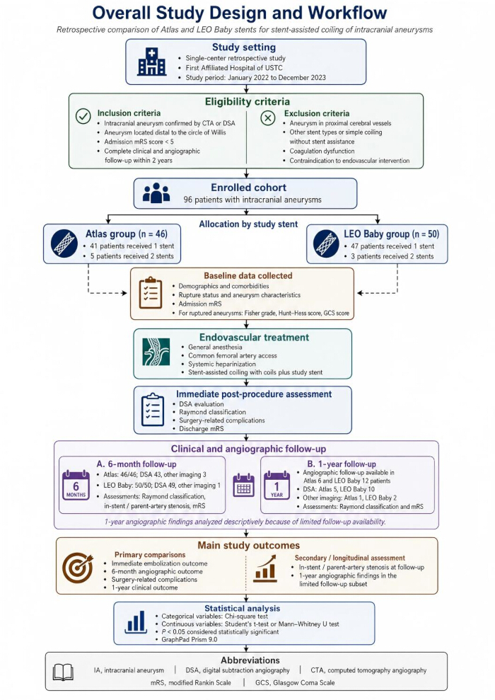
Figure 1: The overall study design and workflow. According to the predefined inclusion and exclusion criteria, a total of 96 patients were enrolled in this study, including 46 in the Atlas group and 50 in the Leo group. Baseline clinical information was collected for all patients. Immediate post‑procedural assessment was performed after treatment, followed by clinical and angiographic follow‑up at 6 months and 12 months post‑treatment. Please click here to view a larger version of this figure.
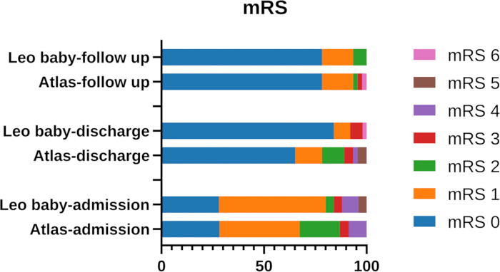
Figure 2: Proportion of patients with modified Rankin Scale (mRS) scores of 0-6 at admission, discharge, and 1-year follow-up. Favorable outcomes (mRS 0–2) were observed in approximately 85% of patients at admission, approximately 90% at discharge, and approximately 92% at 1-year follow‑up in both groups. Please click here to view a larger version of this figure.
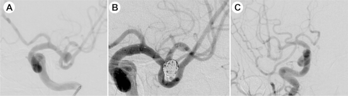
Figure 3: Representative case of favorable outcome following Atlas stent‑assisted coil embolization. (A) Preoperative DSA showing an unruptured irregular aneurysm at the right middle cerebral artery bifurcation, measuring 4.4 × 7.0 mm with a neck size of 6.2 mm. (B) Immediate post‑procedural DSA after Atlas stent (4.0 × 21 mm) deployment and coil embolization, demonstrating dense coil packing with complete occlusion (Raymond grade I). (C) 6-month follow-up DSA confirming complete occlusion and patent parent artery without stenosis. DSA, Digital subtraction angiography. Please click here to view a larger version of this figure.
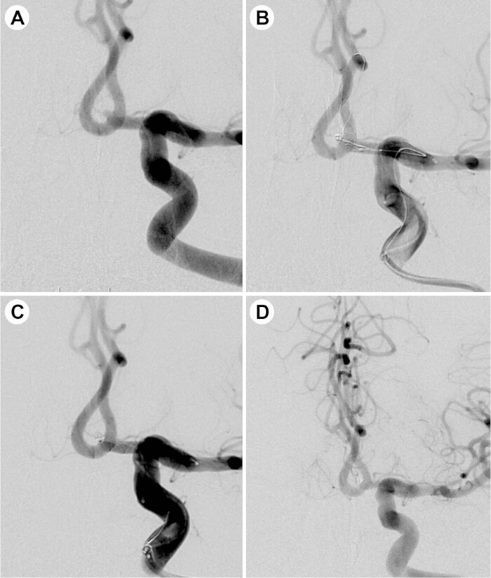
Figure 4: Representative case of favorable outcome following Leo Baby stent‑assisted coil embolization. (A) Preoperative DSA showing a ruptured microaneurysm at the anterior communicating artery, measuring 1.8 × 1.5 mm with a neck size of 1.5 mm. (B) A Leo Baby stent (2.5 × 25 mm) was partially deployed at the aneurysm neck using the dome technique, followed by coil embolization. (C) Immediate post-procedural DSA demonstrating complete stent deployment with good wall apposition, dense coil packing, and complete occlusion (Raymond grade I). (D) 6-month follow-up DSA confirming complete occlusion and patent parent artery without in‑stent stenosis. ACA, Anterior communicating artery; DSA, Digital subtraction angiography. Please click here to view a larger version of this figure.
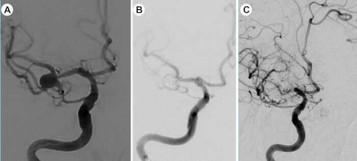
Figure 5: Representative case of in‑stent stenosis following Atlas stent‑assisted coil embolization. (A) Preoperative DSA showing an unruptured saccular aneurysm at the right middle cerebral artery bifurcation, measuring 6.1 × 6.0 mm with a neck width of 6.0 mm, with the neck involving both the superior and inferior trunks of the M2 segment. (B) Immediate post‑procedural DSA after Y‑stent‑assisted coil embolization using two Atlas stents (4.5 × 21 mm each), demonstrating dense coil packing with complete occlusion (Raymond grade I) and patent parent artery. (C) Eight‑month follow‑up DSA revealing significant in‑stent stenosis at the M1 segment (arrow), while the aneurysm remained completely obliterated. Please click here to view a larger version of this figure.
| Variables | Atlas (n = 46) | Leo baby (n = 50) | P value |
| Age (year), Mean ± SD | 61.935 ± 9.202 | 60.380 ± 7.798 | 0.2576 |
| Female, n (%) | 30/46 (65.2) | 32/50 (64.0) | 0.9008 |
| Hypertension, n (%) | 32/46 (69.7) | 29/50 (58.0) | 0.2395 |
| Diabetes, n (%) | 3/46 (6.5) | 5/50 (10.0) | 0.5379 |
| Hyperlipemia, n (%) | 0/46 (0.0) | 0/50 (0.0) | -- |
| Coronary heart disease, n (%) | 2/46 (4.3) | 3/50 (6.0) | 0.7159 |
| Smoking, n (%) | 5/46 (10.9) | 3/50 (6.0) | 0.3885 |
| Drinking, n (%) | 6/46 (13.0) | 1/50 (2.0) | 0.0376 |
| Fisher grade, n (%) | 0.1345 | ||
| 0 | 0/46 (0.0) | 3/50 (6.0) | |
| 1 | 1/46 (2.1) | 3/50 (6.0) | |
| 2 | 8/46 (17.4) | 3/50 (6.0) | |
| 3 | 5/46 (10.9) | 4/50 (8.0) | |
| 4 | 2/46 (4.3) | 4/50 (8.0) | |
| Clinical outcomes, n (%) | 0.6818 | ||
| Good (mRS 0-2) | 40/46 (87.0) | 42/50 (84.0) | |
| Poor (mRS 3-6) | 6/46 (13.0) | 8/50 (16.0) | |
| Rupture H-H score, n (%) | 0.2083 | ||
| 1 | 4/46 (8.7) | 4/50 (8.0) | |
| 2 | 3/46 (6.5) | 8/50 (16.0) | |
| 3 | 8/46 (17.4) | 3/50 (6.0) | |
| 4 | 1/46 (2.2) | 1/50 (2.0) | |
| GCS score, n (%) | 0.3326 | ||
| 3-8 | 3/16 (18.8) | 1/16 (6.3) | |
| 9-12 | 1/16 (6.3) | 0/16 (0.0) | |
| 13-15 | 12/16 (75.0) | 15/16 (93.7) | |
| D-dimer (μg/mL), Mean ± SD | 0.826 ± 0.848 | 0.793 ± 0.841 | 0.8867 |
| INR | 0.975 ± 0.076 | 0.949 ± 0.060 | 0.0682 |
Table 1: Comparison of baseline information of patients. (Atlas group, n = 46; Leo Baby group, n = 50). Variables include age, sex, comorbidities, admission mRS score, Fisher grade, Hunt–Hess grade, GCS score, D-dimer, INR, and drinking status. Continuous variables are shown as mean ± SD, and categorical variables as n (%). INR, International Normalized Ratio; GCS, Glasgow coma scale; mRS, Modified rankin scale.
| Characteristics | Atlas (n = 46) | Leo Baby (n = 50) | P value |
| Ruptured, n (%) | 0.7726 | ||
| Yes | 16/46 (34.8) | 16/50 (32.0) | |
| No | 30/46 (65.2) | 34/50 (68.0) | |
| Location, n (%) | 0.3446 | ||
| ACA | 1/46 (2.2) | 6/50 (12.0) | |
| AcomA | 12/46 (26.1) | 14/50 (28.0) | |
| MCA | 17/46 (37.0) | 19/50 (38.0) | |
| PcomA | 7/46 (15.2) | 2/50 (4.0) | |
| ICA | 3/46 (6.5) | 2/50 (4.0) | |
| BA | 5/46 (10.9) | 6/50 (12.0) | |
| PICA | 1/46 (2.2) | 1/50 (2.0) | |
| Aneurysm number, n (%) | 0.1959 | ||
| 1 | 41/46 (89.1) | 48/50 (96.0) | |
| 2 | 5/46 (10.9) | 2/50 (4.0) | |
| Aneurysm neck width (mm), Mean ± SD | 4.144 ± 2.006 | 3.314 ± 1.674 | 0.061 |
| Maximum aneurysm width (mm), Mean ± SD | 5.172 ± 2.502 | 4.813 ± 5.344 | 0.6643 |
| Aneurysm height (mm), Mean ± SD | 4.715 ± 2.452 | 3.408 ± 1.859 | 0.069 |
| Proximal parent-artery diameter (mm), Mean ± SD | 2.277 ± 0.553 | 2.246 ± 0.815 | 0.8293 |
| Distal parent-artery diameter (mm), Mean ± SD | 2.021 ± 0.531 | 2.057 ± 0.764 | 0.7909 |
| Parent artery atherosclerosis, n (%) | 0.9495 | ||
| Yes | 4/46 (8.7) | 2/50 (4.0) | |
| No | 42/46 (91.3) | 48/50 (96.0) |
Table 2: Comparison of aneurysm characteristics. Morphological measurements were obtained from three-dimensional reconstructed angiographic images. One target aneurysm treated with the study stent was analyzed per patient. Maximum aneurysm width was measured perpendicular to the aneurysm height axis; therefore, aneurysm height could be greater than aneurysm width in elongated, obliquely oriented, or multilobulated aneurysms. Neck-width measurements were reported only for aneurysms with an anatomically definable neck. ACA, Anterior cerebral artery; AcomA, Anterior communicating artery; MCA, Middle cerebral artery; PcomA, Posterior communicating artery; ICA, Internal carotid artery; BA, Basilar artery; PICA, Posterior inferior cerebellar artery.
| Characteristics | Atlas (n=46) | Leo baby (n=50) | P value |
| Stent deployment, n (%) | 0.3885 | ||
| Single stent | 41/46 (89.1) | 47/50 (94.0) | |
| Multiple stents | 5/46 (10.9) | 3/50 (6.0) | |
| Immediate angiographic results | |||
| Raymond class, n (%) | 0.3281 | ||
| 1 | 41/46 (89.1) | 44/50 (88.0) | |
| 2 | 3/46 (6.5) | 1/50 (2.0) | |
| 3 | 2/46 (4.3) | 5/50 (10.0) | |
| Surgery-related complications, n (%) | 0.0857 | ||
| Yes | 13/46 (28.2) | 6/50 (12.0) | |
| No | 33/46 (71.7) | 44/50 (88.0) | |
| Functional outcomes at discharge | |||
| Residual functional impairment, n (%) | 0.2369 | ||
| Yes | 6/46 (13.0) | 3/50 (6.0) | |
| No | 40/46 (87.0) | 47/50 (94.0) | |
| mRS Score at discharge, n (%) | 0.6299 | ||
| Good (mRS 0–2) | 41/46 (89.1) | 46/50 (92.0) | |
| Poor (mRS 3–6) | 5/46 (10.9) | 4/50 (8.0) |
Table 3: Comparison of immediate outcomes and peri‑procedural characteristics. Data are presented as n (%). Favorable discharge outcome defined as mRS 0-2. Post‑hoc power for complication rate difference (28.2% vs. 12.0%) was 42%, suggesting Type II error may explain the non‑significant result (P = 0.0857). mRS, Modified rankin scale.
| Characteristics | Atlas (n=46) | Leo baby (n=50) | P value |
| Angiography, n (%) | 46/46 (100.0) | 50/50 (100.0) | |
| Imaging modality, n (%) | 0.268 | ||
| DSA | 43/46 (93.5) | 49/50 (98.0) | |
| Other | 3/46 (6.5) | 1/50 (2.0) | |
| Raymond class, n (%) | 0.9881 | ||
| 1 | 40/46 (87.0) | 44/50 (88.0) | |
| 2 | 4/46 (8.7) | 4/50 (8.0) | |
| 3 | 2/46 (4.3) | 2/50 (4.0) | |
| In-stent stenosis/Carrier artery stenosis | 0.916 | ||
| Yes | 3/46 (6.5) | 3/50 (6.0) | |
| No | 43/46 (93.5) | 47/50 (94.0) |
Table 4: 6-month angiographic follow-up. All patients were followed up angiographically at 6 months post-procedure. No significant intergroup differences were observed in 6-month angiographic outcomes or in‑stent stenosis rates. DSA, Digital subtraction angiography.
| Characteristics | Atlas (n=46) | Leo baby (n=50) |
| Angiography, n (%) | 6/46 (13.0) | 12/50 (24.0) |
| Imaging modality, n (%) | ||
| DSA | 5/6 (83.3) | 10/12 (83.3) |
| Other | 1/6 (16.7) | 2/12 (16.7) |
| Raymond class, n | ||
| 1 | 5/6 (83.3) | 11/12 (91.7) |
| 2 | 0/6 (0.0) | 0/12 (0.0) |
| 3 | 1/6 (16.7) | 1/12(8.3) |
| mRS Score at last follow-up, n(%) | ||
| Good (mRS 0-2) | 44/46 (95.7) | 46/50 (92.0) |
| Poor (mRS 3-6) | 2/46 (4.3) | 0/50 (0.0) |
Table 5: 1-year angiographic follow-up. Angiographic follow-up at 1 year was available for 6 patients in the Atlas cohort and 12 patients in the Leo Baby cohort. Favorable outcomes (mRS 0–2) were observed in approximately 92%. DSA, Digital subtraction angiography; mRS, Modified Rankin Scale.
Supplementary Table 1: The distribution of stent diameter and length specifications for Atlas and Leo Baby groups. A total of 51 Atlas stents and 53 Leo Baby stents were implanted. Due to five patients in the Atlas group and three patients in the Leo Baby group received two stents.Please click here to download this file.
Supplementary Table 2: Multivariable Firth penalized logistic regression for procedure-related complications and incomplete occlusion. After adjusting for stent type, drinking history, aneurysm neck width, and height, Atlas stent use was not independently associated with procedure‑related complications, immediate incomplete occlusion, or incomplete occlusion at 6‑month follow‑up.Please click here to download this file.
Discussion
The therapeutic efficacy of Neuroform Atlas stents for IAs has been increasingly examined in recent years. Findings from several large-scale prospective studies indicate complete occlusion rates ranging from 82.0% to 89.9%, with ischemic complication rates between 3.3% and 7.6%5,10,11. Consistently, a retrospective analysis by Caragliano and colleagues involving 113 patients reported occlusion and favorable neurological outcome rates of 82.0% and 96.5%, respectively, during follow-up12. In addition, a European multicenter prospective study encompassing 105 patients treated with Neuroform Atlas stents reported an adequate occlusion rate of 95.1% immediately post-procedure, which increased to 98.9% at 1-year DSA follow-up13. In this study, the Atlas cohort achieved a complete occlusion rate of 89.1% on immediate postoperative angiography and 87.0% at 6-month follow-up. Favorable neurological outcomes were found in 89.0% of patients at discharge. These findings are consistent with previously published outcomes.
The Leo Baby stent has been widely adopted in stent-assisted coiling procedures for IAs worldwide, demonstrating favorable safety and durability profiles. A multicenter prospective study conducted in China, encompassing 425 patients with IAs, reported immediate postoperative angiographic occlusion in 357 patients (84.2%), and 372 patients achieved favorable functional outcomes at discharge14. In another study reported by Shen et al., the immediate complete occlusion rate was 82.2%, which increased to 96.2% at the first follow-up (median, 6.8 months), and 91.6% of patients exhibited favorable clinical outcomes during follow-up15. These results are consistent with the findings of the present study, in which Leo Baby stent-assisted coiling yielded complete occlusion rates of 88% both immediately post-procedure and at the six-month follow-up.
Neuroform Atlas and Leo Baby are both low-profile self-expanding stents. Although individual studies have demonstrated favorable clinical outcomes and safety profiles for each device in stent-assisted coiling of IAs, systematic comparative evaluations of these two stent types remain limited. In this retrospective study, we observed that, under comparable baseline conditions, stent-assisted coiling using either the Atlas or Leo Baby stent yielded favorable and comparable angiographic outcomes both immediately post-procedure and at the 6-month follow-up. Furthermore, no statistically significant differences were identified between the two devices in terms of recanalization rate or progressive occlusion. The findings of the present study further corroborate those of previous investigations16,17.
In the Atlas cohort, peri-procedural complications occurred in 13 patients (28.2%), compared with six patients (12.0%) in the Leo Baby cohort. Thromboembolic events represented the predominant complication, a finding consistent with previous reports14. As a laser‑cut, open‑cell stent, the thrombogenic risk associated with Atlas is primarily attributable to flow disturbances secondary to mechanical manipulation. The open‑cell design precludes recapture once partial deployment has occurred; inadvertent microcatheter retrieval or wire entanglement with stent struts may precipitate stent elongation, malapposition, or even fracture. Such structural compromise results in struts protruding into the lumen or the formation of gaps between the stent and the vessel wall, thereby disrupting laminar flow and creating a nidus for platelet aggregation18,19. In contrast, Leo Baby is a braided, closed‑cell stent whose thrombotic propensity is intrinsically linked to the compatibility between its physical characteristics and vascular anatomy. The braided architecture confers higher metal coverage and mesh density, which, while enhancing flow‑diversion effects, also substantially increases the endovascular foreign‑body contact surface area. When deployed in unduly small parent arteries, this high‑density metal mesh is prone to provoke thrombus formation under low‑flow conditions6. Previously reported thromboembolic event rates range from 0% to 14.8% for Atlas20,21 and from 1.2% to 10.3% for Leo Baby22,23; the incidence observed in the present study falls within these published ranges. In this work, the difference in peri-procedural complications between the two groups did not reach statistical significance in our cohort, a finding consistent with several prior comparative studies16,17. However, given the limited statistical power as discussed in the Limitations, this finding should be interpreted with caution and does not imply clinical equivalence between the two devices.
In addition, a significant baseline difference in alcohol use was observed between the two groups (13.0% in the Atlas group vs. 2.0% in the Leo Baby group). Although alcohol consumption may theoretically affect coagulation function and antiplatelet responsiveness, multivariable regression analysis did not identify drinking history as an independent predictor of thromboembolic complications in this cohort (adjusted OR, 1.23; 95% CI, 0.21–7.29; P = 0.817). Nevertheless, given the small number of drinkers (7 patients in total), this finding should be interpreted with caution, and residual confounding cannot be entirely excluded.
Several limitations of this study should be acknowledged. First, although institutional selection criteria were applied consistently, the non‑randomized design inevitably carries a risk of unmeasured confounding and selection bias. Second, the sample size was modest, with post‑hoc power analysis showing only 42% power to detect the observed difference in complication rates (28.2% vs. 12.0%), indicating a high risk of Type II error. Therefore, our findings are strictly exploratory and should serve primarily to generate hypotheses for future investigations. Additionally, 1‑year angiographic follow‑up was available for only 18 of 96 patients, limiting any assessment of long‑term occlusion stability and stenosis risks. Accordingly, further large-scale, multicenter randomized controlled trials are warranted to more definitively evaluate the comparative effectiveness of these stent devices.
In this retrospective comparative study, both the Atlas and Leo Baby stents demonstrated favorable and comparable angiographic outcomes immediately post‑procedure and at 6‑month follow‑up in stent‑assisted coiling of intracranial aneurysms. However, the observed 16.2% absolute difference in peri‑procedural complication rates (28.2% vs. 12.0%) did not reach statistical significance, a finding that may be attributable to insufficient statistical power (42%) rather than true clinical equivalence. Given the non‑randomized design and modest sample size, our results should be considered hypothesis‑generating rather than confirmatory. Larger, adequately powered, multicenter randomized controlled trials are warranted to definitively compare the safety profiles and long‑term efficacy of these two stent systems.
Disclosures
The authors declare no conflicts of interest.
Acknowledgements
The authors have no acknowledgments to declare. And this research received no external funding.
Materials
| Name | Company | Catalog Number | Comments |
|---|---|---|---|
| Aspirin | Bayer; exact legal manufacturer to be verified | u25122311f | 300 mg was the preoperative loading dose |
| Clopidogrel | Sanofi; exact legal manufacturer to be verified | R102511082 | 300 mg was the preoperative loading dose |
| Diagnostic catheter | Cordis Corporation, USA | 5F | None |
| Echelon 10 Micro Catheter | Medtronic, USA | 105-5091-150 | None |
| Excelsior XT-17 Microcatheter | Stryker Neurovascular, USA | Excelsior XT-17 | None |
| GraphPad Prism | GraphPad Software, LLC | Version 9.0 RRID: SCR_002798 | None |
| LEO Baby Stent | Balt, France | leobaby+2.5 | specific stent diameters and lengths are summarized in Table S1 |
| Neuroform Atlas Stent System | Stryker Neurovascular, USA | QC/FC | specific stent diameters and lengths are summarized in Table S1 |
| Navien Intracranial Support Catheter | Medtronic, USA | 6F | None |
| Synchro neurovascular guidewire | Stryker Neurovascular, USA | M00326410 | None |
| Ticagrelor | CSPC Pharmaceutical Group, China | REF H20183320 | Tablet, 90 mg/tablet |
| Tirofiban | Lunan Pharmaceutical Group, China | TL12603001 | Tirofiban hydrochloride injection |
| VASCO Microcatheter | Balt, France | 375555 | None |
| Vascular introducer sheath | Terumo Corporation, Japan | 6F/8F | None |
References
- Almohammad M, et al. Safety and efficacy of stent-assisted coiling with the pEGASUS-HPC stent in wide-necked intracranial aneurysms: a multicenter retrospective analysis. J Neurointerv Surg. 2025.
- Li T, et al. Use of the Neuroform Atlas Stent or LVIS Jr Stent for Treatment of Unruptured Intracranial Aneurysms in Parent Arteries of <2 mm in Diameter: A Multicenter Experience. AJNR Am J Neuroradiol. 2024;45(7):899–905.
- Daou BJ, et al. Stent-assisted coiling of cerebral aneurysms: Head to head comparison between the Neuroform Atlas and EZ stents. Interv Neuroradiol. 2021;27(3):353–61.
- Iwakami T, Fujii N, Son J. Comparison of the Physical Characteristics of Support Stents for Cerebral Aneurysm Embolization. J Neuroendovasc Ther. 2021;15(12):778–86.
- Zaidat OO, et al. Pivotal Trial of the Neuroform Atlas Stent for Treatment of Anterior Circulation Aneurysms: One-Year Outcomes. Stroke. 2020;51(7):2087–94.
- Shen Y, et al. Periprocedural thromboembolic complications of Leo Baby stent in endovascular treatment of intracranial aneurysms: Experience in 149 patients. Interv Neuroradiol. 2024:15910199231217547.
- Strittmatter C, et al. Procedural Outcome Following Stent-Assisted Coiling for Wide-Necked Aneurysms Using Three Different Stent Models: A Single-Center Experience. J Clin Med. 2022;11(12).
- Cho SH, et al. Bench-top Comparison of Physical Properties of 4 Commercially-Available Self-Expanding Intracranial Stents. Neurointervention. 2017;12(1):31–9.
- Zhang L, et al. Clinical and Angiographic Outcomes After Stent-Assisted Coiling of Cerebral Aneurysms With Laser-Cut and Braided Stents: A Comparative Analysis of the Literatures. Front Neurol. 2021;12:666481.
- Akram U, et al. Safety and Efficacy of the Neuroform Atlas Stent for Treatment of Intracranial Aneurysms: A Systematic Review, Meta-Analysis, and Meta-Regression. AJNR Am J Neuroradiol. 2025;46(6):1120–9.
- Jankowitz BT, et al. Neuroform Atlas Stent System for the treatment of intracranial aneurysm: primary results of the Atlas Humanitarian Device Exemption cohort. J Neurointerv Surg. 2019;11(8):801–6.
- Caragliano AA, et al. The low-profile Neuroform Atlas stent in the treatment of wide-necked intracranial aneurysms - immediate and midterm results: An Italian multicenter registry. J Neuroradiol. 2020;47(6):421–7.
- Lefevre PH, et al. Multi-centric European post-market follow-up study of the Neuroform Atlas Stent System: primary results. J Neurointerv Surg. 2022;14(7):694–8.
- Yan Y, et al. Predictors of Perioperative Complications During Leo Baby Stent Treatment for Acutely Ruptured Intracranial Aneurysms: A Retrospective Multicenter Study. Neurosurgery. 2023.
- Shen Y, et al. Initial and mid-term results of Leo Baby stent-assisted coiling of intracranial aneurysms located in small arteries: A single-center experience with 131 consecutive patients. Front Neurol. 2022;13:990532.
- Tang QW, et al. A comparison of Atlas and Leo Baby stents-assisted coiling of intracranial aneurysms with small parent vessels. Ann Med Surg (Lond). 2023;85(8):3783–90.
- Zhang G, et al. Clinical and Angiographic Outcomes of Intracranial Aneurysms Treated with Low-Profile Braided or Laser-Cut Stents: A Propensity Score-Matched Analysis. World Neurosurg. 2025;195:123747.
- Tomio R, Uesugi T, Akaji K. Stent Migration during Coil Embolization with an Open Cell Stent: A Report of Three Cases. J Neuroendovasc Ther. 2020;14(5):188–94.
- Ko HC, Shin HS. Stretched and fractured Neuroform Atlas stent during a stent-assisted coil embolization: A case report. Exp Ther Med. 2023;25(5):207.
- Sweid A, et al. Early Multicenter Experience With the Neuroform Atlas Stent: Feasibility, Safety, and Efficacy. Neurosurgery. 2020;87(3):E321–35.
- Monteiro A, et al. Low-Profile Visualized Intraluminal Support Jr Braided Stent Versus Atlas Self-Expandable Stent for Treatment of Intracranial Aneurysms: A Single Center Experience. Neurosurgery. 2021;88(2):E170–8.
- Luecking H, et al. Stent-Assisted Coiling Using Leo+ Baby Stent : Immediate and Mid-Term Results. Clin Neuroradiol. 2021;31(2):409–16.
- Machi P, et al. Leo Baby Stent Use following Balloon-Assisted Coiling: Single- and Dual-Stent Technique--Immediate and Midterm Results of 29 Consecutive Patients. AJNR Am J Neuroradiol. 2015;36(11):2096–103.