This review highlights the growing potential of modern therapeutic approaches for the management of osteoarthritis (OA). Emerging strategies, including targeted molecular therapies, biologic agents, regenerative medicines, and lifestyle-based interventions, may help reduce inflammation, slow cartilage degeneration, and improve joint function. These advances could contribute to comprehensive long-term treatment strategies.
Review Article
Recent Advancements in the Management of Osteoarthritis: An Updated Review
In This Article
Summary
Abstract
Osteoarthritis (OA) is a long-term progressive degenerative joint disorder that affects a substantial portion of the global population. It is primarily marked by the progressive breakdown of articular cartilage, which results in pain, joint stiffness, and reduced mobility. While the exact mechanisms underlying OA development remain incompletely defined, growing evidence highlights the contribution of chronic low-grade inflammation, metabolic imbalances, and mechanical stress in its progression. The objective of this review is to assess recent therapies for the management of OA. Conventional management approaches for OA, including medications, physiotherapy, and surgical procedures, primarily focus on symptom control and improving patient quality of life. However, these strategies often provide only temporary relief and may be associated with limitations such as side effects, high costs, or limited long-term effectiveness. As a result, there is increasing interest in identifying alternative and more effective therapeutic approaches. Recent advances in research have led to the exploration of innovative treatment modalities, including regenerative medicine techniques, gene therapy, nanoparticles, and nanotherapeutic therapies. These approaches aim to address the underlying causes of OA rather than merely alleviating symptoms. This review provides an overview of recent treatments and a foundation for future studies and supports the development of more effective strategies for osteoarthritis.
Introduction
Osteoarthritis (OA), a chronic, progressive, degenerative joint disorder, is the most common type of arthritis and affects millions worldwide. It is characterized by the gradual degeneration of cartilage of the joint, resulting in stiffness, pain, and functional impairment1. A total of 350 million people (15% of the total population) are affected worldwide2 , and it is prevalent in Asia, including India and China1. By the year 2040, it is projected to have 49% increase in the number of people diagnosed with OA and a 52% rise, totaling 34.6 million in the USA 3. The knee is the most common site of OA4.
OA is classified into primary and secondary types, each differing in origin1,5. Primary OA is the most commonly seen and is associated with aging, genetic factors, and biomechanical stress, and mainly affects joints that are used most often, such as the knees, hands, hips, and spine6,7. It involves progressive cartilage degeneration, leading to pain and reduced joint function. In contrast, secondary OA develops due to identifiable causes such as injury, disease, or joint abnormalities, such as congenital joint anomalies, inflammatory joint diseases (like rheumatoid arthritis), and certain metabolic diseases8,9, and its management focuses on treating the underlying condition to limit progression. The review presents the recent therapies in the management of OA.
Review and Perspective
Risk factors and pathophysiology of OA
The joint shape serves as a major risk factor for OA through its influence on the biomechanical environment of the joint, leading to altered load distribution and increased mechanical stress on the articular cartilage10,11. With age, the joint cartilage results in wear and tear, making one prone to OA, leading to disability among older people12. One in every 3 people over age 65 has OA, and seen more in women than men. Obesity results in increased stress on joints, which will again cause wear and tear of the cartilage in OA13,14,15 and results in systemic inflammation with the mediators of inflammation, such as IL-6 and TNF-α, causing damage to joint cartilage16,17,18. Another risk factor is race, ethnicity, and socioeconomic status19,20. The degenerative process in OA results in pain, inflammation, reduced mobility, and stiffness of the joints. These all adversely affect the quality of life.
The pathophysiology of OA includes the progressive degeneration with damage to articular cartilage, which leads to the rough joint surface and consequently impairs the movement and function of the joint21, as shown in Figure 1. With age, degradation and unusual differentiation of chondrocytes are seen, and this will result in the loss of the extracellular matrix (ECM), which initiates the commencement of OA. This will result in damage to the articular cartilage from the inflammatory mediators such as TNF-α and interleukins, and quicker cartilage degeneration; concomitantly, it results in the proliferation and scarring of synovial tissue, disturbing the production of synovial fluid and affecting the lubrication of the joint, thus compromising the joint function22,23. Similarly, oxidative stress (OS) has an important role in the pathogenesis of OA, where there is more production of reactive oxygen species (ROS)1,24. ROS are mainly produced from the oxidative phosphorylation in mitochondria and the NADPH oxidase (NOX) family enzymes, a group of membrane-bound enzymes that transport electrons across biological membranes to generate ROS25. They mainly affect the subchondral bone, including the synovial tissue. These OS molecules result in direct damage to the ECM, such as proteoglycans and collagen. This will decrease the mechanical stability and function of the joints.
Finally, RNA and DNA also have a role in the progression of OA26. DNA methylation alterations have been linked to aberrant expression of matrix-degrading enzymes, such as matrix metalloproteinases (MMPs) and a disintegrin and metalloproteinase with thrombospondin motifs (ADAMTS), and inflammatory mediators like IL-1β and TNF-α26. MMPs and ADAMTS are families of zinc-dependent enzymes that break down proteins. They form a broader group of proteinases that are critical for tissue remodeling, inflammation, and cellular signaling27. RNA activity regulates key pathways involved in cartilage homeostasis, inflammation, and ECM remodeling. Epigenetic dysregulation contributes to the progression of OA by promoting chondrocyte dysfunction and enhancing catabolic and pro-inflammatory activities in joints28.
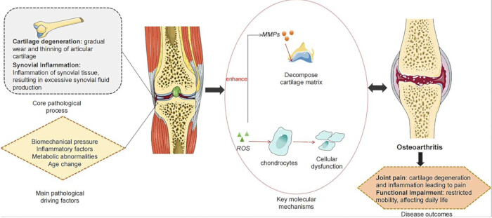
Figure 1: The pathogenesis of osteoarthritis. This adapted diagram shows the pathogenesis and features of osteoarthritis. This is adapted with permission from a study1 under Creative Commons Attribution License 4.0 (CC-BY). Please click here to view a larger version of this figure.
Recent treatments of OA
Management of OA is largely centered on controlling symptoms, with the main goals of reducing pain, restoring joint function, and improving overall quality of life. Current treatment options, such as nonsteroidal anti-inflammatory drugs (NSAIDs), analgesics, physiotherapy, and joint replacement procedures, primarily provide symptomatic relief29. However, these interventions are mostly palliative in nature and do not effectively stop or reverse the underlying progression of the disease.
Regenerative therapy
Recent treatments for OA are moving beyond symptom relief toward modifying disease progression through approaches such as targeted molecular treatments, regenerative medicine, and epigenetic regulation1. Several types of stem cells have potential use in OA. Embryonic stem (ES) cells, which are derived from the inner cell mass of blastocysts, can differentiate into all three germ layers of the embryo30. However, due to ethical concerns, they limit their clinical application31. Mesenchymal stem cells (MSCs) are adult stem cells derived from various tissues, such as bone marrow, adipose tissue, and synovium, and they can differentiate into various cell types, including chondrocytes32. MSCs and growth factors have demonstrated encouraging results as a regenerative therapy—particularly in early-stage OA—with some clinical trials reporting improved joint function, yet concerns about durability and immune responses persist. MSCs also exhibit immunomodulatory and immunosuppressive properties, which can mitigate the inflammatory components of OA33.
Furthermore, transforming growth factor-beta 3 (TGF-β3) can be a potential therapeutic for OA due to its protective effect, which enhances the recruitment of autologous MSCs to damaged cartilage21. TGF-β3 is able to influence the chondrocytes’ life cycle and mediates cellular responses, including proliferation, migration, differentiation, and cell survival, which will ultimately help in cartilage development. Recently, Lee et al.34 studied the hyaluronic acid (HA) gel loaded with TGF-β3 (HAT) for enhanced cartilage regeneration, and they found that HAT is a promising candidate for cartilage regeneration as it provides a rapid and effective clinical approach for cartilage regeneration.
Finally, circadian biology, another emerging area of OA research, influences circadian rhythms, inflammation, and cartilage homeostasis35,36. The disruption of circadian clock genes contributes to OA pathogenesis by disturbing cartilage repair processes and enhancing inflammatory responses.
Gene therapy
Innovative strategies like CRISPR-based gene therapy and 3D bioprinting present exciting possibilities for cartilage regeneration, but are constrained by safety and ethical considerations. Similarly, epigenetic approaches such as HDAC inhibition and miRNA targeting show promise, though challenges in precise delivery and unintended effects remain. Overall, while these emerging therapies offer meaningful progress, combining multiple strategies may be necessary to achieve sustained and effective disease modification.
Nanoparticles and nanotherapeutics
Recently, nanotherapeutics, which use various nanoparticle-based therapies for OA, have been offering a potential benefit in drug delivery, as shown in Figure 226. This nanotherapeutic strategy uses lipids, polymers, and inorganic materials to treat OA through nanoparticles37. This treatment provides the advantages of refining drug delivery and efficacy, reducing cartilage damage, and fostering polarization of M2 macrophages and ECM protein synthesis. In addition, hydrogels, nanoparticles, and nanofibers allow for the sustained release and enhanced bioavailability of therapeutic agents, optimizing treatment efficacy while minimizing systemic side effects38,39.
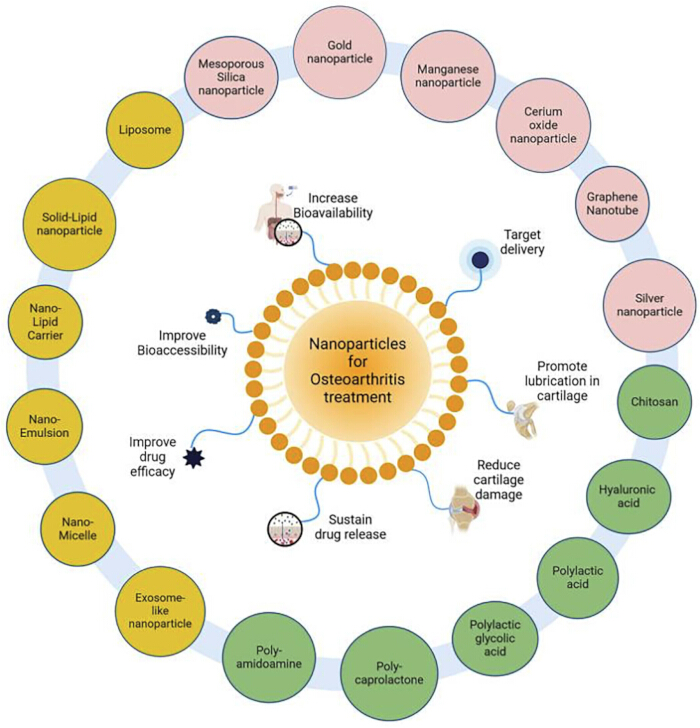
Figure 2: Nanotherapeutics, which uses various nanoparticle-based therapies for OA. This is adapted with permission from a study26 under Creative Commons Attribution License 4.0 (CC-BY). Please click here to view a larger version of this figure.
Figure 3 shows the various Lipid-based nanocarriers (LNs) for drug delivery in OA37. They are liposomes and solid lipid nanoparticles, which are widely used in drug delivery due to their high biocompatibility. Liposomes are especially versatile, as they can encapsulate hydrophilic drugs within their aqueous core and incorporate lipophilic drugs into their lipid bilayer, enabling the delivery of diverse therapeutic agents40. Solid lipid nanoparticles (SLNs) combine the advantages of polymeric carriers and lipid emulsions41. They provide benefits such as controlled drug release, good biocompatibility, and enhanced drug bioavailability. Nanostructured lipid carriers (NLCs) are an improved form of SLNs, offering better physical stability and higher drug loading capacity due to their structured lipid matrix42. They are considered more efficient than traditional lipid-based systems. Nanoemulsions, with droplet sizes below 100 nm, are increasingly used in pharmaceutical and cosmetic applications43. Their large surface area improves drug absorption, and they can be prepared using techniques like ultrasonication and high-pressure homogenization.
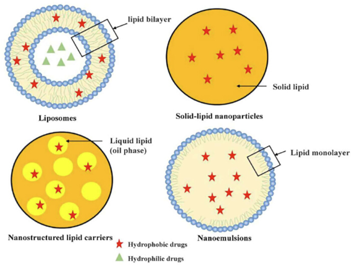
Figure 3: Lipid-based nanocarrier for drug delivery in osteoarthritis. This is adapted with permission from a study37 under Creative Commons Attribution License 4.0 (CC-BY). Please click here to view a larger version of this figure.
Future perspectives
Agents like IL-1β antagonists and ADAMTS-5 inhibitors have been shown to have potential in slowing cartilage breakdown in experimental models, although their long-term clinical benefits remain limited across varied populations. Studies show that there is a strong link between adipocytokine leptin, obesity, and OA44. In obese patients, the leptin levels are increased in synovial fluid, and the leptin receptor (Ob-R) is expressed in cartilage. Leptin causes the production of matrix metalloproteinases (MMPs), pro-inflammatory mediators, and nitric oxide (NO) in chondrocytes. Hence, leptin can be a potential target for disease-modifying drugs for OA in obese patients. Arthroplasty, which is a conventional joint replacement surgery, can be done in advanced OA to replace the damaged joint with an artificial joint. Arthroplasty is most commonly done in the hips, knees, shoulders, and ankles. This is indicated when conservative treatments of OA have failed, causing severe pain and disability45. The goal of arthroplasty is to relieve pain, restore function without restoring damaged cartilage, and improve the patient's quality of life.
Furthermore, improvements in drug delivery systems, particularly through nanotechnology, could enhance therapeutic precision while reducing adverse effects. Regenerative approaches such as stem cell therapy and gene editing also need robust, large-scale clinical studies to establish their long-term safety and economic viability. Furthermore, tailoring treatments based on individual molecular profiles may improve clinical outcomes. Finally, the safety and ethical evaluation of nanotechnology and nanocarriers should be conducted for safety evaluation. Finally, public health interventions are important steps to reduce the prevalence of obesity, and also, this could reduce health inequalities20.
Over the next 5–10 years, OA treatment is expected to move beyond simple pain management toward therapies that can slow disease progression and promote tissue repair. Future approaches are likely to focus on disease-modifying osteoarthritis drugs (DMOADs), regenerative therapies, and personalized medicine. Stem cells, platelet-rich plasma (PRP), exosomes, and tissue engineering techniques may help reduce inflammation and support cartilage regeneration. Advances in nanotechnology could also improve targeted drug delivery within the joint while minimizing side effects. In addition, artificial intelligence and digital health technologies may improve early diagnosis, patient monitoring, and individualized treatment planning. However, challenges such as high costs, limited long-term clinical evidence, and regulatory concerns still need to be addressed before these therapies become widely available. Overall, future OA management is expected to become more personalized, regenerative, and focused on slowing disease progression rather than only controlling symptoms. Future treatments should consider the limitations of available treatments for OA and focus on the development of disease-modifying osteoarthritis drugs (DMOADs). Important considerations should be made on OA’s mechanisms, such as protease activity, OS, and epigenetic modifications. Exploring combination therapies that act on multiple pathways may provide greater benefits than single-target strategies.
Conclusions
The main treatment objectives in OA are to reduce pain, restore joint function, improve overall quality of life, and delay disease progression as much as possible. Due to its complex and multifactorial nature, effective management often involves a multidisciplinary approach. Treatment planning requires careful evaluation of symptom severity, the joints involved, and the patient’s general health, along with consideration of lifestyle, activity level, and individual responses to different therapeutic options. Various potential new treatments include regenerative medicine, gene therapy, targeted molecular therapies, nanoparticles, and nanotherapeutic therapies. Finally, OA treatment is expected to move beyond simple pain management toward therapies that can slow disease progression and promote tissue repair over the next 5–10 years.
Disclosures
The authors disclose no competing interests.