This protocol evaluates postoperative systemic immune-inflammatory responses and perioperative recovery outcomes after robotic and laparoscopic proximal gastrectomy in patients with upper gastric cancer using systemic immune-inflammatory index, neutrophil-to-lymphocyte ratio, and platelet-to-lymphocyte ratio measurements.
A subscription to JoVE is required to view this content. Sign in or start your free trial.
Method Article
Systemic Immune-Inflammatory Index For Evaluating Robotic and Laparoscopic Proximal Gastrectomy in Upper Gastric Cancer
In This Article
Summary
Abstract
Tumor resection is a commonly used treatment method in clinical practice. With the increasing refinement of tumor surgery, the application of robotic-assisted surgery in clinical procedures has expanded. However, effective indicators for evaluating surgical efficacy and postoperative prognosis remain limited. This study introduced the systemic immune-inflammatory index (SII) to evaluate perioperative inflammatory responses and postoperative recovery following robotic and conventional laparoscopic surgery. A total of 81 patients who underwent robotic-assisted gastrointestinal tumor resection and 81 patients who underwent conventional laparoscopic gastrointestinal tumor resection were included. SII was calculated using the formula: platelet count × (neutrophil count/lymphocyte count). Postoperative SII, neutrophil-to-lymphocyte ratio (NLR), and platelet-to-lymphocyte ratio (PLR) increased after surgery, peaked on postoperative day 3, and gradually returned toward baseline by postoperative day 7. From postoperative day 3 onward, SII and NLR values in the robotic surgery group were significantly lower than those in the laparoscopic surgery group (P < 0.05), and these differences remained significant on postoperative days 5 and 7 (P < 0.05). PLR values were significantly lower in the robotic surgery group on postoperative day 3 only (P < 0.05), whereas no significant differences were observed on postoperative days 5 or 7 (P > 0.05). Patients in the robotic surgery group also demonstrated improved postoperative recovery indicators, including earlier ambulation, earlier postoperative exhaust and feeding, lower average drainage volume and shorter postoperative hospital stay. These findings suggest that SII may serve as a useful indicator for evaluating postoperative inflammatory status and perioperative recovery following minimally invasive gastric cancer surgery.
Introduction
Gastric cancer remains one of the leading causes of cancer-related mortality worldwide and represents a major public health burden in China1. The development and progression of gastric cancer are associated with multiple factors, including dietary habits, environmental exposure, and lifestyle-related risk factors2,3,4. Current treatment strategies for gastric cancer commonly involve multimodal management, including neoadjuvant therapy, surgical resection, and adjuvant chemotherapy, depending on tumor stage and patient condition5,6. Surgical resection remains the primary curative treatment for resectable gastric cancer. With the continued advancement of minimally invasive techniques, laparoscopic and robotic-assisted surgical approaches have been increasingly applied in gastric cancer surgery7,8,9. These techniques may improve surgical precision and perioperative recovery while reducing operative trauma.
The Systemic Immune-Inflammatory Index (SII) is a comprehensive biomarker used to evaluate systemic inflammatory and immune status10. Previous studies have demonstrated that postoperative inflammatory responses are associated with clinical recovery and prognosis in patients with gastric cancer11. Conventional inflammatory indicators, including the Neutrophil-to-Lymphocyte Ratio (NLR) and Platelet-to-Lymphocyte Ratio (PLR), have also been used to evaluate postoperative inflammatory status12,13. However, these indicators may not fully reflect the combined immune and inflammatory response of patients after surgery. SII integrates platelet, neutrophil, and lymphocyte counts and may therefore provide a more comprehensive assessment of postoperative inflammatory status14.
In this study, postoperative immune-inflammatory responses were evaluated by analyzing perioperative changes in SII, calculated as follows15:
 (1)
(1)
Here, PLT is the platelet count, N is the neutrophil count, and L is the lymphocyte count.
Differences in SII, NLR, PLR, and postoperative recovery indicators were compared between patients undergoing robotic-assisted surgery and conventional laparoscopic surgery for upper gastric cancer. This study aimed to evaluate the potential clinical value of SII in assessing postoperative inflammatory response and perioperative recovery following minimally invasive gastric cancer surgery.
Access restricted. Please log in or start a trial to view this content.
Protocol
The testing data of all patients pathologically diagnosed with gastric adenocarcinoma were obtained from the laboratory database collected between 2024 and 2025 at the Affiliated Huai’an No.1 People’s Hospital of Nanjing Medical University. Upper gastric cancer was defined as tumors located in the upper third of the stomach or esophagogastric junction. We retrospectively analyzed the collected data. The samples were deidentified prior to use and were processed in compliance with approved institutional protocols approved by the Ethics Committee of the Affiliated Huai’an No.1 People’s Hospital of Nanjing Medical University (KY-2024-250-01). Written informed consent was obtained from all patients prior to enrollment. Inclusion criteria included patients diagnosed with gastric adenocarcinoma by surgery and pathological examination who had not undergone preoperative radiotherapy or chemotherapy and whose preoperative routine hematological test results were within normal ranges. Exclusion criteria included conditions inconsistent with the inclusion criteria, including incomplete clinical or laboratory data. Tumor staging was performed according to the 8th edition of the AJCC/UICC TNM staging system. According to the surgical approach, patients were assigned to the laparoscopic surgery group or robotic-assisted surgery group.
Comparison of surgical steps and surgical details between the conventional laparoscopic surgery group and the robotic-assisted surgery group
The indications for proximal gastrectomy (PG) in this study included: (1) adenocarcinoma of the esophagogastric junction or upper third of the stomach; (2) the possibility of achieving R0 resection while preserving at least 5 cm of the distal stomach; and (3) the absence of bulky lymph node metastasis along the celiac axis (No. 12a, 8a, or 11). For patients with advanced-stage (Stage III) disease, PG was performed following multidisciplinary team (MDT) consensus, prioritizing the balance between oncological safety and postoperative nutritional preservation. Among the enrolled patients, 85.2% (n = 69) in the robotic-assisted surgery group and 82.7% (n = 67) in the laparoscopic surgery group underwent curative-intent R0 resection. The remaining patients underwent palliative surgery to alleviate life-threatening symptoms, including tumor-related hemorrhage or gastric outlet obstruction. Negative surgical margins were confirmed by postoperative pathological examination.
Reconstruction was performed using the modified side-overlap with fundoplication by Yamashita (mSOFY) technique. First, a small incision was created on the right side of the esophageal stump and on the anterior wall of the gastric remnant. A 45 mm linear cutting stapler was then inserted into the prepared anastomotic openings of the esophagus and remnant stomach. The esophagus was rotated 90° counterclockwise along its axis to facilitate anastomosis between the right wall of the esophagus and the remnant stomach, after which the gastric wall was sutured to the left side of the esophagus. The common opening between the esophagus and remnant stomach was subsequently closed using inverted sutures. The left and lower sides of the esophagus were then sutured to the remnant stomach to maintain close apposition of the esophagus against the gastric wall. After opening the sutured common opening, the posterior wall of the lower esophageal segment was compressed into a valve-like structure by the pressure generated from the pseudo-fornix configuration. Intracorporeal anastomosis was performed in all patients undergoing the mSOFY procedure.
After re-establishing pneumoperitoneum, both diaphragmatic crura were incised, and the two corners of the gastric remnant were sutured and fixed to the corresponding diaphragmatic crura to facilitate the subsequent reconstruction procedure. For laparoscopic proximal gastrectomy with mSOFY reconstruction, a five-port configuration was used. Patients were placed in the supine split-leg position with a 20° reverse Trendelenburg (head-up) tilt and a 10° right lateral tilt to facilitate exposure of the upper abdomen and esophagogastric junction. Pneumoperitoneum pressure was maintained between 10–15 mmHg according to the patient’s individual condition.
The trocar layout was established using the five-port method. A 12 mm infraumbilical trocar was inserted as the camera port to provide panoramic laparoscopic visualization. A 12 mm trocar placed at the left anterior axillary line below the costal margin served as the main operating port for vessel ligation, lymphadenectomy, and anastomosis. A 5 mm trocar placed at the left midclavicular line approximately 3 cm above the umbilicus served as the left auxiliary port to facilitate esophageal stump management and anastomosis. Two assistant ports were established on the right side: one 5 mm trocar at the right anterior axillary line below the costal margin for tissue retraction and exposure, and another 5 mm trocar at the right midclavicular line approximately 3 cm above the umbilicus to assist with delicate procedures, including anastomotic reinforcement and anti-reflux fundoplication.
According to the 5th edition of the Gastric Cancer Treatment Guidelines published by the Japanese Gastric Cancer Association, standard lymph node dissection included D1+ and D2 lymphadenectomy. D1+ lymph node dissection was generally performed for patients with early upper gastric cancer staged as cT1N0. D2 lymph node dissection was performed for patients with cT2 or higher tumors or clinically lymph node-positive (cN+) disease. D1+ lymph node dissection included lymph node stations No. 1, 2, 3a, 4sa, 4sb, 7, 8a, 9, and 11p, whereas D2 lymph node dissection included stations No. 1, 2, 3a, 4sa, 4sb, 7, 8a, 9, 10, 11p, and 11d.
Indocyanine green fluorescence imaging was not used during surgery. Multiple surgical teams participated in the procedures; however, all participating surgeons were professionally trained and experienced in minimally invasive gastric cancer surgery, ensuring consistent surgical quality and operative standards across cases. Surgical procedures and operative details were compared between the laparoscopic surgery group and robotic-assisted surgery group to evaluate differences between the two minimally invasive surgical approaches.
Performing blood routine tests on the collected two-year specimens
Fasting peripheral blood samples were collected from all patients in EDTA-K2 anticoagulant tubes at 8:00 a.m. on the day of surgery and postoperative days 3, 5, and 7. The collected specimens were sealed in sterile transport bags and transferred to the laboratory for analysis at room temperature. Peripheral blood samples were analyzed using an automated hematology analyzer. Laboratory personnel were blinded to surgical grouping, and all samples were tested under standardized quality control conditions. Platelet count (×109/L), lymphocyte count (×109/L), and neutrophil count (×109/L) were recorded for each patient. The corresponding SII was calculated using Equation 1, and the NLR16 and PLR17 were calculated using the following Equations 2 and 3, respectively:
 (2)
(2)
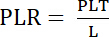 (3)
(3)
Collection of patient data from the laparoscopic surgery group and the robotic-assisted surgery group
To achieve the study objectives, comprehensive preoperative and postoperative clinical data were collected and analyzed for patients in both the laparoscopic surgery group and robotic-assisted surgery group. The collected parameters included demographic characteristics (sex and age), hematological profiles (complete blood count results), and perioperative recovery indicators. Key postoperative recovery indicators were defined as follows: (1) time to first flatus, defined as the interval between completion of surgery and the first passage of gas per rectum; (2) time to first ambulation, defined as the interval between completion of surgery and the patient’s first out-of-bed activity, either assisted or independent; and (3) postoperative hospital stay, defined as the total number of days from surgery to discharge according to standardized clinical discharge criteria. Postoperative complications, including anastomotic leakage, reflux esophagitis, anastomotic stenosis, reoperation, and readmission, were assessed during hospitalization. Complication severity was graded according to the Clavien–Dindo classification system. All patients underwent standardized postoperative management and rehabilitation protocols. Perioperative antibiotic administration and thromboprophylaxis protocols were also standardized between groups. The present study focused on perioperative and in-hospital postoperative outcomes; therefore, long-term postoperative follow-up was not included in the study design.
Statistical analysis
All statistical analyses were performed using statistical analysis software and graphing software. Student’s t-tests, chi-square tests, and repeated-measures analysis of variance (ANOVA) were used for statistical comparisons between groups and across postoperative time points. Data are presented as mean ± standard deviation. The chi-square test was used for statistical analysis of categorical clinical data. Normality testing was performed before parametric analyses. A P value <0.05 was considered statistically significant. Missing or incomplete data were excluded from the analysis.
Access restricted. Please log in or start a trial to view this content.
Results
Comparison of surgical steps and surgical details between the conventional laparoscopic surgery group and the robotic-assisted surgery group
As shown in Figure 1, statistical analysis of operative parameters demonstrated that the robotic-assisted surgery group required a longer reconstruction time than the laparoscopic surgery group (P < 0.05). However, the robotic-assisted approach was associated with significantly lower estimated int...
Access restricted. Please log in or start a trial to view this content.
Discussion
Gastric cancer is associated with high morbidity and mortality worldwide, and its incidence remains a major public health concern19,20. With the continuous development of diagnostic and therapeutic technologies, particularly advances in surgical treatment, the management of gastric cancer has improved substantially21. Therefore, further evaluation of minimally invasive surgical approaches for gastric cancer remains clinically important. In...
Access restricted. Please log in or start a trial to view this content.
Disclosures
Conflict of Interests:
The authors declare no competing interests.
Acknowledgements
The authors thank all team members for their support and contributions to this study.
Access restricted. Please log in or start a trial to view this content.
Materials
| Name | Company | Catalog Number | Comments |
|---|---|---|---|
| Absorbable sutures | Ethicon, Cincinnati, OH, USA | SXMD1B405 | Anastomotic reinforcement and fixation |
| Automated hematology analyzer | Sysmex, Kobe, Japan | XN-2800 | Peripheral blood cell analysis |
| Carbon dioxide insufflator | Olympus, Tokyo, Japan | UHI system | Pneumoperitoneum establishment |
| Circular stapler | Johnson & Johnson, New Brunswick, NJ, USA | EEA | Esophagogastric anastomosis |
| EDTA-K2 anticoagulant blood collection tubes | WEGO, Weihai, China | 20260101 | Peripheral blood sample collection |
| Endoscopic linear stapler | Ethicon, Cincinnati, OH, USA | ECH60C | Gastric transection and reconstruction |
| Energy device for tissue dissection | Ethicon, Cincinnati, OH, USA | Harmonic 1100 | Tissue dissection and hemostasis |
| Presentation software | Microsoft, Redmond, WA, USA | PowerPoint | Figure preparation, annotation, and data visualization |
| Laparoscopic camera system | Karl Storz, Tuttlingen, Germany | TC200 | Intraoperative visualization |
| Laparoscopic surgical system | Karl Storz, Tuttlingen, Germany | 26605AA | Conventional minimally invasive surgery |
| Nasogastric tube | Huacheng, Guangzhou, China | 16 Fr | Gastric decompression |
| Needle holder | Olympus, Tokyo, Japan | A5690 | Intracorporeal suturing |
| Nonabsorbable sutures | Ethicon, Cincinnati, OH, USA | Mersilke | Tissue fixation |
| Pathology specimen container | Cidabio, Guangzhou, China | Bio-CD2492 | Surgical specimen collection |
| Personal protective equipment | 3M, St. Paul, MN, USA | 4565 | Surgical safety and biosafety compliance |
| Robotic-assisted surgical system | Intuitive Surgical, Sunnyvale, CA, USA | Da Vinci Xi IS4000 | Robotic-assisted minimally invasive surgery |
| Statistical analysis software | GraphPad Software, San Diego, CA, USA | GraphPad Prism 8.0 | Statistical analysis |
| Statistical analysis software | IBM Corp., Armonk, NY, USA | SPSS 17.0 | Statistical analysis |
| Surgical forceps and graspers | Olympus, Tokyo, Japan | A63010S | Tissue manipulation |
| Surgical trocars | AOFO/Intuitive Surgical | FQ/Cannula | Port placement during surgery |