This retrieval-based protocol combines scanning electron microscopy, qualitative comparison with simplified reference fractures, and energy-dispersive X-ray spectroscopy. Body-region fractures predominantly showed bucco-lingually oriented features, whereas angle-region fractures showed superior-inferior orientation, suggesting that the mechanical environment of plate failure may vary by anatomical subsite.
Research Article
Retrieval Analysis of Fractured Mandibular Reconstruction Plates Using Scanning Electron Microscopy
In This Article
Summary
Abstract
Mandibular reconstruction plates are widely used following segmental mandibulectomy, but plate fracture remains a significant complication. This study presents a retrieval-based protocol that uses scanning electron microscopy (SEM) to characterize fracture morphology and to infer site-specific mechanical failure directions in titanium mandibular reconstruction plates. We hypothesized that fracture-surface orientation would differ by anatomical fracture location and would exhibit qualitative similarities to reference fractures produced under different loading directions. Seven patients with fractured plates were identified, yielding nine fracture sites. Retrieved plates were cleaned, mounted, and examined by SEM to identify crack initiation sites, fatigue striations, beach marks, and final rupture zones. Energy-dispersive X-ray spectroscopy was performed to assess elemental composition and exclude corrosion-related abnormalities. Two unused reference plates were manually fractured under simplified bucco-lingual and superior-inferior loading, and their fracture-surface features were qualitatively compared with those of the clinical specimens. All six body-region fracture sites showed predominantly bucco-lingually oriented fracture-surface features, whereas the three mandibular-angle fracture sites showed predominantly superior-inferior orientation. Body-region fractures shared selected morphological features with the bucco-lingual reference fracture, while angle-region fractures showed similarities to the superior-inferior reference fracture. Energy-dispersive X-ray spectroscopy detected primarily titanium and oxygen, with no marked unexpected deposits or compositional abnormalities. These findings suggest that the mechanical environment of mandibular reconstruction plate failure may differ by anatomical subsite. However, because the reference experiments used one specimen per loading condition, relied on manual, unquantified loading, and included no cyclic fatigue testing, the observed similarities should be considered qualitative and hypothesis-generating rather than definitive evidence of causal loading mechanisms. Further quantitative biomechanical and computational studies are needed before subsite-specific plate design or reinforcement strategies can be recommended.
Introduction
Mandibular reconstruction after segmental mandibulectomy is essential for restoring continuity, facial contour, mastication, and speech in patients with oncologic or infectious bone defects1,2. Load-bearing titanium reconstruction plates remain widely used because they provide immediate stability, are surgically practical, and can serve either as definitive treatment or as an interim solution before secondary reconstruction3,4. Despite these advantages, complications such as plate exposure, loosening, infection, and fracture continue to limit long-term success5,6,7. Plate fracture is particularly important because it may necessitate revision surgery and is often associated with functional deterioration and soft tissue compromise5,8,9,10,11. Clinical and biomechanical studies have suggested that factors such as radiation exposure, defect length, unsupported span, plate contouring, and fixation strategy influence failure risk12,13,14,15,16. However, the exact directionality of the stresses causing fracture at different mandibular subsites remains insufficiently defined3,5,8,12.
Fractographic analysis offers a direct way to investigate implant failure at the material level. Scanning electron microscopy (SEM)-based fractography enables detailed identification of fatigue striations, presumed crack-initiation sites, beach-mark-like patterns, and final rupture morphology, thereby providing qualitative information regarding the mechanical conditions associated with failure3,8,12. Unlike biomechanical testing, which evaluates structural behavior under controlled loading conditions, or finite element analysis, which computationally estimates stress and strain distributions, SEM-based fractography directly examines morphological features preserved on retrieved fracture surfaces13,14,15,16. However, this approach is primarily qualitative and cannot independently quantify the magnitude of in vivo loading or establish a unique causal loading mechanism; therefore, it is most appropriately used as an exploratory method for identifying failure patterns and generating biomechanical hypotheses.
Although prior studies have examined fracture surfaces of failed mandibular plates3,8,12, the relationship between fracture morphology and anatomical site has not been systematically clarified. The aim of this exploratory retrieval study was to characterize the fracture-surface morphology of clinically retrieved mandibular reconstruction plates and to compare the observed features with those of reference plates fractured under simplified bucco-lingual and superior-inferior loading conditions. We hypothesized that fracture-surface features would differ by the anatomical location of plate failure and would exhibit qualitative similarities to reference fractures produced under different loading directions.
Protocol
All procedures involving human-derived materials were performed in accordance with the Declaration of Helsinki and institutional guidelines. The study protocol was approved by the Institutional Review Board of Seoul National University Dental Hospital (IRB No. CDE19001). Because this was a retrospective study using de-identified clinical data and explanted hardware retrieved from the explant database, the requirement for informed consent was waived by the board. The overall study workflow consisted of four stages: identification and clinical classification of retrieved fractured plates, SEM-based fracture-surface assessment, EDS analysis, and qualitative comparison with simplified generated fractures. An overall study workflow is provided in Figure 1. The reagents and the equipment used are listed in the Table of Materials.
Identification of eligible cases and documenting clinical, defect, and plate characteristics
This retrospective retrieval study included patients who underwent mandibular reconstruction with load-bearing titanium plates and later developed plate fracture requiring removal. Clinical charts, operative records, and radiographic images were reviewed to document patient age, sex, primary diagnosis, reconstructed side, defect location, radiation history, postoperative infection, dentition, and residual occlusal units, time from reconstruction to fracture, and anatomical fracture location. Seven patients contributed seven retrieved plates. Two plates had two distinct fracture lines; therefore, nine fracture sites were included in the descriptive fractographic analysis. The patient and plate were considered the clinical sampling units, whereas the individual fracture site was used as the unit of fracture-surface description. Fracture locations were categorized as posterior body, anterior body, or mandibular angle. Plate-related variables included plate system, nominal plate thickness, hole width, number and distribution of fixation screws, and the length of the unsupported plate span. The exact number of intraoperative bending and rebending maneuvers could not be determined because these details were not consistently documented in the operative records. Panoramic radiographs were used to confirm fracture location and correlate imaging findings with the retrieved specimens.
SEM-based analysis of fracture morphology
After removal, each fractured plate was handled carefully to preserve the fracture surface. Gross biological debris was gently reduced by rinsing the specimens with distilled water without mechanical scraping of the fracture surface. Each plate specimen was placed in a 15-mL conical tube containing sufficient 100% ethanol to fully immerse the specimen. The closed tube was placed in an ultrasonic bath and sonicated for 3 min at 37 kHz, 100W, and 37 °C. After sonication, the specimen was removed from the tube and dried in a clean-air chamber. The same preparation procedure was applied to all clinical and reference specimens. No polishing, grinding, or conductive metal coating was performed on the fracture surfaces. After drying in a clean-air chamber, the specimens were mounted on aluminum stubs using conductive carbon tape. Both opposing fracture surfaces were initially examined at low magnification whenever available. The surface selected for detailed imaging was required to show visible peripheral and central fracture regions, minimal debris obscuring morphological features, no prominent retrieval-related scratches or instrument marks, and no substantial crushing, bending, or post-fracture deformation. When both surfaces met these criteria, both were retained for analysis. Anatomical orientation was determined by matching the plate contour, screw-hole configuration, and fracture location with operative records and panoramic radiographs. The bone-facing surface was designated as the lingual aspect and the opposite surface as the buccal aspect, while the superior and inferior directions were determined from the plate position relative to the mandibular border. These directions were marked and maintained during mounting on the aluminum stub.
Mounted specimens were examined using secondary electron imaging with a secondary electron detector at magnifications of 80×, 200×, 400×, and 1,000×. At approximately 80× magnification, the investigators first confirmed that the entire fracture surface, peripheral margins, and central rupture region were adequately visualized. The presumed crack-initiation region and final overload zone were then identified before targeted imaging at 200×, 400×, and 1,000×. Overview images were acquired at approximately 80× magnification using an accelerating voltage of 10.0 kV, a probe current of 0.10 nA, a dwell time of 5.0 µs, and a working distance of 40.0 mm. Each fracture surface was reviewed systematically from the peripheral margin to the central rupture zone. Each fracture surface was assessed using predefined morphological categories, including the presumed crack-initiation margin, predominant crack-propagation axis, striation-like markings, beach-mark-like patterns, ductile tearing, brittle-appearing regions, and the presumed final overload zone. Crack-propagation orientation was classified as predominantly bucco-lingual, predominantly superior-inferior, mixed, or indeterminate. Special attention was given to the orientation of fatigue features relative to the buccal, lingual, superior, and inferior aspects of the plate in order to infer the predominant direction of mechanical stress. The investigators independently reviewed the images before conducting a consensus review.
Comparison of clinical fractures with reference fractures under simplified loading conditions
Unused reconstruction plates of the same system and nominal dimensions as the clinically retrieved plates were used to generate reference fractures. One plate was tested under bucco-lingual loading, and another under superior-inferior loading. For each plate, the intended loading point was marked in advance. The plate was then manually supported at locations corresponding to two holes away from the marked loading point on each side.
Loading was applied manually at the marked location until a gross plate fracture occurred. For the bucco-lingual condition, force was applied in the bucco-lingual direction. For the superior-inferior condition, force was applied in the superior-inferior direction.
These reference fractures were generated to provide simplified morphological comparisons rather than to reproduce the complex cyclic and multidirectional loading environment experienced by mandibular reconstruction plates in vivo. After fracture, all reference specimens underwent the same cleaning, mounting, and SEM imaging procedures used for the retrieved clinical plates.
Clinical and reference fractures were compared using the following predefined features: presumed crack-initiation margin, predominant fracture-plane orientation, crack-propagation axis, orientation of striation-like or beach-mark-like features, distribution of ductile tearing, and location of the presumed final overload zone. Each clinical fracture was classified as more similar to the bucco-lingual reference pattern, more similar to the superior-inferior reference pattern, mixed, or indeterminate. Disagreements between the investigators were resolved by joint review and consensus. Similarity assessment was qualitative and was not intended to establish a unique causal loading mechanism.
Assessment of elemental composition by energy-dispersive X-ray spectroscopy
Energy-dispersive X-ray spectroscopy was performed during electron microscopy to assess elemental composition and screen for evidence of corrosion-related degradation or contamination. Energy-dispersive X-ray spectroscopy was performed using an SEM-integrated silicon-drift detector. Spectra were acquired at an accelerating voltage of 20 kV and a working distance of 10.0 mm at 1,000× magnification. A rectangular region of interest was positioned over a relatively flat portion of the fracture surface while avoiding the specimen edge and gross surface debris. Elemental composition was quantified from the acquired spectrum and reported as weight percentage (wt%) and atomic percentage (at%).
Protocol completion and compilation of final outputs
Complete the procedure after confirming that each evaluable fracture site has an anatomically oriented SEM image set, a completed morphological assessment, a crack-propagation orientation classification, a qualitative reference-pattern classification, and an EDS composition report expressed as wt% and at%. Integrate these outputs with the corresponding clinical and radiographic data to generate the final specimen-level analysis dataset.
Results
Identification of eligible cases and documenting clinical, defect, and plate characteristics
Seven patients with fractured mandibular reconstruction plates were identified for retrieval-based analysis. The cohort included five males and two females, with a mean age of 60.6 years (range, 30 years to 79 years) at the time of fracture. The underlying diagnoses were squamous cell carcinoma in four patients, osteomyelitis in two patients, and osteosarcoma in one patient. The mean interval from reconstruction surgery to plate fracture was 14 months, ranging from 2 months to 47 months. Reconstruction had been performed on the left mandible in three patients and on the right mandible in four patients. Fracture sites were evenly distributed among three anatomical regions: posterior body (n = 3), anterior body (n = 3), and mandibular angle (n = 3). Four reconstruction plate systems were represented. The plates contained a median of 16 holes (range, 12–16 holes). The median total number of fixation screws was 9 (range, 7–14), with 2–8 screws placed on the proximal segment and 4–6 screws on the distal segment. The unsupported plate span had a median length of 6 plate holes (range, 2–7 holes). The number of functional occlusal units ranged from four to eight, with a median of six units. Four patients had received radiotherapy, whereas three had not. No patient had documented postoperative infection or nonunion at the time of plate fracture. Detailed specifications for each plate, including the manufacturer and plate system, nominal thickness, hole width, fixation details, and unsupported span, are provided in Figure 2 and Table 1.
SEM-based analysis of fracture morphology
SEM revealed site-associated differences in the observed fracture-surface morphology. In the mandibular body region, all six body-region fracture sites showed a predominantly buccolingual orientation of the observed fracture-surface features (Figure 3 and Figure 4).
Striation-like markings and beach-mark-like patterns were observed at the buccal or lingual margins in body-region fractures. These features were considered compatible with progressive crack growth under repeated loading, although the magnitude and direction of the in vivo loading could not be determined from morphology alone. As crack propagation advanced across the plate, the peripheral regions often transitioned to brittle fracture morphology. In the mandibular angle group, the observed fracture-surface features were predominantly oriented along the superior-inferior axis (Figure 5).
The presumed crack-initiation region was located at the superior or inferior plate margin in 3/3 specimens. Vertically oriented ductile-tearing features and brittle-appearing regions extending through the plate thickness were observed in 3/3 specimens. These observations were compatible with site-associated differences in fracture morphology; however, fracture morphology alone could not establish the magnitude or causal direction of in vivo loading (Table 2).
Representative high-magnification SEM micrographs and the corresponding raw image dataset for each fracture site are provided in Supplementary File 1.
Comparison of clinical fractures with reference fractures under simplified loading conditions
Loading was applied manually; therefore, force, displacement, loading rate, fracture load, and fatigue-related parameters were not measured. Two reference plates were fractured, with one plate assigned to each simplified loading direction. Because only one plate was tested under each condition, experimental reproducibility could not be assessed. Bucco-lingual loading produced fractures characterized predominantly by crack initiation at the lateral margins of the plate and fracture-surface features oriented along the bucco-lingual axis. In contrast, superior-inferior loading produced fractures with crack initiation at the superior or inferior plate margin and features oriented predominantly along the superior-inferior axis.
On qualitative comparison, the body-region clinical fractures more frequently shared morphological characteristics with the fractures generated under bucco-lingual loading. These shared features included lateral-margin crack initiation and a predominantly transverse orientation of the fracture-surface features. The clinical fractures at the mandibular angle more frequently resembled the superior-inferior reference fractures, particularly with respect to crack initiation at the superior or inferior margin and the vertical orientation of the observed fracture features.
These comparisons showed selected qualitative morphological similarities between the clinical and reference fractures. However, because the reference experiments used simplified loading conditions and did not reproduce the cyclic, multidirectional forces acting on reconstruction plates in vivo, the observed similarities were interpreted as qualitative and hypothesis-generating rather than as definitive evidence of the causal loading mechanism (Figure 6).
The reference experiments were exploratory qualitative comparisons and were not intended to provide biomechanical validation.
Assessment of elemental composition by energy-dispersive X-ray spectroscopy
Energy-dispersive X-ray spectroscopy demonstrated that titanium and oxygen accounted for the largest detected elemental proportions in the analyzed regions. Small amounts of carbon were detected in five patients, and sodium was detected in one patient only. No marked unexpected elemental deposits or compositional abnormalities were detected at the analyzed sites. Values represent a single area measurement from each selected fracture-surface region (Table 3). The complete EDS spectra and raw quantitative outputs are provided in Supplementary File 1.
Conclusion
This exploratory retrieval study identified differences in fracture-surface morphology among anatomical failure sites of mandibular reconstruction plates. Body-region fractures more frequently showed features oriented along the bucco-lingual axis, whereas angle-region fractures more frequently showed features oriented along the superior-inferior axis. Manually generated reference fractures showed selected qualitative similarities to the corresponding clinical specimens but served only as exploratory comparators and were not intended as biomechanical validation. Because no quantitative force, displacement, loading-rate, or fatigue-testing parameters were obtained, and because the experimental model did not reproduce the complex cyclic and multidirectional loading environment of the mandible in vivo, these observations should be interpreted as hypothesis-generating rather than causal. These findings generate the possibility that the mechanical environment associated with plate failure may vary by anatomical subsite. Further studies incorporating standardized cyclic fatigue testing, quantitative force and displacement measurements, larger homogeneous cohorts, and finite element analysis are required before site-specific reinforcement strategies can be recommended.
DATA AVAILABILITY:
All data generated or analyzed in this study are included in the manuscript and its supplemental files. Representative high-magnification SEM images, raw image datasets, energy-dispersive X-ray spectroscopy spectra, and raw quantitative outputs are provided as supplemental materials. The raw SEM images, EDS spectra, quantitative outputs, and de-identified analytical dataset are publicly available in Zenodo at DOI: 10.5281/zenodo.20744888.
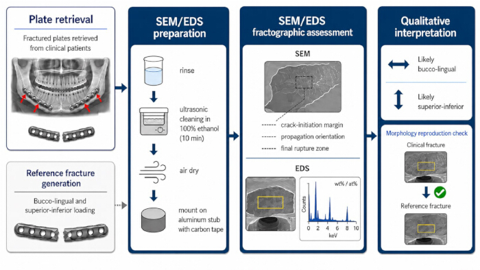
Figure 1: Overall study workflow of the retrieval-based fractographic study. Clinical cases with fractured mandibular reconstruction plates were identified, and the retrieved plates were characterized according to fracture location, plate and fixation configuration, unsupported span, functional occlusal units, radiotherapy exposure, and infection or nonunion status. Retrieved specimens were cleaned, mounted, and examined using SEM to assess fracture-surface morphology and energy-dispersive X-ray spectroscopy to characterize elemental composition. Two unused reference plates were manually fractured under simplified bucco-lingual and superior-inferior loading conditions. Clinical and reference fracture surfaces were qualitatively compared using predefined morphological criteria, including the presumed crack-initiation margin, fracture-plane orientation, propagation axis, striation- or beach-mark-like features, ductile tearing, and the final overload zone. The comparison was intended to generate hypotheses regarding possible site-dependent mechanical environments and was not considered a quantitative biomechanical validation. Please click here to view a larger version of this figure.
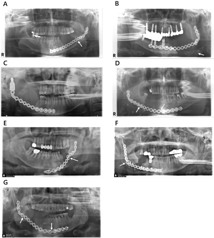
Figure 2: Anatomical location of clinical fractures in retrieved mandibular reconstruction plates. Panoramic radiographs from seven patients (A–G) showing the anatomical locations of mandibular reconstruction plate fractures. Fracture sites were identified in the posterior body (A,D,E), anterior body (C,D,G), and mandibular angle (B,F,G). Please click here to view a larger version of this figure.
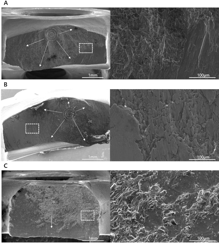
Figure 3: Scanning electron micrographs of clinical fractures in posterior mandibular body fractures. (A,B) Fracture surfaces from Patients 1 and 5 showing buccal-to-lingual crack propagation with concentric fatigue striations. (C) Fracture surface from Patient 4 showing bucco-lingually oriented features with ductile-tearing and brittle-appearing regions. Please click here to view a larger version of this figure.
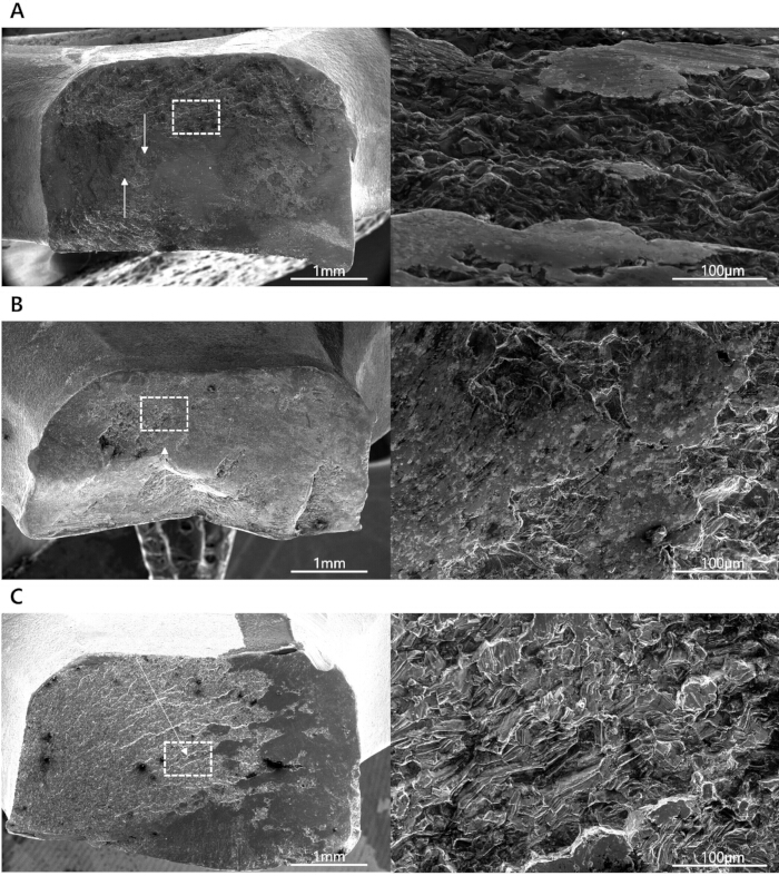
Figure 4: Scanning electron micrographs of clinical fractures in anterior mandibular body fractures. (A) Fracture surface from Patient 3 showing mixed buccal-to-lingual and lingual-to-buccal directional features with striation-like markings and beach-mark-like patterns. (B) Fracture surface from Patient 4 showing lingual-buccally oriented features with ductile-tearing and brittle-appearing regions. (C) Fracture surface from Patient 7 showing bucco-lingually oriented features with beach-mark-like patterns. Please click here to view a larger version of this figure.
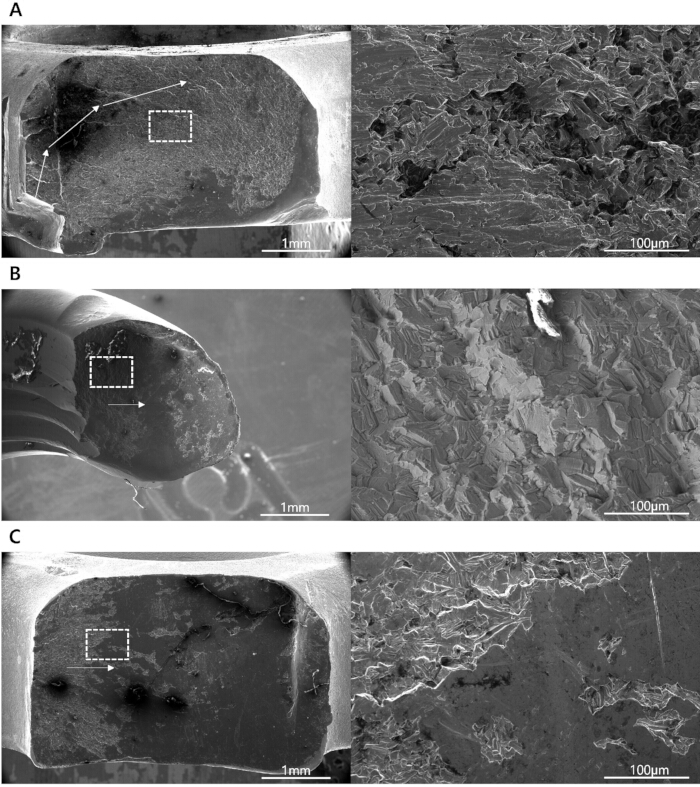
Figure 5: Scanning electron micrographs of clinical fractures in mandibular angle fractures. (A) Fracture surface from Patient 2 showing inferior-superiorly oriented features with striation-like markings. (B,C) Fracture surfaces from Patients 6 and 7 showing inferior-superiorly oriented features with combined ductile-tearing and brittle-appearing regions. Please click here to view a larger version of this figure.
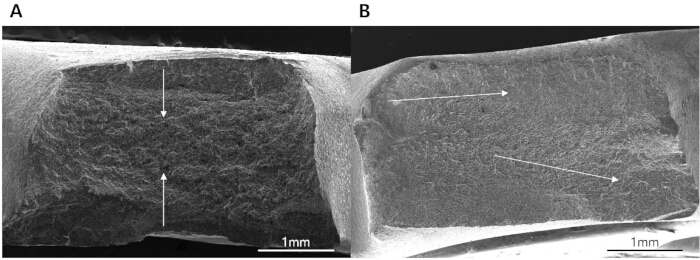
Figure 6: Reference fracture surfaces generated under simplified loading conditions. (A) Reference fracture generated under bucco-lingual loading and (B) reference fracture generated under superior-inferior loading. Arrows indicate the predominant orientation of the observed fracture-surface features. Please click here to view a larger version of this figure.
| Patients | Sex | Age | Diagnosis | Side | Defect region | Plate system | Thickness (mm) | Screw hole (mm) | Total plate holes | Fixation screws, total (proximal/distal) | Un supported span (number of holes) | Funct ional occlusal units, n | Radio therapy | Infection or nonunion | Time of plate fracture (months) | |
| 1 | M | 36 | SCC | Lt | Posterior body | Titanium MatrixMANDIBLE Reconstruction Plate (DePuy Synthes) | 2.8 | 2.4 | 15 | 8 (4/4) | 7 | 6 | yes | no | 20 | |
| 2 | M | 76 | SCC | Lt | Angle | Universal 2 Mandible Reconstruction Plate (Stryker Leibinger) | 2.8 | 2.3 | 16 | 10 (5/5) | 6 | 4 | yes | no | 10 | |
| 3 | F | 30 | Osteosarcoma | Rt | Anterior body | Universal 2 Mandible Reconstruction Plate (Stryker Leibinger) | 2.8 | 2.3 | 13 | 7 (2/5) | 6 | 8 | yes | no | 47 | |
| 4 | M | 70 | SCC | Rt | Posterior body | Universal 2 Mandible Reconstruction Plate (Stryker Leibinger) | 2.8 | 2.3 | 16 | 11 (6/5) | 5 | 6 | yes | no | 12 | |
| Anterior body | ||||||||||||||||
| 5 | M | 74 | Osteomyelitis | Lt | Posterior body | Titanium MatrixMANDIBLE Reconstruction Plate (DePuy Synthes) | 2.8 | 2.4 | 12 | 7 (3/4) | 5 | 8 | no | no | 2 | |
| 6 | F | 79 | Osteomyelitis | Rt | Angle | OPTIMUS Mandible Reconstruction Plate (Osteonic) | 2.6 | 2.4 | 16 | 14 (8/6) | 2 | 8 | no | no | 3 | |
| 7 | M | 59 | SCC | Rt | Angle | Universa 2 Mandible Reconstruction Plate (Stryker) | 2.8 | 2.3 | 16 | 9 (4/5) | 7 | 6 | no | no | 4 | |
| Anterior body | ||||||||||||||||
Table 1: Clinical, reconstructive, and implant characteristics of the retrieved mandibular reconstruction plates. Summary of patient sex, age, diagnosis, plate manufacturer and system, nominal plate profile (thickness, screw hole), reconstructed mandibular side, fracture location, total number of plate holes, number and distribution of fixation screws, unsupported plate span, functional occlusal units, radiotherapy history, infection or nonunion status, and time from reconstruction to plate fracture. Functional occlusal units were defined as opposing maxillary–mandibular contacts in the molar, premolar, and anterior regions, with each contacting unit counted once.
| Patient No. | Anatomical fracture site | Direction of fracture propagation | Fracture morphology |
| 1 | Posterior body | Buccal à Lingual | Concentric striations |
| 2 | Angle | Inferior à Superior | Striations and beach marks |
| 3 | Anterior body | Buccal à Lingual | Beach marks |
| Lingual à Buccal | |||
| 4 | Posterior body | Buccal à Lingual | Ductile and brittle |
| Anterior body | Lingual à Buccal | Ductile and brittle | |
| 5 | Posterior body | Lingual à Buccal | Concentric striations |
| 6 | Angle | Inferior à Superior | Ductile and brittle |
| 7 | Angle | Inferior à Superior | Ductile and brittle |
| Anterior body | Buccal à Lingual | Beach marks |
Table 2: Fractographic findings of fractured mandibular reconstruction plates observed by scanning electron microscopy. Summary of anatomical fracture site, direction of crack propagation, and dominant fracture morphology for each analyzed fracture surface.
| Ti | O | C | Na | ||||||
| Patient No. | Mass Norm % | Atom % | Mass Norm % | Atom % | Mass Norm % | Atom % | Mass Norm % | Atom % | |
| 1 | 75.85 | 51.2 | 24.15 | 48.8 | |||||
| 2 | 90.25 | 75.58 | 9.75 | 24.42 | |||||
| 3 | 57.77 | 30.29 | 24.95 | 39.15 | 11.72 | 24.5 | 5.56 | 6.07 | |
| 4 | 81.45 | 58.77 | 16.94 | 36.58 | 1.62 | 4.65 | |||
| 5 | 84.79 | 62.52 | 9.87 | 21.78 | 5.34 | 15.7 | |||
| 6 | 84.4 | 62.8 | 12.29 | 27.37 | 3.31 | 9.83 | |||
| 7 | 74.8 | 47.09 | 16.53 | 31.14 | 8.67 | 21.76 | |||
Table 3: Elemental composition of fractured mandibular reconstruction plates determined by energy-dispersive X-ray spectroscopy. Elemental composition is reported as normalized weight percentage (wt%) and atomic percentage (at%) obtained by EDS area analysis at 20 kV and a working distance of 10.0 mm.
Supplementary File 1: Energy-dispersive X-ray spectroscopy analysis from Patients 1–7. Scanning electron microscopy images demonstrate the selected regions of interest for EDS analysis, with corresponding spectra and elemental composition tables. Titanium was the predominant element in all specimens, accompanied by oxygen, consistent with titanium oxide formation on the plate surface. Carbon was detected in Patients 3–7, and sodium was detected in Patient 3.Please click here to download this file.
Discussion
This exploratory study proposes a retrieval-based protocol that combines SEM, comparative reference loading, and energy-dispersive X-ray spectroscopy to characterize fracture-surface morphology and to explore site-associated directional patterns in fractured mandibular reconstruction plates. Previous studies have identified fatigue as a major mechanism of mandibular reconstruction plate failure through direct material and fractographic analyses3,8,12. Other studies have focused primarily on clinical risk factors, plate design, or computational and bench-top biomechanical evaluation9,10,11,13,14,15,16,17. In the present study, fracture-surface features differed according to anatomical fracture location. Body-region fractures more frequently showed features oriented along the bucco-lingual axis, whereas angle-region fractures more frequently showed features oriented along the superior-inferior axis. Simplified reference loading experiments yielded selected morphological features qualitatively similar to those observed in the corresponding clinical specimens. These findings suggest that the predominant mechanical environment of plate failure may vary according to anatomical subsite, although they do not establish a unique causal loading mechanism.
The observed differences provide a possible extension of the general concept of fatigue-related plate failure. In the mandibular body, fractures predominantly propagated in the buccolingual direction, suggesting vulnerability to lateral bending stress. However, the reference fracture experiments were designed solely for qualitative, exploratory morphological comparisons and were not intended for biomechanical validation. Because loading was applied manually, no quantitative measurements of force, displacement, loading rate, fracture load, or fatigue-testing parameters were obtained. Moreover, the model did not reproduce the complex cyclic, multidirectional, and patient-specific loading environment experienced by mandibular reconstruction plates in vivo. Accordingly, the observed similarities should be interpreted only as hypothesis-generating patterns requiring confirmation through standardized biomechanical testing.
If confirmed in larger and more controlled studies, anatomical variation in the mechanical environment may have implications for reconstruction plate design. Potential strategies could include regional modification of plate stiffness, cross-sectional geometry, plate thickness, or supplemental reinforcement. Nevertheless, the present findings are insufficient to recommend a particular subsite-specific plate configuration. Such recommendations would require comparative testing that accounts for defect length, unsupported plate span, fixation method, plate contouring, residual bony support, and patient-specific loading conditions11,13,14,15,16,17.
Energy-dispersive X-ray spectroscopy detected titanium and oxygen as the dominant elements, with small and inconsistent amounts of carbon and sodium in a subset of specimens. These minor signals are more likely attributable to surface oxidation, adsorbed contaminants, or physiologic residues than to true alloy alteration18,19,20,21. No marked unexpected elemental deposits or compositional abnormalities were identified in the analyzed regions. However, energy-dispersive X-ray spectroscopy provides localized elemental information and cannot independently exclude localized corrosion, fretting, tribocorrosion, or other surface-degradation processes18,19,20,21. Therefore, the energy-dispersive X-ray spectroscopy findings should not be interpreted as definitive evidence that chemical degradation played no role in plate failure. Rather, the fractographic findings were more compatible with progressive mechanical failure, while the relative contribution of corrosion-related processes remains uncertain.
Retrieval-based fractography has several strengths. It directly examines implants that failed in clinical practice and enables identification of potential crack-initiation regions, propagation features, and final rupture zones. It can also relate fracture morphology to clinical and anatomical information without relying entirely on assumed loading conditions. However, retrieval analysis cannot reconstruct the complete loading history of an implant or quantify the magnitude, frequency, and direction of forces acting before failure. The observed fracture surface may also reflect the combined effects of plate contouring, repeated loading, local stress concentration, biological exposure, explantation, and specimen preparation.
Several protocol steps are critical to the quality and reliability of fractographic analysis. The fracture surface should be protected from scraping and instrument contact during retrieval and cleaned gently without mechanical abrasion, because contamination and secondary damage may obscure fatigue-related features. Anatomical orientation should be established before mounting by correlating plate contour and screw-hole configuration with operative and radiographic findings; specimens with uncertain orientation should be classified as indeterminate. Low-magnification examination of the entire fracture surface should precede targeted high-magnification imaging to preserve the spatial relationship among the presumed initiation region, propagation features, and final rupture zone. Secure conductive mounting and appropriate adjustment of the working distance and scan conditions can reduce charging and focusing artifacts. For EDS analysis, regions near specimen edges, carbon tape, or visible debris should be avoided to minimize misleading elemental signals18. Consistent imaging conditions, predefined morphological categories, and independent review followed by consensus are important for reducing interpretive variability, and fractures with obscured or ambiguous features should not be assigned to a directional reference category.
Practical advantages of the protocol include the use of clinically retrieved implants, minimal destructive specimen preparation, integration of clinical, radiographic, morphological, and elemental data, and a standardized low-to-high magnification workflow. The method can be implemented in laboratories with access to conventional SEM and EDS equipment without requiring a dedicated mechanical testing system. Predefined orientation, imaging, and classification procedures may also improve reproducibility across specimens and investigators. However, accessibility remains dependent on the availability of SEM expertise and appropriate retrieval handling.
Finite element analysis and standardized fatigue testing provide complementary information. Finite element analysis can estimate stress and strain distributions and evaluate the effects of defect geometry, plate design, fixation configuration, and loading assumptions13,16,17. However, its results are sensitive to assumptions regarding muscle forces, occlusal contact, material properties, boundary conditions, and bone–implant interaction13,16. Cyclic bench-top fatigue testing can provide quantitative measurements such as construct stiffness, cycles to failure, fatigue strength, and failure load under standardized conditions13,15. Nevertheless, such experiments necessarily simplify the variable biological, anatomical, and multidirectional loading environment encountered in vivo. Retrieval-based fractography, finite element analysis, and fatigue testing should therefore be considered complementary rather than interchangeable approaches.
This protocol may be useful in multiple areas of craniomaxillofacial research. It may facilitate comparison between conventional and patient-specific plates, support validation of finite element predictions using retrieved implants, and help identify which mandibular subsites are most vulnerable in different reconstructive settings11,13,14,15,16,17. In addition, it may aid in the development of hybrid reconstruction strategies that combine plates with vascularized bone grafts, load-sharing segments, or supplemental bars in mechanically demanding regions1,2,13,14,15.
Several limitations should be acknowledged. First, this was a small retrospective retrieval study of seven patients and nine fracture sites, with two plates contributing two fracture lines each; therefore, some observations were not statistically independent, and the findings may not be generalizable. The cohort was also heterogeneous with respect to diagnosis, defect location, radiotherapy exposure, functional occlusal units, unsupported plate span, fixation configuration, and plate system. Differences in plate thickness, cross-sectional profile, hole geometry, material processing, and manufacturer-specific design may have affected stress concentration and fracture-surface morphology11,13,14,15,16. Because of the small and heterogeneous cohort, the effects of anatomical site could not be separated from those of plate-system design.
Second, the number, location, and extent of intraoperative bending or rebending maneuvers were not consistently documented. Repeated contouring may introduce residual stress or local mechanical weakening and could therefore have contributed to plate failure16.
Third, the reference fracture model was highly simplified. Thus, the reference experiments should not be interpreted as biomechanical validation of the loading directions inferred from the clinical fracture surfaces. Only one unused plate was tested for each loading direction, and loading was applied manually and monotonically without quantitative measurement of force, displacement, loading rate, or fracture load. Reproducibility could not be assessed, and the model did not reproduce the cyclic, multidirectional, and patient-specific forces present in vivo. Accordingly, the experimental fractures served only as qualitative morphological references.
Fourth, comparison between clinical and reference fractures was based on qualitative assessment of predefined fractographic features. Although images were independently reviewed and disagreements were resolved by consensus, the interpretation remained partly subjective, and fracture morphology alone cannot uniquely determine the magnitude or direction of the causative loading.
Finally, SEM and energy-dispersive X-ray spectroscopy provide localized structural and elemental information but cannot reconstruct the complete mechanical, biological, or chemical history of the implants18. The absence of marked compositional abnormalities does not exclude localized corrosion, fretting, tribocorrosion, or interactions between surface degradation and fatigue19,20,21. These findings should therefore be regarded as exploratory and hypothesis-generating and require confirmation through larger retrieval cohorts, standardized cyclic fatigue testing, quantitative mechanical measurements, and patient-specific finite element analysis.
Future studies should integrate retrieval-based fractography with patient-specific geometry, functional loading data, and reconstructive design variables. Larger multicenter retrieval cohorts may clarify whether particular defect patterns or fixation strategies predispose to body-dominant or angle-dominant failure. Overall, these observations provide a basis for future investigation of whether subsite-specific reinforcement may be beneficial, but they do not directly support a particular plate-design strategy.
Disclosures
The authors declare no competing financial interests.
Acknowledgements
This work was supported by the New Faculty Startup Fund from Seoul National University (No. 860-20250053). The authors thank the staff of Seoul National University Dental Hospital for assistance with specimen handling and imaging support.
Materials
| Name | Company | Catalog Number | Comments |
|---|---|---|---|
| Energy-dispersive X-ray spectroscopy detector (Apreo 2) | Thermo Fisher Scientific | Part No. 1229004 | Integrated detector used for elemental analysis during scanning electron microscopy |
| OPTIMUS Mandible Reconstruction Plate | Osteonic | MST326M17 | Retrieved fractured clinical specimens analyzed in this study |
| Scanning electron microscope (Apreo 2) | Thermo Fisher Scientific | Part No. 1229004 | Used for fractographic examination of fracture surfaces |
| Titanium MatrixMANDIBLE Reconstruction Plate | DePuy Synthes | 04.503.739 | Retrieved fractured clinical specimens analyzed in this study |
| Ultrasonic bath | SHARP | UT-106 | Ethanol cleaning of the plates |
| Universal 2 Mandible Reconstruction Plate | Stryker Leibinger | 55-28922 55-28920 | Retrieved fractured clinical specimens analyzed in this study |
References
- Paré A, et al. Reconstruction of segmental mandibular defects: current procedures and perspectives. Laryngoscope Investig Otolaryngol. 2019;4:587-596.
- Cordeiro PG, Henderson PW, Matros E. A 20-year experience with 202 segmental mandibulectomy defects: a defect classification system, algorithm for flap selection, and surgical outcomes. Plast Reconstr Surg. 2018;141:571e–581e.
- Almansoori AA, et al. Fracture of standard titanium mandibular reconstruction plates and preliminary study of three-dimensional printed reconstruction plates. J Oral Maxillofac Surg. 2020;78:153-166.
- Ryu JK, et al. Mandibular reconstruction using a titanium plate: the impact of radiation therapy on plate preservation. Int J Radiat Oncol Biol Phys. 1995;32:627-634.
- Shibahara T, Noma H, Furuya Y, Takaki R. Fracture of mandibular reconstruction plates used after tumor resection. J Oral Maxillofac Surg. 2002;60:182-185.
- Seol GJ, et al. Reconstruction plates used in the surgery for mandibular discontinuity defect. J Korean Assoc Oral Maxillofac Surg. 2014;40:266-271.
- Fanzio PM, et al. Plate exposure after anterolateral thigh free-flap reconstruction in head and neck cancer patients with composite mandibular defects. Ann Surg Oncol. 2015;22:3055-3060.
- Martola M, Lindqvist C, Hänninen H, Al-Sukhun J. Fracture of titanium plates used for mandibular reconstruction following ablative tumor surgery. J Biomed Mater Res B Appl Biomater. 2007;80:345-352.
- Petrides GA, Fox RJ, Winters R. Factors associated with plate fractures in mandibular reconstruction. Curr Opin Otolaryngol Head Neck Surg. 2025;33:242-248.
- Ritschl LM, et al. Retrospective analysis of complications in 190 mandibular resections and simultaneous reconstructions with free fibula flap, iliac crest flap or reconstruction plate: a comparative single centre study. Clin Oral Investig. 2021;25:2905-2914.
- Kreutzer K, et al. Patient-specific 3D-printed mini- versus reconstruction plates for free flap fixation at the mandible: retrospective study of clinical outcomes and complication rates. J Craniomaxillofac Surg. 2023;51:621-628.
- Katakura A, Shibahara T, Noma H, Yoshinari M. Material analysis of AO plate fracture cases. J Oral Maxillofac Surg. 2004;62:348-352.
- Gutwald R, Jaeger R, Lambers FM. Customized mandibular reconstruction plates improve mechanical performance in a mandibular reconstruction model. Comput Methods Biomech Biomed Engin. 2017;20:426-435.
- Zeller AN, Neuhaus MT, Weissbach LVM, et al. Patient-specific mandibular reconstruction plates increase accuracy and long-term stability in immediate alloplastic reconstruction of segmental mandibular defects. J Maxillofac Oral Surg. 2020;19:609–615.
- Kasper R, Winter K, Pietzka S, Schramm A, Wilde F. Biomechanical in vitro study on the stability of patient-specific CAD/CAM mandibular reconstruction plates: a comparison between selective laser melted, milled, and hand-bent plates. Craniomaxillofac Trauma Reconstr. 2021;14:135-143.
- Wan B, et al. On effect of residual stress on fracture behavior of mandibular reconstruction plates. Eng Fract Mech. 2024;305:110158.
- Do J, Han JJ, Lee JH, Kwon IJ. Clinical outcomes and mechanical evaluation of patient-specific mandibular reconstruction following tumor resection: a retrospective study with finite element analysis. World J Surg Oncol. 2026;24:167.
- Newbury DE, Ritchie NWM. Performing elemental microanalysis with high accuracy and high precision by scanning electron microscopy/silicon drift detector energy-dispersive X-ray spectrometry (SEM/SDD-EDS). J Mater Sci. 2015;50:493-518.
- Kuphasuk C, et al. Electrochemical corrosion of titanium and titanium-based alloys. J Prosthet Dent. 2001;85:195-202.
- Zaffe D, Bertoldi C, Consolo U. Element release from titanium devices used in oral and maxillofacial surgery. Biomaterials. 2003;24:1093-1099.
- Rodrigues DC, Valderrama P, Wilson TG Jr, et al. Titanium corrosion mechanisms in the oral environment: a retrieval study. Materials (Basel). 2013;6:5258-5274.
Reprints and Permissions
This article has been published
Video Coming Soon