This review summarizes the role of DNA methylation in immune regulation and transplant rejection. DNA methylation regulates immune cell development and differentiation, modulates immune cell function, and may serve as a biomarker and therapeutic target for improving graft survival.
Review Article
Immunological Regulation of DNA Methylation and its Implication in Allograft Rejection
64 views
⸱
September 11th, 2026
In This Article
Summary
Abstract
Transplant rejection remains a major barrier to long-term graft survival and is driven by complex interactions between immune responses and molecular regulatory mechanisms. DNA methylation, a major epigenetic modification, plays an important role in regulating immune cell development, differentiation, maturation, and function. Aberrant DNA methylation can alter the expression of immune-related genes and influence the nature and intensity of immune responses. In transplantation, these changes may affect T cells, B cells, and innate immune cells involved in graft rejection. DNA methylation also contributes to the regulation of signaling pathways associated with immune activation, tolerance, and inflammatory responses. Changes in methylation patterns may therefore influence both the progression of rejection and the maintenance of graft function. This review summarizes current knowledge of DNA methylation in immune regulation and examines its implications for transplant rejection, with particular emphasis on thymic development, helper and cytotoxic T cells, regulatory T cells, B cells, innate immune cells, and signaling pathways. It also considers the potential relevance of DNA methylation as a biomarker and therapeutic target in transplantation. Understanding these epigenetic mechanisms may support the development of more precise approaches for assessing rejection risk, modulating immune responses, and improving long-term graft outcomes following solid organ transplantation.
Introduction
Immunology is the scientific discipline concerned with the structure and function of the immune system, including the mechanisms underlying immune responses and the maintenance of homeostasis following recognition of self and non-self components. The field also encompasses pathological processes arising from dysregulated immune function. The immune system performs three fundamental functions: defense, surveillance, and homeostasis. Together, these functions prevent the invasion of external pathogens and harmful substances, eliminate non-self components within the body, and maintain a stable internal immune environment. Based on the characteristics of immune responses, immunity is classified as innate or adaptive. Innate immunity provides a rapid, non-specific response to foreign pathogens, whereas adaptive immunity provides a highly specific response directed against particular antigens1,2. During an immune response, external and internal factors regulate the expression of genes associated with immune function. Such regulation involves classical genetic mechanisms as well as epigenetic modifications, forming a complex and dynamic transcriptional regulatory network3.
Traditional genetics focuses on changes in gene function resulting from alterations in DNA sequences, whereas epigenetics examines reversible and heritable changes in gene expression that occur without modifying the underlying DNA sequence. Environmental stimuli, including infections, chemical exposures, and pharmaceuticals, as well as internal factors such as cytokines and hormones, can alter the epigenetic landscape of genes. These changes regulate gene expression and ultimately shape cellular phenotypes and biological functions. Major epigenetic mechanisms controlling gene expression include DNA methylation, histone modification, chromatin remodeling, and non-coding RNA-mediated regulation. During early embryonic development, DNA methylation marks are almost completely erased and subsequently re-established during differentiation4,5. Precise removal and re-establishment of these epigenetic modifications are essential for normal cellular development, differentiation, and maturation. In contrast, aberrant epigenetic alterations can disrupt gene function and contribute to disease onset and progression6. DNA methylation patterns also change dynamically during the development and differentiation of T cells and B cells7,8. These findings highlight the critical role of epigenetic regulation in directing the differentiation of bone marrow hematopoietic stem cells into diverse immune cell lineages. Histone modifications regulate the differentiation and functional plasticity of monocytes/macrophages and helper T cells9,10,11,12,13,14 and influence key signaling pathways, including Toll-like receptor pathways15,16. The chromatin remodeler Mi-2beta is highly expressed in thymocytes and directly regulates CD4 gene expression and T cell development17. Non-coding RNAs also function as important regulators of immune responses. For example, interferon-γ (IFN-γ) plays a central role in host defense against intracellular bacterial infections, whereas miR-21 can suppress IFN-γ production by directly targeting IFN-γ mRNA18. Collectively, these findings emphasize the central role of epigenetic mechanisms in regulating immune responses.
Immune cells are central to immune function and represent key drivers of transplant rejection, a major determinant of graft survival19,20,21. Although extensive immunological studies have examined the mechanisms underlying transplant rejection, many aspects remain poorly understood22. Growing evidence also demonstrates critical roles for epigenetic mechanisms in immune cell development, differentiation, and maturation23,24. Examination of the molecular basis of transplant rejection from an epigenetic perspective may therefore support earlier intervention and prevention of transplant rejection and chronic allograft nephropathy following organ transplantation, with potential benefits for long-term graft survival. Among the various epigenetic modifications, DNA methylation represents the most stable mechanism regulating transcription in the mammalian genome25. Accordingly, this review examines the role of DNA methylation in immune cell development, differentiation, and function, as well as its involvement in acute rejection following organ transplantation.
Review and Perspective
1. DNA methylation and sequencing technology
DNA methylation uses S-adenosylmethionine as a methyl donor, and DNA methyltransferases catalyze the covalent addition of a methyl group to the fifth carbon of cytosine within CpG dinucleotides (cytosine–phosphate–guanine), thereby converting cytosine to 5-methylcytosine (5mC). This modification adds an additional regulatory layer to gene expression beyond the DNA sequence itself. In general, regions with low levels of methylation permit protein binding and support a chromatin structure that favors active gene transcription26. In contrast, methylated DNA regions can suppress gene transcription and expression by recruiting methyl-CpG-binding domain (MBD) proteins, promoting histone modifications and chromatin remodeling complexes, or directly blocking the binding of transcription factors and other DNA-binding proteins27,28,29. However, DNA methylation can also be associated with gene activation, particularly within gene body regions, where it positively correlates with gene expression. Although this relationship occurs less frequently, gene body methylation is common in ubiquitously expressed genes30,31.
The biological function of DNA methylation must be interpreted within its genomic context. CpG dinucleotides often cluster in regions called CpG islands, which are defined as DNA segments longer than 200 base pairs (bp), with a G+C content of at least 50% and an observed/expected (Obs/Exp) ratio of at least 0.632. In the mammalian genome, CpG dinucleotides are relatively rare and account for only about 1% of the genome. Approximately 60% of human gene promoters are associated with CpG islands, and these regions are typically unmethylated in normal cells. Only a small proportion (~6%) becomes methylated during early development or tissue differentiation33,34. CpG shores refer to regions within 2,000 bp upstream and downstream of CpG islands, and their methylation status closely correlates with transcriptional silencing. Approximately 70% of differentially methylated regions occur in CpG shores during methylation reprogramming23,35. In addition, most tissue-specific methylation occurs in shores rather than in CpG islands34,35. CpG shelves are located within 2,000 bp upstream and downstream of CpG shores, whereas the term “open sea” refers to all remaining genomic regions outside CpG islands, shores, and shelves.
DNA methylation occurs through two main processes: maintenance methylation and de novo methylation. Maintenance methylation primarily occurs during semiconservative DNA replication, when maintenance methyltransferases copy existing methylation patterns onto the daughter strand. DNMT1 serves as the principal maintenance methyltransferase; it preferentially targets hemimethylated DNA, represents the most abundant methyltransferase in cells, and is actively transcribed during the S phase of the cell cycle36. In contrast, de novo methylation establishes new methylation patterns independently of DNA replication by targeting previously unmethylated sites. DNMT3A and DNMT3B function as the primary de novo methyltransferases. They are highly expressed in embryonic stem cells and play essential roles in establishing methylation patterns during embryonic development, but their expression decreases as cells differentiate27. The DNMT3 family also includes DNMT3L, which lacks catalytic activity but is critical for establishing maternal genomic imprinting37. DNMT3L enhances the activity of DNMT3A and DNMT3B, thereby improving the efficiency of DNA methylation38. Across the genome, many methylated cytosines undergo DNA demethylation, particularly within gene body regions. Demethylation occurs through active and passive mechanisms. Active demethylation involves the oxidation of 5mC into its oxidized derivatives by TET enzymes39. In contrast, passive demethylation results from the gradual dilution of methylated cytosines during DNA replication when maintenance methyltransferase activity is absent40.
Because DNA methylation plays a central role in regulating gene transcription and expression, it has become a major focus of research over the past decade, driven in part by advances in methylation detection technologies. Based on differences in sample pretreatment, DNA methylation detection approaches can be broadly classified into three categories41. The first approach relies on methylation-sensitive restriction endonucleases, which selectively fail to cleave methylated DNA regions and thus generate fragments of different sizes depending on methylation status. The second approach enriches methylated DNA fragments using methylation-binding proteins or antibodies that specifically recognize 5-methylcytosine. The third approach uses bisulfite treatment to convert unmethylated cytosines into uracils while leaving methylated cytosines unchanged, enabling precise discrimination between methylated and unmethylated sites. Currently, bisulfite conversion combined with next-generation sequencing represents the most widely used strategy for analyzing DNA methylation. Table 1 summarizes the major methylation-detection approaches, including their principles, advantages, limitations, and potential applications in transplantation research. Continued advances in DNA methylation detection technologies expand the technical toolkit for epigenetic research and support the development of novel epigenetic therapeutic strategies41,42,43,44,45.
2. Methylation characteristics of early thymic cell development and differentiation
Transplant rejection involves complex mechanisms that include both cell-mediated and antibody-mediated immune responses. Current evidence in transplant immunology indicates that T cell-mediated immunity plays a central role in graft rejection46,47. The thymus functions as the primary organ for T cell development, differentiation, and maturation48. During thymocyte development, the precursor stage of T cells, DNA methylation patterns undergo continuous and dynamic remodeling. These changes include both demethylation and methylation at genomic regions that regulate T cell transcription, although these processes do not occur in a fully balanced manner. During early development, genes encoding key T cell receptor (TCR) components and essential regulatory factors, including CD3, RUNX3, RORC, RAG1, and LCK, predominantly undergo demethylation7. More broadly, DNA demethylation occurs more frequently than methylation during immune cell differentiation49,50. In contrast, de novo methylation is relatively limited and mainly occurs during the early commitment stage of thymocyte development7. De novo methylation is also critical for silencing genes associated with the pluripotent potential of stem cells, thereby enabling lineage commitment and differentiation7,51. Another important feature of thymocyte DNA methylation is that most methylation changes remain stable and largely irreversible once established during differentiation. However, some genes, such as the RAG genes, display dynamic regulation: they undergo demethylation during early immune cell development and are subsequently remethylated at later stages7.
3. DNA methylation in immune cell differentiation and function
DNA methylation plays a crucial role in regulating the differentiation, function, and plasticity of diverse immune cell populations, thereby influencing the outcome of transplant immune responses.
- Adaptive immune cells
- Helper T cells (CD4+ T Cells)
CD4+ T cells act as the primary effector cells in acute organ transplant rejection52,53. Naive T cells (Tn) differentiate into multiple CD4+ T cell subsets through coordinated genetic, epigenetic, and additional regulatory mechanisms. Upon stimulation by specific signals, the epigenetic landscape of naive T cells can change, thereby altering gene transcription and expression. These changes can ultimately influence both the type and intensity of immune rejection following transplantation. After T cell activation, the IL2 gene undergoes rapid demethylation and remains in an unmethylated state. This epigenetic change is essential for enhancing the transcription and expression of genes that support T cell function54.
DNA methylation also plays a critical role in directing the differentiation of naive T cells into Th1 and Th2 subsets55,56. In naive CD4+ T cells, the IFNG gene is highly methylated; it becomes demethylated specifically during Th1 differentiation, while remaining heavily methylated in Th2 cells57. In contrast, the IL4 gene is strongly methylated in naive T cells and Th1 cells but undergoes partial demethylation in Th2 cells58. External factors can further regulate these methylation patterns by modulating the activity of DNA methyltransferases (DNMTs). For example, vitamin D deficiency increases DNMT activity and promotes hypermethylation of the IFNG gene, whereas vitamin D supplementation can reverse these changes59. Biological aging also influences DNA methylation patterns. Studies, including those by Yu, show that DNMT3a mRNA expression and methylation of the IL4 gene promoter increase with age, whereas methylation of the IFNG gene promoter decreases60. These findings suggest that age-related changes in DNA methylation may shift the balance between Th1 and Th2 cell responses.
The differentiation of naive T cells also involves two other important CD4+ T cell subsets: Th17 and regulatory T (Treg) cells61,62. DNA methylation tightly regulates this process. The expression of the Th17-specific transcription factor RORγt and the cytokine IL-17 depends on the methylation status of their respective gene promoter regions63,64,65. Th17 cells within graft tissues can aggravate transplant rejection and promote vascular injury66. DNA methylation also influences the functional plasticity of Th17 cells. Under conditions of chronic inflammation, the IFNG gene in Th17 cells remains demethylated, allowing these cells to produce IFN-γ. As a result, Th17 cells can acquire features of both Th1 and Th17 phenotypes67. Moreover, in vitro studies show that specific stimulation conditions can drive Th17 cells to differentiate into functional Treg cells with strong immunosuppressive activity68.
In contrast, Treg cells play a critical role in maintaining immune tolerance and preserving homeostasis within grafts69. These cells can originate in the thymus or differentiate in the periphery under the influence of TGF-β; regardless of their origin, Treg cells consistently express the transcription factor Foxp370. Epigenetic regulation, particularly DNA methylation, plays a key role in controlling the expression of genes associated with Treg function, including Foxp3 and Ctla4, thereby directly influencing their transcriptional activity71,72,73,74. During thymic Treg cell development, the Foxp3 gene undergoes progressive demethylation. This demethylated state persists after the cells migrate to peripheral tissues and requires sustained TCR signaling of appropriate strength62,75,76,77. In the absence of DNMT1, TCR stimulation can effectively induce Foxp3 expression in Foxp3-negative T cells in both the thymus and peripheral tissues. However, if TCR stimulation fails to induce sufficient epigenetic remodeling, cells cannot maintain stable Foxp3 expression, which is essential for preserving the Treg phenotype62,75,78. Stable expression of Foxp3 is largely controlled by epigenetic modifications at cis-regulatory elements, particularly conserved non-coding sequences (CNS)71,79,80. Among these, the methylation status of the Treg-specific demethylated region (TSDR) serves as a reliable marker for distinguishing stable from unstable Treg populations71,81,82,83,84,85,86. In naturally occurring naive Treg cells (nTregs), the TSDR remains unmethylated, allowing the recruitment of transcription factors such as Stat5, NFAT, Runx1, and CREB, which support stable Foxp3 expression. In contrast, in induced Treg cells (iTregs), the TSDR is typically methylated, leading to less stable Foxp3 expression72,74,87,88,89. In addition, loss of demethylation enzymes such as TET1 or TET2 leads to hypermethylation of the Foxp3 locus, which impairs Treg function and promotes the development of autoimmune diseases90. Clinical studies further demonstrate that higher levels of Foxp3 demethylation in renal allograft biopsy samples with subclinical rejection are associated with improved graft outcomes82,84.
Treg cells have increasingly been investigated as a means of preventing allograft rejection and promoting immune tolerance91. A sufficient number of Treg cells must persist within the graft to effectively suppress effector T cells that drive transplant rejection, thereby maintaining immune tolerance92. However, Treg cells exhibit a degree of phenotypic instability. Under certain conditions, they can undergo epigenetic changes, including demethylation of the RORC gene, which leads to expression of the pro-inflammatory cytokine IL-1793,94. This shift highlights the plasticity of Treg cells and poses a challenge for their therapeutic use. Experimental studies further suggest that epigenetic modulation can enhance Treg function. For example, Guo et al. showed in animal models that combining DNA methylation inhibitors with IL-35 increases Foxp3 expression, promotes Treg cell differentiation, and improves graft survival95. Despite these advances, the understanding of Treg cell-related mechanisms remains incomplete. Future studies should define the conditions that promote Treg differentiation and stabilize the Treg phenotype, with the goal of applying these findings to the treatment of immune-mediated diseases. - Cytotoxic T cells (CD8+ T Cells)
After activation, naive CD8+ T cells differentiate into cytotoxic effector cells known as cytotoxic T lymphocytes (CTLs). These cells are commonly classified into two subsets: Tc1 cells, which produce IFN-γ, and Tc2 cells, which produce IL-4 and IL-596. Clinical studies show that the proportion of CD69+/CD8+ T cells in the peripheral blood of heart transplant patients correlates with severe rejection. In addition, CD8+ T cells within myocardial tissue display a CD69+ activated phenotype and exhibit perforin-mediated cytotoxic activity97,98. Experimental evidence further supports the role of CD8+ T cells in transplant rejection. Jones et al. demonstrated that, in T cell-depleted mice generated through thymectomy and anti-CD4/CD8 treatment, transfer of TCR antigen-specific CD8+ T cells alone is sufficient to induce rejection of fully mismatched allogeneic heart grafts99. These findings highlight the critical contribution of CD8+ T cells to immune-mediated graft rejection. DNA methylation also regulates CD8+ T cell development and function. In the absence of DNMT1, thymocyte survival is impaired100. In addition, deletion of DNMT3a in mature CD8+ T cells promotes memory cell differentiation while reducing terminal effector differentiation. This effect is partly mediated by increased expression of T cell factor 1 (Tcf1), as DNMT3a targets the promoter region of the Tcf7 gene and induces de novo methylation in early effector CD8+ T cells101. Furthermore, in a mouse model of acute viral infection, conditional deletion of TET2 in T cells enhances the formation of memory CD8+ T cells102. Together, these findings demonstrate that DNA methylation critically regulates the development, differentiation, and functional responses of CD8+ T cells. - B cells
T cells play a central role in transplant rejection; however, B cells also contribute to both cellular and humoral rejection responses103,104. Humoral rejection is primarily mediated by B lymphocytes. Upon activation, B cells differentiate into plasma cells that produce and secrete large amounts of specific antibodies, which can trigger inflammatory damage in the graft105. DNA methylation further regulates B-cell immune responses. Lee et al. showed that extensive DNA demethylation occurs during early B cell development, particularly within gene body regions and non-CpG island regions. In contrast, the regulatory influence of DNA methylation becomes less pronounced at later stages of B cell maturation106. In addition, Shaknovich et al. identified significant differences in DNA methylation profiles between germinal center B cells and naive B cells107. Germinal center B cells undergo demethylation, which alters the expression of functional genes and affects key signaling pathways, including NF-κB and MAPK pathways107,108. Functional studies further demonstrate that DNA methylation constrains B-cell activity. Deletion of the de novo methyltransferases Dnmt3a and Dnmt3b (Dnmt3 deficiency) promotes B cell development and maturation. At the same time, it enhances B-cell activation and supports the expansion of germinal center B cells and plasma cells. These findings indicate that de novo DNA methylation normally acts to limit B cell activation and restrain plasma cell differentiation109.
- Helper T cells (CD4+ T Cells)
- Innate immune cells
The immune response following solid organ transplantation involves coordinated interactions between innate and adaptive immunity. The innate immune system includes hematopoietic and non-hematopoietic cells such as dendritic cells, macrophages, natural killer cells, and neutrophils, all of which can contribute to post-transplant immune rejection110.
Dendritic cells act as key regulators that bridge innate and adaptive immunity, with the capacity to promote either immune activation or tolerance111,112. During differentiation from monocytes, dendritic cells undergo widespread changes in DNA methylation, characterized by a marked reduction in methylation levels across many genomic regions. These changes occur primarily at enhancer regions and transcription factor-binding sites that are essential for lineage specification and immune activation113,114,115. A hallmark of this differentiation process is the downregulation of CD14 and upregulation of CD209 on the cell surface. This shift correlates with demethylation of CpG sites within the promoter region of the CD209 gene116. In addition, treatment with DNA methylation inhibitors significantly increases the expression of co-stimulatory molecules such as CD40 and CD86, thereby enhancing antigen presentation and promoting the functional maturation of dendritic cells. These findings indicate that DNA methylation directly regulates dendritic cell function and immune activity117.
Monocytes and macrophages are essential components of the immune system because they mediate the phagocytosis and clearance of abnormal cells and invading pathogens. Extensive evidence shows that macrophages contribute not only to the inflammatory damage observed in early acute transplant rejection but also to the progression of fibrosis during late chronic rejection118,119. The balance between M1 macrophages (classically activated) and M2 macrophages (alternatively activated) critically regulates immune responses. A shift toward M1 macrophage dominance promotes inflammation and tissue injury. In contrast, reducing DNMT1 expression, either through DNA methylation inhibitors or gene knockout, lowers methylation at the peroxisome proliferator-activated receptor γ1 (PPARγ1) promoter. This change promotes M2 macrophage activation and helps limit immune-mediated damage120. Differences in DNA methylation patterns further highlight the role of epigenetic regulation in macrophage function. Studies have identified significant variation in promoter methylation of specific genes in peripheral blood monocytes/macrophages between individuals with and without coronary artery disease121. In addition, differentiation of monocytes into macrophages involves substantial changes in methylation at transcription factor-binding sites, indicating that DNA methylation in key regulatory regions plays a precise and critical role in macrophage differentiation122.
Natural killer (NK) cells exert both effector and regulatory functions in transplant rejection. They can promote graft rejection while also contributing to the development of immune tolerance. During the differentiation of bone marrow hematopoietic stem cells into NK cells, reduced methylation of the KIR gene leads to increased KIR expression, which plays an important role in immune regulation. In addition, treatment with DNA methylation inhibitors induces demethylation of the KIR gene and promotes its expression in NK cells123,124. These findings indicate that DNA methylation is a key regulator of NK cell function.
Neutrophils also participate actively in transplant rejection. Activated neutrophils release a range of mediators that recruit and activate antigen-presenting cells, thereby initiating antigen-specific T cell responses and amplifying immune rejection125,126,127. PRV-1, an anchor protein expressed on neutrophils, is regulated by DNA methylation at its promoter region under both physiological and pathological conditions128.
4. DNA methylation in key signaling pathways
DNA methylation not only directly regulates the transcription of functional genes but also influences cellular behavior by modulating key signaling pathways. For example, hypermethylation of the promoter region of the secreted frizzled-related protein (SFRP) gene leads to gene silencing. This silencing disrupts normal regulation of the Wnt/β-catenin signaling pathway, resulting in its aberrant activation and promoting the development of colorectal cancer129. Similarly, hypermethylation of upstream negative regulators of the mTOR signaling pathway in immune cells reduces their expression, leading to excessive mTOR activation and contributing to inflammatory injury in diabetic kidneys. Interventions such as decitabine or RNA interference can reduce methylation of these regulatory genes by inhibiting DNMT1, thereby attenuating the progression of diabetic nephropathy130. In addition, DNA methyltransferase inhibitors can suppress cell proliferation by regulating signaling pathways such as NF-κB and JAK/STAT, thereby influencing disease development and progression131,132 (Figure 1).
5. Clinical implications: immunosuppressive drugs and DNA methylation interactions
Standard-of-care immunosuppressive agents have been shown to influence DNA methylation patterns in immune cells, although the effects vary across different drugs and genomic contexts. Studies of IFNγ promoter methylation in T cells demonstrated that mycophenolic acid counteracts demethylation induced by T cell activation, whereas tacrolimus does not exert the same effect at this specific locus133. In contrast, sirolimus has been shown to modulate global methylation profiles through its effects on mTOR signaling pathways134, and DNA methylation itself has been demonstrated to regulate the mTOR pathway, thereby affecting allograft survival and acute rejection after renal transplantation135. Furthermore, glucocorticoids such as dexamethasone induce widespread DNA methylation remodeling within the peripheral immune compartment. The neutrophil dexamethasone methylation index serves as a sensitive pharmacodynamic marker of glucocorticoid exposure and is associated with immunosuppressive immune profiles136. ATG induction therapy has also been reported to alter the DNA methylome of kidney transplant recipients, and methylation-based models have been shown to predict infection risk more effectively than transcriptomic profiles137. Individual epigenetic variability may influence patient responses to these therapies, suggesting that methylation biomarkers could potentially guide personalized immunosuppressive regimens136,137. These drug–epigenome interactions warrant further investigation because they may have significant implications for optimizing long-term graft outcomes.
6. Challenges and perspectives: Balancing epigenetic modulation with immune safety
Despite their promise in transplantation, DNMT inhibitors (DNMTis), such as azacitidine and decitabine, pose infectious and oncogenic risks. Treatment with these agents has been associated with severe septic complications138,139, and systemic hypomethylating-agent exposure has also been associated with an increased risk of secondary acute myeloid leukemia transformation, with affected patients showing a median overall survival of less than 5 months140. Beyond drug-related toxicities, the lack of cell-type specificity represents an important challenge. Systemic demethylation may destabilize the Treg compartment while activating effector T cells, potentially exacerbating rejection141, and may compromise CD8+ memory T cell protection against opportunistic pathogens such as CMV101,102.
To overcome these limitations, emerging strategies aim to achieve cell-type- and locus-specific modulation. Nanoparticle platforms can deliver DNMT is preferentially to graft-infiltrating cells142,143. CRISPR/dCas9 epigenome editing enables targeted demethylation of Foxp3-TSDR to stabilize Tregs without global effects144,145. Biomarker-guided monitoring may also support safer clinical translation146.
Conclusions
Improving graft survival remains a primary goal in organ transplantation research. Transplant rejection is a complex immunological process in which epigenetic modifications from both the donor organ and the recipient interact dynamically to shape the overall epigenetic landscape. The recipient's immune system is a major driver of graft rejection. Epigenetic mechanisms critically regulate the development, differentiation, and function of immune cells, thereby influencing rejection responses and graft survival after transplantation. In addition, factors such as immunosuppressive drugs, infections, and the post-transplant environment further complicate the relationship between epigenetic regulation and immune responses. These factors can alter epigenetic patterns and modify immune cell behavior, adding another layer of complexity to transplant outcomes. Further investigation of the relationship between epigenetic mechanisms and transplant rejection is therefore needed to identify novel biomarkers and therapeutic targets and to improve diagnostic approaches and treatment strategies in organ transplantation.
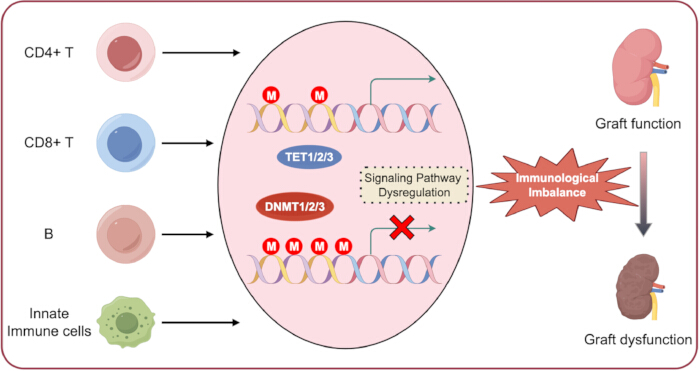
Figure 1. DNA methylation in transplant immunity. Immune cells maintain distinct intrinsic DNA methylation patterns throughout their lifespan. Disruption of this physiological process can induce aberrant DNA methylation and dysregulation of downstream signaling pathways, ultimately resulting in impaired immune homeostasis and allograft dysfunction.Please click here to view a larger version of this figure.
| Method | Principle | Advantages | Limitations | Applications in Transplantation |
| MeDIP-seq⁴¹ | Anti-5mC antibody enrichment + NGS | Cost-effective for genome-wide analysis; suitable for large sample cohorts | Low resolution (~100–300 bp); cannot distinguish 5mC from 5hmC | Screening of global methylation changes in PBMCs from transplant recipients |
| WGBS⁴² | Bisulfite conversion + NGS | Single-base resolution; genome-wide coverage | High cost; requires large data volume | Comprehensive methylome mapping of graft biopsies; identification of novel rejection-related DMRs |
| RRBS⁴³ | Restriction enzyme enrichment + bisulfite conversion + NGS | Cost-effective; high coverage of CpG-rich regions | Limited coverage of non-CpG and CpG-poor regions | Targeted methylome analysis in graft tissue with reduced sequencing cost |
| Methylation-specific PCR⁴⁴ | Bisulfite conversion + PCR with methylation-specific primers | High sensitivity; simple, rapid, and low cost | Targets only specific loci; qualitative or limited quantitative analysis | Validation of candidate DMRs from discovery cohorts; clinical biomarker development |
| Pyrosequencing⁴⁵ | Bisulfite conversion + sequencing-by-synthesis | Quantitative; single-base resolution at targeted loci | Moderate throughput; requires prior target identification | Precise quantification of methylation levels at specific CpG sites, such as FOXP3 TSDR in Treg cells |
Table 1: Summary of major DNA methylation detection approaches. The table summarizes commonly used DNA methylation detection methods, their principles, advantages, limitations, and potential applications in transplantation research.
Disclosures
The authors declare no competing interests or potential conflicts of interest.
Acknowledgements
The authors thank Figdraw for assistance with figure preparation.
References
- Güell-Alonso R, Cabrera-Pérez R, Vives J. Immunocompatibility in transplantation: adapting to a changing therapeutic landscape. Front Immunol. 2025;16:1648534.
- Li XQ, Ding RD, Cai JZ. Organ transplantation: current status, challenges, and future prospects. MedComm. 2026;7(1):e70567.
- Medzhitov R, Horng T. Transcriptional control of the inflammatory response. Nat Rev Immunol. 2009;9:692–703.
- Bagci H, Fisher AG. DNA demethylation in pluripotency and reprogramming: the role of tet proteins and cell division. Cell Stem Cell. 2013;13:265–269.
- Gu TP, et al. The role of Tet3 DNA dioxygenase in epigenetic reprogramming by oocytes. Nature. 2011;477:606–610.
- Cedar H, Bergman Y. Programming of DNA methylation patterns. Annu Rev Biochem. 2012;81:97–117.
- Rodriguez RM, et al. Regulation of the transcriptional program by DNA methylation during human αβ T-cell development. Nucleic Acids Res. 2015;43:760–774.
- Rauch TA, et al. A human B cell methylome at 100-base pair resolution. Proc Natl Acad Sci U S A. 2009;106:671–678.
- Hardbower DM, et al. Ornithine decarboxylase regulates M1 macrophage activation and mucosal inflammation via histone modifications. Proc Natl Acad Sci U S A. 2017;114:E751–E760.
- De Santa F, et al. The histone H3 lysine-27 demethylase Jmjd3 links inflammation to inhibition of polycomb-mediated gene silencing. Cell. 2007;130:1083–1094.
- Antignano F, Zaph C. Regulation of CD4 T-cell differentiation and inflammation by repressive histone methylation. Immunol Cell Biol. 2015;93:245–252.
- Wei G, et al. Global mapping of H3K4me3 and H3K27me3 reveals specificity and plasticity in lineage fate determination of differentiating CD4+ T cells. Immunity. 2009;30:155–167.
- Liu Z, et al. The histone H3 lysine-27 demethylase Jmjd3 plays a critical role in specific regulation of Th17 cell differentiation. J Mol Cell Biol. 2015;7:505–516.
- He H, et al. Histone methylation mediates plasticity of human FOXP3(+) regulatory T cells by modulating signature gene expressions. Immunology. 2014;141:362–376.
- Menden H, et al. Histone deacetylase 6 regulates endothelial MyD88-dependent canonical TLR signaling, lung inflammation, and alveolar remodeling in the developing lung. Am J Physiol Lung Cell Mol Physiol. 2019;317:L332–L346.
- Irizarry-Caro RA, et al. TLR signaling adapter BCAP regulates inflammatory to reparatory macrophage transition by promoting histone lactylation. Proc Natl Acad Sci U S A. 2020;117:30628–30638.
- Williams CJ, et al. The chromatin remodeler Mi-2beta is required for CD4 expression and T cell development. Immunity. 2004;20:719–733.
- Yang F, et al. MiR-21 is remotely governed by the commensal bacteria and impairs anti-TB immunity by down-regulating IFN-γ. Front Microbiol. 2020;11:512581.
- Nankivell BJ, Kuypers DR. Diagnosis and prevention of chronic kidney allograft loss. Lancet. 2011;378:1428–1437.
- Zununi Vahed S, et al. Genetics and epigenetics of chronic allograft dysfunction in kidney transplants. Iran J Kidney Dis. 2016;10:1–9.
- Goldberg RJ, Weng FL, Kandula P. Acute and chronic allograft dysfunction in kidney transplant recipients. Med Clin North Am. 2016;100:487–503.
- Wood KJ, Bushell A, Hester J. Regulatory immune cells in transplantation. Nat Rev Immunol. 2012;12:417–430.
- Ji H, et al. Comprehensive methylome map of lineage commitment from haematopoietic progenitors. Nature. 2010;467:338–342.
- Suarez-Alvarez B, Rodriguez RM, Fraga MF, Lopez-Larrea C. DNA methylation: a promising landscape for immune system-related diseases. Trends Genet. 2012;28:506–514.
- Eckhardt F, et al. DNA methylation profiling of human chromosomes 6, 20 and 22. Nat Genet. 2006;38:1378–1385.
- Thomson JP, et al. CpG islands influence chromatin structure via the CpG-binding protein Cfp1. Nature. 2010;464:1082–1086.
- Esteller M. Epigenetic gene silencing in cancer: the DNA hypermethylome. Hum Mol Genet. 2007;16(Spec No 1):R50–R59.
- Lopez-Serra L, Esteller M. Proteins that bind methylated DNA and human cancer: reading the wrong words. Br J Cancer. 2008;98:1881–1885.
- Kuroda A, et al. Insulin gene expression is regulated by DNA methylation. PLoS One. 2009;4:e6953.
- Hellman A, Chess A. Gene body-specific methylation on the active X chromosome. Science. 2007;315:1141–1143.
- Jones PA. Functions of DNA methylation: islands, start sites, gene bodies and beyond. Nat Rev Genet. 2012;13:484–492.
- Esteller M. Epigenetics in evolution and disease. Lancet. 2008;372:S90–S96.
- Straussman R, et al. Developmental programming of CpG island methylation profiles in the human genome. Nat Struct Mol Biol. 2009;16:564–571.
- Irizarry RA, et al. The human colon cancer methylome shows similar hypo- and hypermethylation at conserved tissue-specific CpG island shores. Nat Genet. 2009;41:178–186.
- Doi A, et al. Differential methylation of tissue- and cancer-specific CpG island shores distinguishes human induced pluripotent stem cells, embryonic stem cells and fibroblasts. Nat Genet. 2009;41:1350–1353.
- Portela A, Esteller M. Epigenetic modifications and human disease. Nat Biotechnol. 2010;28:1057–1068.
- Bourc'his D, et al. Dnmt3L and the establishment of maternal genomic imprints. Science. 2001;294:2536–2539.
- Gowher H, et al. Mechanism of stimulation of catalytic activity of Dnmt3A and Dnmt3B DNA-(cytosine-C5)-methyltransferases by Dnmt3L. J Biol Chem. 2005;280:13341–13348.
- Wu X, Zhang Y. TET-mediated active DNA demethylation: mechanism, function and beyond. Nat Rev Genet. 2017;18:517–534.
- Bochtler M, Kolano A, Xu GL. DNA demethylation pathways: additional players and regulators. Bioessays. 2017;39:1–13.
- Laird PW. Principles and challenges of genomewide DNA methylation analysis. Nat Rev Genet. 2010;11:191–203.
- Lister R, et al. Human DNA methylomes at base resolution show widespread epigenomic differences. Nature. 2009;462:315–322.
- Meissner A, et al. Reduced representation bisulfite sequencing for comparative high-resolution DNA methylation analysis. Nucleic Acids Res. 2005;33:5868–5877.
- Herman JG, et al. Methylation-specific PCR: a novel PCR assay for methylation status of CpG islands. Proc Natl Acad Sci U S A. 1996;93:9821–9826.
- Colella S, et al. Sensitive and quantitative universal pyrosequencing methylation analysis of CpG sites. Biotechniques. 2003;35:114–120.
- Nankivell BJ, Alexander SI. Rejection of the kidney allograft. N Engl J Med. 2010;363:1451–1462.
- Gelman AE, et al. CD4+ T lymphocytes are not necessary for the acute rejection of vascularized mouse lung transplants. J Immunol. 2008;180:4754–4762.
- Seo W, Taniuchi I. Transcriptional regulation of early T-cell development in the thymus. Eur J Immunol. 2016;46:531–538.
- Calvanese V, et al. A promoter DNA demethylation landscape of human hematopoietic differentiation. Nucleic Acids Res. 2012;40:116–131.
- Lee ST, et al. A global DNA methylation and gene expression analysis of early human B-cell development reveals a demethylation signature and transcription factor network. Nucleic Acids Res. 2012;40:11339–11351.
- Challen GA, et al. Dnmt3a is essential for hematopoietic stem cell differentiation. Nat Genet. 2011;44:23–31.
- Chitilian HV, et al. The strength of cell-mediated xenograft rejection in the mouse is due to the CD4+ indirect response. Xenotransplantation. 1998;5:93–98.
- Pierson RN, et al. Xenogeneic skin graft rejection is especially dependent on CD4+ T cells. J Exp Med. 1989;170:991–996.
- Bruniquel D, Schwartz RH. Selective, stable demethylation of the interleukin-2 gene enhances transcription by an active process. Nat Immunol. 2003;4:235–240.
- Sanders VM. Epigenetic regulation of Th1 and Th2 cell development. Brain Behav Immun. 2006;20:317–324.
- Wilson CB, Rowell E, Sekimata M. Epigenetic control of T-helper-cell differentiation. Nat Rev Immunol. 2009;9:91–105.
- Schoenborn JR, et al. Comprehensive epigenetic profiling identifies multiple distal regulatory elements directing transcription of the gene encoding interferon-gamma. Nat Immunol. 2007;8:732–742.
- Santangelo S, et al. Chromatin structure and DNA methylation of the IL-4 gene in human T(H)2 cells. Chromosome Res. 2009;17:485–496.
- Jiao X, et al. Vitamin D deficiency during pregnancy affects the function of Th1/Th2 cells and methylation of IFN-γ gene in offspring rats. Immunol Lett. 2019;212:98–105.
- Xia Y, et al. Age-related changes in DNA methylation associated with shifting Th1/Th2 balance. Inflammation. 2016;39:1892–1903.
- Muranski P, Restifo NP. Essentials of Th17 cell commitment and plasticity. Blood. 2013;121:2402–2414.
- Ohkura N, et al. T cell receptor stimulation-induced epigenetic changes and Foxp3 expression are independent and complementary events required for Treg cell development. Immunity. 2012;37:785–799.
- Janson PCJ, et al. Profiling of CD4+ T cells with epigenetic immune lineage analysis. J Immunol. 2011;186:92–102.
- Thomas RM, Sai H, Wells AD. Conserved intergenic elements and DNA methylation cooperate to regulate transcription at the il17 locus. J Biol Chem. 2012;287:25049–25059.
- Huang Y, et al. Decipherer manifestations and Treg/Th17 imbalance in multi-staging rheumatoid arthritis and correlation with TSDR/RORC methylation. Mol Immunol. 2020;127:1–11.
- Yuan X, et al. A novel role of CD4 Th17 cells in mediating cardiac allograft rejection and vasculopathy. J Exp Med. 2008;205:3133–3144.
- Cohen CJ, et al. Human Th1 and Th17 cells exhibit epigenetic stability at signature cytokine and transcription factor loci. J Immunol. 2011;187:5615–5626.
- Ye J, et al. Human tumor-infiltrating Th17 cells have the capacity to differentiate into IFN-γ+ and FOXP3+ T cells with potent suppressive function. Eur J Immunol. 2011;41:936–951.
- Issa F, Chandrasekharan D, Wood KJ. Regulatory T cells as modulators of chronic allograft dysfunction. Curr Opin Immunol. 2011;23:648–654.
- Josefowicz SZ, Rudensky A. Control of regulatory T cell lineage commitment and maintenance. Immunity. 2009;30:616–625.
- Baron U, et al. DNA demethylation in the human FOXP3 locus discriminates regulatory T cells from activated FOXP3(+) conventional T cells. Eur J Immunol. 2007;37:2378–2389.
- Kim HP, Leonard WJ. CREB/ATF-dependent T cell receptor-induced FoxP3 gene expression: a role for DNA methylation. J Exp Med. 2007;204:1543–1551.
- Morikawa H, et al. Differential roles of epigenetic changes and Foxp3 expression in regulatory T cell-specific transcriptional regulation. Proc Natl Acad Sci U S A. 2014;111:5289–5294.
- Polansky JK, et al. Methylation matters: binding of Ets-1 to the demethylated Foxp3 gene contributes to the stabilizationization of Foxp3 expression in regulatory T cells. J Mol Med (Berl). 2010;88:1029–1040.
- Toker A, et al. Active demethylation of the Foxp3 locus leads to the generation of stable regulatory T cells within the thymus. J Immunol. 2013;190:3180–3188.
- Jordan MS, et al. Thymic selection of CD4+CD25+ regulatory T cells induced by an agonist self-peptide. Nat Immunol. 2001;2:301–306.
- Kawahata K, et al. Generation of CD4(+)CD25(+) regulatory T cells from autoreactive T cells simultaneously with their negative selection in the thymus and from nonautoreactive T cells by endogenous TCR expression. J Immunol. 2002;168:4399–4405.
- Josefowicz SZ, Wilson CB, Rudensky AY. Cutting edge: TCR stimulation is sufficient for induction of Foxp3 expression in the absence of DNA methyltransferase 1. J Immunol. 2009;182:6648–6652.
- Floess S, et al. Epigenetic control of the foxp3 locus in regulatory T cells. PLoS Biol. 2007;5:e38.
- Polansky JK, et al. DNA methylation controls Foxp3 gene expression. Eur J Immunol. 2008;38:1654–1663.
- Barzaghi F, et al. Demethylation analysis of the FOXP3 locus shows quantitative defects of regulatory T cells in IPEX-like syndrome. J Autoimmun. 2012;38:49–58.
- Bestard O, et al. Intragraft regulatory T cells in protocol biopsies retain foxp3 demethylation and are protective biomarkers for kidney graft outcome. Am J Transplant. 2011;11:2162–2172.
- Liu J, et al. T regulatory cells in cord blood—FOXP3 demethylation as reliable quantitative marker. PLoS One. 2010;5:e13267.
- Wieczorek G, et al. Quantitative DNA methylation analysis of FOXP3 as a new method for counting regulatory T cells in peripheral blood and solid tissue. Cancer Res. 2009;69:599–608.
- Zhuo C, et al. Higher FOXP3-TSDR demethylation rates in adjacent normal tissues in patients with colon cancer were associated with worse survival. Mol Cancer. 2014;13:153.
- Lal G, et al. Epigenetic regulation of Foxp3 expression in regulatory T cells by DNA methylation. J Immunol. 2009;182:259–273.
- Zhou L, Chong MM, Littman DR. Plasticity of CD4+ T cell lineage differentiation. Immunity. 2009;30:646–655.
- Burchill MA, et al. IL-2 receptor beta-dependent STAT5 activation is required for the development of Foxp3+ regulatory T cells. J Immunol. 2007;178:280–290.
- Kanamori M, et al. Induced regulatory T cells: their development, stability, and applications. Trends Immunol. 2016;37:803–811.
- Yang R, et al. Hydrogen sulfide promotes Tet1- and Tet2-mediated Foxp3 demethylation to drive regulatory T cell differentiation and maintain immune homeostasis. Immunity. 2015;43:251–263.
- Issa F, Robb RJ, Wood KJ. The where and when of T cell regulation in transplantation. Trends Immunol. 2013;34:107–113.
- Issa F, Wood KJ. CD4+ regulatory T cells in solid organ transplantation. Curr Opin Organ Transplant. 2010;15:757–764.
- Suarez-Alvarez B, Baragano Raneros A, Ortega F, Lopez-Larrea C. Epigenetic modulation of the immune function: a potential target for tolerance. Epigenetics. 2013;8:694–702.
- Schmidl C, et al. Epigenetic reprogramming of the RORC locus during in vitro expansion is a distinctive feature of human memory but not naive Treg. Eur J Immunol. 2011;41:1491–1498.
- Guo H, et al. Inhibiting cardiac allograft rejection with interleukin-35 therapy combined with decitabine treatment in mice. Transpl Immunol. 2013;29:99–104.
- Betts RJ, Kemeny DM. CD8+ T cells in asthma: friend or foe? Pharmacol Ther. 2009;121:123–131.
- Schwengerdt KO, Fricker FJ, Bahjat KS, Kuntz ST. Increased expression of the lymphocyte early activation marker CD69 in peripheral blood correlates with histologic evidence of cardiac allograft rejection. Transplantation. 2000;69:2102–2107.
- Rutella S, et al. Induction of CD69 antigen on normal CD4+ and CD8+ lymphocyte subsets and its relationship with the phenotype of responding T-cells. Cytometry. 1999;38:95–101.
- Jones ND, et al. T-cell activation, proliferation, and memory after cardiac transplantation in vivo. Ann Surg. 1999;229:570–578.
- Lee PP, et al. A critical role for Dnmt1 and DNA methylation in T cell development, function, and survival. Immunity. 2001;15:763–774.
- Ladle BH, et al. De novo DNA methylation by DNA methyltransferase 3a controls early effector CD8+ T-cell fate decisions following activation. Proc Natl Acad Sci U S A. 2016;113:10631–10636.
- Carty SA, et al. The loss of TET2 promotes CD8(+) T cell memory differentiation. J Immunol. 2018;200:82–91.
- Schiffer L, et al. Chemokine CXCL13 as a new systemic biomarker for B-cell involvement in acute T cell-mediated kidney allograft rejection. Int J Mol Sci. 2019;20(10):2552. doi:10.3390/ijms20102552.
- Xu H, He X, Xu R. B cell activating factor, renal allograft antibody-mediated rejection, and long-term outcome. J Immunol Res. 2018;2018:5251801.
- Schmitz R, et al. B cells in transplant tolerance and rejection: friends or foes? Transpl Int. 2020;33:30–40.
- Lee ST, et al. A global DNA methylation and gene expression analysis of early human B-cell development reveals a demethylation signature and transcription factor network. Nucleic Acids Res. 2012;40:11339–11351.
- Shaknovich R, et al. DNA methyltransferase 1 and DNA methylation patterning contribute to germinal center B-cell differentiation. Blood. 2011;118:3559–3569.
- Caron G, et al. Cell-cycle-dependent reconfiguration of the DNA methylome during terminal differentiation of human B cells into plasma cells. Cell Rep. 2015;13:1059–1071.
- Barwick BG, et al. B cell activation and plasma cell differentiation are inhibited by de novo DNA methylation. Nat Commun. 2018;9:1900.
- Kaur BP, Secord E. Innate immunity. Pediatr Clin North Am. 2019;66:905–911.
- Steinman RM, Banchereau J. Taking dendritic cells into medicine. Nature. 2007;449:419–426.
- Merad M, Collin M, Bromberg J. Dendritic cell homeostasis and trafficking in transplantation. Trends Immunol. 2007;28:353–359.
- Zhang X, et al. DNA methylation dynamics during ex vivo differentiation and maturation of human dendritic cells. Epigenetics Chromatin. 2014;7:21.
- Pacis A, et al. Bacterial infection remodels the DNA methylation landscape of human dendritic cells. Genome Res. 2015;25:1801–1811.
- Klug M, et al. Active DNA demethylation in human postmitotic cells correlates with activating histone modifications, but not transcription levels. Genome Biol. 2010;11:R63.
- Bullwinkel J, Lüdemann A, Deberry J, Singh PB. Epigenotype switching at the CD14 and CD209 genes during differentiation of human monocytes to dendritic cells. Epigenetics. 2011;6:45–51.
- Frikeche J, et al. Impact of the hypomethylating agent 5-azacytidine on dendritic cells function. Exp Hematol. 2011;39:1056–1063.
- van den Bosch TP, et al. CD16+ monocytes and skewed macrophage polarization toward M2 type hallmark heart transplant acute cellular rejection. Front Immunol. 2017;8:346.
- Wang YY, et al. Macrophage-to-myofibroblast transition contributes to interstitial fibrosis in chronic renal allograft injury. J Am Soc Nephrol. 2017;28:2053–2067.
- Wang X, et al. Epigenetic regulation of macrophage polarization and inflammation by DNA methylation in obesity. JCI Insight. 2016;1:e87748.
- Bakshi C, Vijayvergiya R, Dhawan V. Aberrant DNA methylation of M1-macrophage genes in coronary artery disease. Sci Rep. 2019;9:1429.
- Dekkers KF, et al. Human monocyte-to-macrophage differentiation involves highly localized gain and loss of DNA methylation at transcription factor binding sites. Epigenetics Chromatin. 2019;12:34.
- Li G, Yu M, Weyand CM, Goronzy JJ. Epigenetic regulation of killer immunoglobulin-like receptor expression in T cells. Blood. 2009;114:3422–3430.
- Schenk A, Bloch W, Zimmer P. Natural killer cells—an epigenetic perspective of development and regulation. Int J Mol Sci. 2016;17:326.
- Scapini P, et al. The neutrophil as a cellular source of chemokines. Immunol Rev. 2000;177:195–203.
- Chertov O, Yang D, Howard OM, Oppenheim JJ. Leukocyte granule proteins mobilize innate host defenses and adaptive immune responses. Immunol Rev. 2000;177:68–78.
- Hirayama S, et al. Prevention of neutrophil migration ameliorates rat lung allograft rejection. Mol Med. 2006;12:208–213.
- Jelinek J, et al. Epigenetic control of PRV-1 expression on neutrophils. Exp Hematol. 2007;35:1677–1683.
- Suzuki H, et al. Epigenetic inactivation of SFRP genes allows constitutive WNT signaling in colorectal cancer. Nat Genet. 2004;36:417–422.
- Chen G, et al. Aberrant DNA methylation of mTOR pathway genes promotes inflammatory activation of immune cells in diabetic kidney disease. Kidney Int. 2019;96:409–420.
- Shang D, Han T, Xu X, Liu Y. Decitabine induces G2/M cell cycle arrest by suppressing p38/NF-κB signaling in human renal clear cell carcinoma. Int J Clin Exp Pathol. 2015;8:11140–11148.
- Zhu Z, et al. STAT3 signaling pathway is involved in decitabine induced biological phenotype regulation of acute myeloid leukemia cells. Am J Transl Res. 2015;7:1896–1907.
- Peters F, et al. Interferon-gamma DNA methylation is affected by mycophenolic acid but not by tacrolimus after T-cell activation. Front Immunol. 2017;8:822.
- Kraig E, et al. A randomized control trial to establish the feasibility and safety of rapamycin treatment in an older human cohort: immunological, physical performance, and cognitive effects. Exp Gerontol. 2018;105:53–69.
- Zhu C, et al. DNA methylation modulates allograft survival and acute rejection after renal transplantation by regulating the mTOR pathway. Am J Transplant. 2021;21(2):567–581.
- Wiencke JK, et al. DNA methylation as a pharmacodynamic marker of glucocorticoid response and glioma survival. Nat Commun. 2022;13(1):5537.
- Hsu FM, et al. DNA methylation predicts infection risk in kidney transplant recipients. Life Sci Alliance. 2025;8(7):e202403124. doi:10.26508/lsa.202403124.
- Bai JF, et al. Efficacy and safety of venetoclax and azacitidine for acute myeloid leukemia in China: a real-world single-center study. BMC Cancer. 2025;25(1):990.
- Arora S, et al. Tumor lysis syndrome and infectious complications during treatment with venetoclax combined with azacitidine or decitabine in patients with acute myeloid leukemia. Leuk Res. 2022;117:106844.
- Batra A, et al. Characteristics and survival of secondary acute myeloid leukemia from myelodysplastic syndromes in older adults: a population analysis. Clin Lymphoma Myeloma Leuk. 2025;25(6):e417–e423.
- Bai L, Hao X, Keith J, Feng Y. DNA methylation in regulatory T cell differentiation and function: challenges and opportunities. Biomolecules. 2022;12(9):1282.
- Trinh A, Huang Y, Shao H, et al. Targeting the ADPKD methylome using nanoparticle-mediated combination therapy. APL Bioeng. 2023;7(2):026111.
- Yuan N, Ma Z, Xue Z, et al. DNMT1-targeted macrophages delivering drug-loaded nanoparticles attenuate heart allograft fibrosis via PTPRD-STAT3/6 axis. Cell Death Discov. 2026. doi:10.1038/s41420-026-03295-5.
- Cameron J, Martino P, Nguyen L, Li X. Cutting edge: CRISPR-based transcriptional regulators reveal transcription-dependent establishment of epigenetic memory of Foxp3 in regulatory T cells. J Immunol. 2020;205(11):2953–2958.
- Liu XS, et al. Rescue of fragile X syndrome neurons by DNA methylation editing of the FMR1 gene. Cell. 2018;172:979–992.e6.
- Velut Y, Poulet G, Bersez T, et al. Epigenetic signatures on plasma cell-free DNA to detect kidney allograft rejection in a non-invasive way: development of a 10-plex digital PCR assay. Biomark Res. 2025;13:118.