This narrative review evaluates current evidence, technical requirements, and interpretation limits for three- and four-dimensional pelvic floor ultrasound in chronic pelvic pain, and proposes an explicitly review-derived, unvalidated acquisition and reporting framework for future clinical research.
A subscription to JoVE is required to view this content. Sign in or start your free trial.
Review Article
Three- and Four-Dimensional Pelvic Floor Ultrasound in Chronic Pelvic Pain: Evidence, Technical Considerations, and a Proposed Framework
In This Article
Summary
Abstract
Chronic pelvic pain (CPP) is a heterogeneous condition in which visceral, musculoskeletal, neuropathic, and pain-processing contributors often overlap. Pelvic floor ultrasound can depict morphology and movement, but its CPP-specific evidence base remains limited. This targeted narrative review summarizes direct CPP evidence; evidence from endometriosis, dyspareunia, myofascial pain, and methodological pelvic floor cohorts; and the technical distinctions among transperineal/translabial and transvaginal routes, static three-dimensional (3D) volumes, real-time four-dimensional (4D) assessment, multiplanar reconstruction, and tomographic ultrasound imaging (TUI). Direct evidence includes one reliability study in 49 women with CPP, one small uncontrolled treatment cohort, and a recent cross-sectional study of 747 women; none establishes a diagnostic threshold or validates an imaging phenotype. We therefore propose, rather than validate, a structured acquisition and reporting framework that specifies patient preparation, probe pressure, rest-contraction-Valsalva maneuvers, quality control, pain-limited modifications, measurements, and uncertainty language. Reduced hiatal excursion, asymmetry, or discontinuity should be reported as observations, not as proof of increased tone or a pain mechanism. Clinical interpretation requires symptom reproduction, pelvic floor examination, and, when indicated, manometry, electromyography, magnetic resonance imaging, or disease-specific imaging. Prospective CPP-specific validation and reliability studies are required before routine diagnostic use.
Introduction
Chronic pelvic pain (CPP) is persistent or recurrent pain perceived in pelvic structures and is commonly operationalized as lasting at least 6 months, although contemporary guidance emphasizes functional impact and overlapping pain mechanisms rather than duration alone1,2. Gynecologic, urologic, gastrointestinal, musculoskeletal, neurologic, and psychosocial contributors frequently coexist. Consequently, an imaging abnormality may be relevant, incidental, secondary to guarding, or only one component of a broader pain phenotype. Any pelvic floor imaging framework must therefore separate description from diagnosis and must state how findings are integrated with clinical examination.
Pelvic floor involvement is clinically plausible but cannot be inferred from pain alone. In a population-based controlled study, women with CPP showed higher resting tone, reduced relaxation capacity, and greater pressure-pain sensitivity on standardized intravaginal examination; pelvic muscle tenderness was also markedly more frequent in CPP than in pain-free controls3,4. However, a systematic review of 24 studies found that the association between pain and increased tone depended on the assessment method: digital palpation showed an association, whereas other objective measures did not consistently do so5. These findings support examination of the pelvic floor while warning against treating high tone as a self-evident imaging diagnosis.
Ultrasound offers real-time observation of pelvic floor anatomy and movement at rest and during contraction and straining. Three-dimensional (3D) volumes permit offline reconstruction of the axial plane of minimal hiatal dimensions, whereas four-dimensional (4D) ultrasound denotes real-time volume acquisition during a maneuver. Tomographic ultrasound imaging (TUI) displays parallel reconstructed slices and is best established for structural assessment of puborectalis insertion and levator trauma, not for measuring tone or identifying a pain generator6,7. These terms describe different acquisition or display operations and should not be used interchangeably.
The CPP-specific ultrasound literature is small. A reliability study evaluated stored 3D/4D volumes in 49 women with CPP, a prospective uncontrolled cohort followed 11 women with clinically diagnosed levator hypertonicity during magnetic-stimulation treatment, and a 2026 cross-sectional study compared translabial 4D morphometry in 747 women with and without persistent pelvic pain8,9,10. These studies show feasibility and group-level associations, but they do not validate a diagnostic threshold, demonstrate disease specificity, or prove that the measured ultrasound feature causes pain.
This review therefore has two aims: first, to critically distinguish direct CPP evidence from adjacent and methodological evidence; and second, to propose a reproducible acquisition, measurement, interpretation, and reporting framework for research and service evaluation. The framework is author-proposed and review-derived. It is not a consensus statement, clinical practice guideline, or validated diagnostic pathway.
Access restricted. Please log in or start a trial to view this content.
Review and Perspective
Review methodology and evidence classification
A targeted narrative literature search was selected because the question spans clinical CPP studies, endometriosis and dyspareunia cohorts, pelvic floor myofascial pain, imaging-method studies, reliability studies, and terminology reports. Narrative methods are appropriate for mapping heterogeneous evidence, but transparent scope and critical appraisal remain necessary11,12.
The submitted draft reported searches of PubMed/MEDLINE, Embase, Scopus, and Web of Science for English-language publications from January 2010 through March 25, 2026. Because the original exports and complete line-by-line search histories were unavailable, those searches could not be retrospectively reconstructed or assigned record counts. During revision, a targeted PubMed update through July 31, 2026, and Crossref metadata verification were used to identify newly available direct evidence and validate citations. Concepts combined terms for (1) chronic or persistent pelvic pain, dyspareunia, endometriosis, or pelvic floor myofascial pain; (2) pelvic floor, transperineal, translabial, transvaginal, 3D, 4D, or tomographic ultrasound; and (3) levator ani, puborectalis, levator hiatus, contraction, relaxation, distensibility, avulsion, or reliability. Database-specific scope and the purpose of each concept block are provided in Table 1. Reference lists of key articles and consensus documents were also checked.
Eligible evidence was classified by directness rather than being pooled. Category A comprised studies enrolling women with CPP or persistent pelvic pain and reporting pelvic floor ultrasound. Category B comprised endometriosis, dyspareunia, or clinically defined pelvic floor myofascial pain cohorts with pain-relevant ultrasound outcomes. Category C comprised postpartum, prolapse, incontinence, or other pelvic floor populations, included only to support acquisition, measurement, terminology, reliability, or structural TUI methods. Category D comprised guidelines, consensus statements, and reviews. Studies were excluded when they lacked a pain-relevant or transferable technical contribution, reported only organ-specific pelvic pathology, or did not permit separation of the imaging method from a broader intervention.
For study-level appraisal, we charted the design, sample size, population, and pain definition; examination route; acquisition mode; equipment or software, when reported; maneuvers; parameters; comparator; principal findings; reliability; limitations; and evidence directness (Table 2). Claims were calibrated to design: cross-sectional associations were not described as causal; treatment-linked imaging change was not treated as a diagnostic validation; and postpartum or prolapse evidence was not generalized to CPP without an explicit methodological extrapolation label. Figure 1 summarizes this evidence architecture.
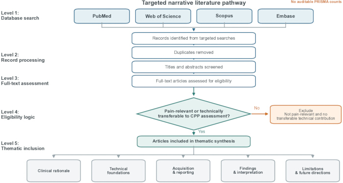
Figure 1: Targeted narrative literature pathway. The original top-to-bottom structure is retained: searching four databases, duplicate removal, title and abstract screening, full-text assessment, pain-relevant or technically transferable eligibility, and organization into five thematic domains. No PRISMA-style counts are shown because an auditable screening log was unavailable. Evidence is tagged as direct CPP, adjacent pain-related, methodological extrapolation, or guidance. CPP, chronic pelvic pain. Please click here to view a larger version of this figure.
Table 1: Transparent scope and eligibility framework for the targeted narrative literature search. Please click here to download this Table.
Direct CPP evidence and adjacent pain-related evidence
The clinical rationale begins with evidence from examination rather than imaging. Standardized intravaginal assessment demonstrates that pelvic floor dysfunction and tenderness are more common in women with CPP, but the magnitude and meaning of increased tone vary by measurement method3,4,5. Ultrasound can complement this assessment by documenting geometry and movement; it cannot replace symptom history, palpation, or evaluation of overlapping visceral and nociplastic contributors. The most defensible role is therefore to provide reproducible observations that can be compared with examination findings and followed over time.
The strongest direct CPP reliability evidence comes from a prospective cross-sectional study of 49 women in which two raters assessed 147 transperineal 3D volumes8. Anteroposterior diameter, hiatal area, and urethra-anus distance showed useful intra-rater performance, whereas transverse diameter, levator thickness, and levator-urethra gap were less reliable, particularly between raters. The study did not relate measurements to pain intensity and included parous women with dyspareunia of unclear cause. It supports feasibility and selective measurement reliability, not diagnostic accuracy.
A larger 2026 study compared translabial 4D morphometry among 747 gynecology patients, including 469 with persistent pelvic pain and 278 without pain9. At Valsalva, the mean levator hiatal area was smaller in the pain group (14.4 versus 18.6 cm²; p = 0.009), with additional differences among women currently receiving pain treatment. The investigators emphasized the overlapping distributions and explicitly stated that no diagnostic measurement or cutoff could be derived. This is important direct evidence of a group-level association, but its cross-sectional design cannot distinguish pre-existing reduced distensibility from guarding, maneuver performance, treatment selection, or consequences of pain.
The only treatment-linked CPP ultrasound report in the identified literature followed 11 women with clinically diagnosed levator hypertonicity before and after eight magnetic-stimulation sessions10. Symptoms and two midsagittal ultrasound measures changed; however, the cohort was uncontrolled, nearly half of the participants received concomitant medication, and the imaging markers were not independently validated. Endometriosis studies provide larger and sometimes randomized adjacent evidence: women with deep endometriosis have shown smaller hiatal dimensions or reduced maneuver-related change, and a 34-participant trial reported greater Valsalva hiatal-area change after pelvic floor physiotherapy13,14,15,16,17. These findings support research hypotheses but do not justify extrapolating endometriosis-specific effects to all CPP populations. Study-level details and directness labels are provided in Table 2.
Table 2: Study-level evidence map for pelvic floor assessment and ultrasound in chronic pelvic pain and adjacent pain-related populations. Directness categories and design-specific limitations are reported for every study. Please click here to download this Table.
Modality-specific technical foundations and inference boundaries
The examination route should be named explicitly. The terms transperineal and translabial are commonly used for examinations performed with an external probe placed on the perineum or labia; they provide a broad midsagittal field, dynamic compartment motion, and 3D/4D volume acquisition with relatively little tissue 6,7,18,19,20 or endovaginal imaging uses an internal higher-frequency probe and may depict focal muscle morphology at higher spatial resolution, but insertion depth and probe pressure can alter anatomy and may reproduce pain. Route-specific values are therefore not interchangeable, and pain responses, consent, and tolerability must be documented.
Acquisition dimensionality is a separate issue. A static 3D volume captures one state and permits offline reconstruction. Real-time 4D acquisition records sequential volumes during contraction, Valsalva, coughing, or relaxation; it is more sensitive to coaching, frame rate, motion, and the participant's ability to complete the maneuver. A two-dimensional midsagittal cine loop may be adequate for bladder-neck or anorectal motion, whereas an axial 3D volume is required for the levator hiatal area. The label should not substitute for reporting what was actually acquired.
Multiplanar reconstruction displays orthogonal sagittal, coronal, and axial planes from one stored volume. The axial plane of minimal hiatal dimensions is located by using the inferior pubic symphysis and anorectal angle as landmarks and then checking that the puborectalis sling is represented consistently6,21. Hiatal area, anteroposterior diameter, and transverse diameter are measurements of geometry in a defined state. Differences between ultrasound and magnetic resonance imaging (MRI) are expected because of posture, loading, temporal resolution, and landmark selection; values should not be treated as directly interchangeable.
TUI is a display method, not a separate acquisition route. Parallel axial slices, commonly centered on the plane of minimal hiatal dimensions, allow systematic inspection of the puborectalis insertion across adjacent levels. This approach has been validated mainly for structural avulsion or discontinuity in obstetric populations. A visible insertion gap is therefore a structural observation. It does not measure resting activity, tenderness, relaxation, or symptom causation, and it should not be relabeled as ‘hypertonicity’ or ‘painful muscle’ without independent clinical evidence.
Functional assessment requires comparison across correctly performed states. Contraction should produce a cranio-ventral lift and reduction in selected hiatal dimensions; Valsalva should generate caudal displacement and hiatal enlargement when the maneuver is adequate. Ultrasound-based contraction scales and comparisons with palpation or perineometry show moderate rather than perfect concordance in non-CPP populations26,27,28. A small change may reflect reduced excursion, weak effort, guarding, pain-limited performance, poor coaching, breath holding, or an acquisition-plane error. It should be described before it is interpreted.
MRI can provide a wider field, tissue contrast, and evaluation of extra-pelvic or deep soft-tissue pathology, whereas ultrasound offers dynamic assessment, portability, and repeated maneuvers. Comparative studies show clinically useful agreement for some hiatal measures but also systematic differences21,29,30,31,32. MRI is not a universal reference standard for muscle tone, and neither modality establishes a pain generator on anatomy alone. Modality selection should follow the clinical question: ultrasound is suited to dynamic pelvic floor behavior and targeted morphology, whereas MRI or disease-specific pelvic imaging is appropriate when broader structural characterization is required.
Figure 2 separates route, acquisition mode, reconstruction, target, and defensible inference. Figure 3 provides published examples of a midsagittal acquisition plane, axial hiatal measurements at rest, contraction, and Valsalva, and normal and abnormal TUI appearances. These examples are from pregnancy or postpartum methodological cohorts, not CPP cohorts; they are included to clarify technique and structural terminology and must not be used as CPP-specific diagnostic exemplars (Table 3).
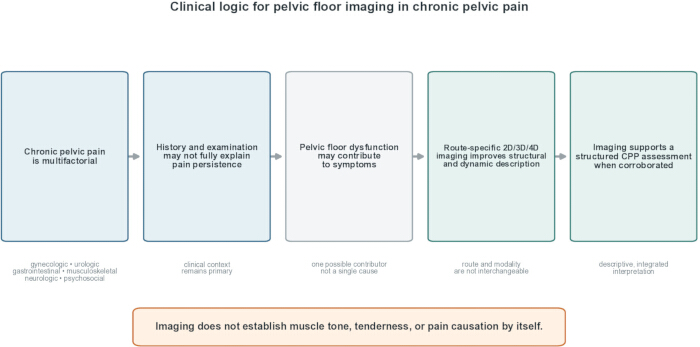
Figure 2: Clinical logic for integrating pelvic floor imaging into CPP assessment. The original five-step left-to-right pathway is retained: multifactorial CPP, limits of history and examination, pelvic floor dysfunction as one possible contributor, route-specific 2D/3D/4D structural and dynamic description, and corroborated imaging within a structured assessment. Imaging alone does not establish muscle tone, tenderness, or pain causation. CPP, chronic pelvic pain; 2D, two-dimensional; 3D, three-dimensional; 4D, four-dimensional. Please click here to view a larger version of this figure.
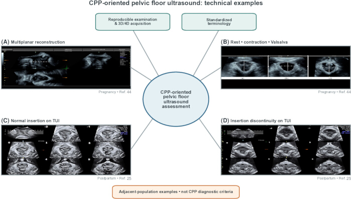
Figure 3: Published technical examples of pelvic floor ultrasound planes, maneuvers, and TUI. (A) Multiplanar translabial volume with midsagittal, coronal, and axial views; the dashed line marks axial-plane selection. (B) Axial levator hiatus at rest, maximal contraction, and Valsalva, with area and diameters outlined. Panels A and B are adapted from Mart n-Arias et al.44. (C) Normal TUI series and (D) bilateral puborectalis insertion discontinuity indicated by arrows. Panels C and D are adapted from Yan et al.25. Images were proportionally cropped or resized and globally annotated; no selective local enhancement was applied. These pregnancy and postpartum examples illustrate technique and are not CPP-specific diagnostic criteria. TUI, tomographic ultrasound imaging; CPP, chronic pelvic pain. Please click here to view a larger version of this figure.
Table 3: Technical distinctions among examination route, acquisition dimensionality, reconstruction method, anatomical target, and defensible inference. Please click here to download this Table.
Proposed review-derived acquisition and reporting protocol
The following framework operationalizes the review findings for prospective validation. It is deliberately divided into core, optional, and experimental elements (Table 4). 'Core' denotes the minimum information required to reproduce and interpret an examination; 'optional' denotes a clinically indicated extension; and 'experimental' denotes a measure that should remain within a predefined research protocol. None of these labels implies that the framework has been validated for CPP.
Preparation and consent are core. Record the referral question, pain locations, symptom triggers, relevant pelvic surgical and obstetric histories, and whether examination is intended for morphology, dynamic function, or both. Explain the external or internal route, obtain consent, provide privacy and a chaperone according to local policy, and agree on a stop signal. Record bladder status using the local protocol, body position, and hip and knee positions, probe orientation, and whether pain, anxiety, or mobility limited positioning. For transvaginal imaging, document insertion tolerance and whether probe pressure reproduced the patient's typical pain6,7,18,33.
Equipment documentation is core for research and recommended for clinical audit. Report the ultrasound system, transducer type, frequency range, volume angle, acquisition quality setting, focal zone, depth, gain, harmonic or speckle-reduction settings, and the analysis software and its version. Use the minimum probe pressure required to maintain acoustic contact, and record any departure from this principle. Presets may be used, but a named preset alone is insufficient because settings can change between machines or software versions.
Acquire a stable midsagittal reference image before volume capture. The pubic symphysis, urethra, bladder neck, vagina, anorectal junction, and anal canal should be identifiable where the route permits. Save at least one rest cine loop or image after visible movement has settled. Repeat rest acquisition if recent contraction, coughing, or probe repositioning has produced guarding. If the landmarks cannot be visualized without excessive pressure, record the limitation rather than forcing the examination.
The core maneuver sequence is rest, maximal voluntary contraction, return to rest, and maximal Valsalva. Provide standardized coaching and one practice attempt. Acquire at least three technically adequate attempts for contraction and Valsalva when tolerated, with sufficient recovery to observe return toward baseline. Record the selected attempt and the selection rule, such as the best landmark visibility and an adequate maneuver, rather than the largest or smallest measurement alone. Optional maneuvers include cough or symptom-provoking tasks when clinically justified and safe.
Volume acquisition should include the entire levator hiatus and puborectalis insertion within the region of interest. For dynamic 4D acquisition, report frame or volume rate and maneuver duration. For TUI, report the reference plane, number of slices, interslice distance, and state in which the volume was acquired. Avoid applying structural avulsion criteria to a volume with incomplete coverage, marked motion artifact, or a nonstandard reference plane. Representative normal and suboptimal images should be retained for training and audit.
Quality control is a decision loop. Before measuring, verify landmark visibility, minimal distortion from probe pressure, correct state labeling, adequate contraction or Valsalva, absence of levator coactivation when relaxation is expected, full volume coverage, and reproducible plane selection. If a criterion fails, repeat once with revised coaching or probe position if the participant agrees. If pain or fatigue prevents repetition, stop and label the examination 'pain-limited' or 'maneuver-limited'; do not classify the failed maneuver as abnormal physiology.
Core measurements should be limited to those linked to the question and supported by acceptable reliability: hiatal anteroposterior diameter and area in a defined axial plane, and maneuver-related absolute and percentage change when repeated states are available8,27. Transverse diameter, levator thickness, levator-urethra gap, and route-specific focal features may be optional or experimental because reliability is population-, route-, and operator-dependent. Report the units, side, plane, state, number of attempts, selected attempt, whether the analysis was performed online or offline, and whether the reader was blinded.
The structured report should contain: indication and symptom context; consent and tolerability; route and equipment; position and bladder status; acquisition and maneuver adequacy; measurements by state; morphology and TUI observations; technical limitations; comparison with prior studies; and an interpretation statement with explicit uncertainty. Figure 4 converts these elements into a CPP-oriented checklist. The recommended conclusion is descriptive—for example, 'reduced hiatal enlargement during a pain-limited Valsalva attempt'—followed by a recommendation for clinical correlation rather than a categorical label of muscle overactivity.
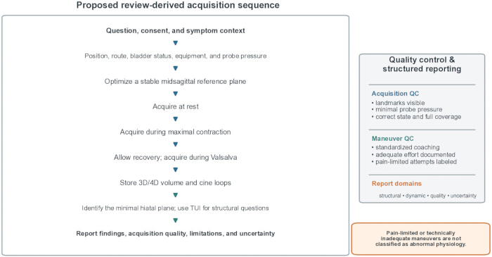
Figure 4: Proposed review-derived acquisition and reporting workflow. Core steps include consent and symptom context, route and equipment documentation, standardized rest-contraction-Valsalva acquisition, maneuver-quality checks, reproducible plane selection, and descriptive reporting. Optional or experimental extensions are used only for a defined question. Pain-limited or technically inadequate maneuvers are labeled and are not classified as abnormal physiology. Please click here to view a larger version of this figure.
Table 4: Proposed review-derived acquisition, quality-control, measurement, and reporting checklist. Elements are labeled core, optional, or experimental. Please click here to download this Table.
Measurement, reporting, and non-causal interpretation
Interpretation begins by separating an observation from a clinical construct. 'Reduced distensibility' means that a prespecified dimension or area increases less than expected across an adequately performed maneuver; it does not itself mean high tone. 'Clinically confirmed increased resting tone' requires an examination method capable of assessing resting activity or resistance, such as standardized palpation, manometry, or electromyography, with the method and threshold clearly stated5,26. 'Guarding' is a context-dependent protective response, and 'inadequate or paradoxical maneuver' describes performance, not baseline physiology.
Smaller hiatal dimensions or reduced Valsalva enlargement have been reported in persistent pelvic pain and deep-endometriosis cohorts9,13,14. These are group-level differences with overlapping individual distributions. Potential explanations include reduced distensibility, pain-related guarding, fear of symptom provocation, incomplete pressure generation, coactivation, prior obstetric injury, prolapse status, age, or measurement error. A report should therefore name the state and maneuver quality and avoid phrases such as 'ultrasound confirms hypertonicity' unless an independent validated assessment supports that diagnosis.
Contraction-related change is also multidimensional. Reduced hiatal narrowing may indicate limited excursion, weakness, poor task understanding, pain inhibition, or measurement instability. Increased resting activity does not necessarily prevent a stronger voluntary contraction: an endometriosis study using electromyography found no association between resting activity and pain and found greater rather than lower activation during maximal contraction in participants with higher resting activity15,27,28,34. Ultrasound and palpation should be treated as complementary observations rather than interchangeable gold standards.
Asymmetry, focal discontinuity, or an enlarged levator-urethra gap may indicate structural injury when acquired and interpreted with validated TUI methods8,22,25. Structural avulsion should be distinguished from generalized levator complex appearance, focal puborectalis morphology, and functional movement. A unilateral insertion defect may coexist with, contribute to, or be unrelated to pain. Reports should state whether the criteria were applied at the recommended slices and whether the finding was reproduced by a second trained reader when used for research.
Posterior-compartment motion can be described when the field and maneuver are technically adequate, but diagnostic criteria require established definitions and the appropriate clinical context17,35. Reduced rectal emptying, rectocele, intussusception, or paradoxical movement should not be inferred from a limited CPP-focused pelvic floor volume. When bowel symptoms are prominent, a dedicated dynamic examination and standardized interpretation template are more appropriate than opportunistic labeling.
The association between pain and tone is especially vulnerable to circular reasoning. If pain is used to infer high tone and a small hiatal change is then taken as confirmation, the imaging result adds no independent information. The systematic review evidence shows method-dependent and sometimes absent associations5. The framework, therefore, requires documentation of symptom reproduction, tenderness, relaxation after contraction, and, when clinically indicated, manometry or electromyography. Figure 5 uses bidirectional, dashed associations and competing explanations rather than a causal arrow from sonographic pattern to pain.
Myofascial pelvic pain studies provide relevant but heterogeneous adjacent evidence. A case-control study of 49 women with myofascial pelvic pain and 41 controls combined electrophysiology with 2D/3D ultrasound, while a transvaginal study of 220 postpartum women associated puborectalis deficiency or focal thickening with palpation-defined pain36,37. The latter population was postpartum, the route was transvaginal, and the diagnostic reference was palpation; its findings should not be generalized to nonpostpartum CPP or to transperineal measurements.
The pain phenotype must remain explicit in the report. Endometriosis-associated pain may include myofascial, visceral, neuropathic-like, and nociplastic components, and clinical examination may identify greater symptom burden even when imaging does not explain it16,38,39. A technically normal pelvic floor ultrasound does not exclude clinically important dysfunction or pain. Conversely, an abnormal image does not establish clinical relevance without concordant symptoms, examination, and an anatomically plausible question.
Table 5 provides permissible wording, competing explanations, required corroboration, and wording to avoid. The proposed interpretive hierarchy comprises: (1) a technically adequate descriptive finding; (2) an association supported by symptom or examination concordance; and (3) a hypothesis requiring prospective confirmation. No current CPP ultrasound marker meets the standard for a stand-alone diagnostic criterion.
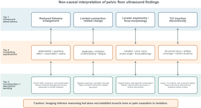
Figure 5: Three-tier non-causal interpretation pathway for pelvic floor ultrasound in CPP. Tier 1 lists observations; Tier 2 lists competing explanations; Tier 3 states required corroboration and permissible descriptive wording. Dashed bidirectional connectors indicate association and uncertainty rather than causation. Ultrasound findings do not establish muscle tone or pain causation in isolation. CPP, chronic pelvic pain; TUI, tmaging. Please click here to view a larger version of this figure.
Table 5: Interpretation boundaries, competing explanations, required corroboration, permissible report wording, and wording to avoid. Please click here to download this Table.
Reproducibility, limitations, and research priorities
The first limitation is evidence directness. Only a small number of studies enrolled broadly defined CPP or persistent pelvic pain cohorts and performed pelvic floor ultrasound8,9,10. Much of the apparent technical maturity comes from endometriosis, postpartum, prolapse, or incontinence populations. Those studies are valuable for defining planes, measurements, or reliability, but they cannot establish CPP-specific prevalence, diagnostic thresholds, or treatment-response markers.
The second limitation is reproducibility. Studies differ in bladder preparation, position, probe route and pressure, acquisition angle, coaching, attempt selection, software, reader training, and whether pain limited the maneuver. Reliability can also differ by parameter: in the direct CPP study, some dimensions were reproducible while levator thickness and transverse measures were not8. Future studies should preregister acquisition rules, blind readers to symptoms, report intra- and inter-rater intraclass correlation coefficients with 95% confidence intervals, and provide limits of agreement rather than correlation alone.
The third limitation is route and target heterogeneity. Transperineal hiatal morphometry, transvaginal focal puborectalis imaging, and midsagittal dynamic compartment assessment do not sample the same tissue or generate interchangeable measurements. Combining them under 'pelvic floor ultrasound' obscures both strengths and risks. Multicenter studies should select one primary route and target, define route-specific secondary measures, and test cross-platform reproducibility before pooling.
The fourth limitation concerns TUI. TUI has a defensible role in standardized structural assessment of puborectalis insertion and levator trauma, but the evidence base is predominantly obstetric22,23,25. CPP studies should not use avulsion scoring as a proxy for muscle tone, spasm, or nociceptive relevance. If TUI is included, the analysis plan should specify a structural hypothesis, reader training, slice interval, diagnostic criteria, and a clinical correlation that is independent of the image.
Selection and spectrum bias also require attention. Tertiary pain clinics overrepresent severe, treatment-experienced, and overlapping disorders, while obstetric cohorts overrepresent parity-related anatomy40,41. A useful validation cohort should state CPP duration and phenotype, recruit consecutive participants, include pain-free and clinically relevant symptomatic comparators, and record endometriosis, prolapse, parity, surgery, menopause, and ongoing therapy. Without these variables, an apparent imaging association may reflect population composition rather than pain biology.
Quantitative reporting remains incomplete. Future studies should provide the number analyzed for each state; reasons for missing maneuvers; means and standard deviations or medians and interquartile ranges, as appropriate; effect estimates with 95% confidence intervals; prespecified primary comparisons; and multiplicity control. Diagnostic research should report sensitivity and specificity only against an independently defined reference standard and should include calibration rather than reporting only an optimal cutoff derived and tested in the same sample.
Comparative imaging research should be question-specific. Ultrasound-MRI comparisons are useful for geometry and structural lesions, but MRI should not be treated as a gold standard for resting tone or pain causation21,29,30. A stronger design would combine ultrasound with standardized examination, symptom reproduction, manometry or electromyography, and disease-specific imaging where indicated, then test whether ultrasound adds incremental and reproducible information beyond those measures.
Prospective validation should proceed in stages: technical repeatability in pain-limited and pain-free participants; blinded reader reliability; construct validity against prespecified examination or physiologic measures; discrimination across relevant clinical groups; longitudinal responsiveness; and external validation across different systems and sites. The proposed checklist and report template can serve as study tools, but their feasibility, patient acceptability, completion rate, and inter-rater reliability must be measured before routine adoption.
Within the broader context of nonpharmacological conservative therapies for CPP, rehabilitative ultrasound imaging may be useful as visual biofeedback; however, this application is distinct from diagnosis42,43. Research should specify whether the target is motor learning, relaxation, contraction, or symptom self-management and should compare ultrasound feedback with equivalent therapist contact. Improvements in task performance should not be assumed to mediate pain reduction unless formally tested.
No original patient ultrasound images were supplied with this review. To address the need for technical examples without fabricating patient data, Figure 3 adapts images from two open-access publications: a prenatal exercise study showing multiplanar and rest-contraction-Valsalva views and a postpartum TUI study showing intact and avulsed puborectalis insertions25,44. These images are explicitly labeled as adjacent-population technical examples; no new patient data were acquired, and no CPP-specific image phenotype is claimed.
Table 6 and Figure 6 summarize the research roadmap: standardized acquisition; CPP-specific cohorts; independent reference measures; reliability and agreement; transparent missing-data reporting; prospective outcome linkage; and external validation. Until these steps are completed, 3D/4D pelvic floor ultrasound and TUI should be used as structured descriptive components of a broader assessment, not as stand-alone diagnostic tests.
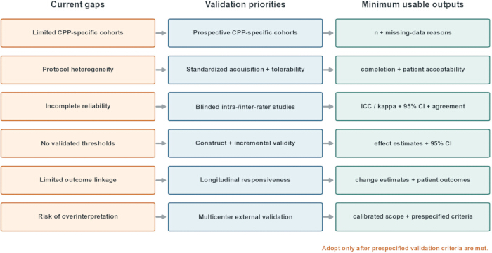
Figure 6: Research roadmap for validating pelvic floor ultrasound in CPP. Present gaps lead to staged studies of technical repeatability, blinded reader reliability, construct and incremental validity, longitudinal responsiveness, and external validation. Minimum outputs include sample size, missing-data reasons, effect estimates and 95% confidence intervals, intra- and inter-rater reliability, limits of agreement, patient acceptability, and prespecified failure criteria. CPP, chronic pelvic pain; TUI, tomographic ultrasound imaging. Please click here to view a larger version of this figure.
Table 6: Minimum research priorities for CPP-specific validation, reliability, comparative imaging, longitudinal follow-up, and external validation Please click here to download this Table.
Access restricted. Please log in or start a trial to view this content.
Conclusions
Three- and four-dimensional pelvic floor ultrasound can reproducibly describe selected aspects of pelvic floor morphology and movement, but CPP-specific validation is incomplete. Direct studies show feasibility and group-level associations, not a diagnostic threshold or causal imaging phenotype. The review-derived framework proposed here standardizes preparation, acquisition, maneuver quality, measurement, reporting, and uncertainty language for future evaluation. Reduced hiatal change should be described as reduced excu...
Access restricted. Please log in or start a trial to view this content.
Disclosures
The authors have nothing to disclose.
Acknowledgements
The authors thank Sir Run Run Shaw Hospital for administrative and technical support and colleagues who contributed to manuscript preparation.
Access restricted. Please log in or start a trial to view this content.
References
- Allaire C, et al. Guideline No. 445: Management of chronic pelvic pain. J Obstet Gynaecol Can. 2024;46(1):102283.
- Khatri G, Khan A, Raval G, Chhabra A. Diagnostic evaluation of chronic pelvic pain. Phys Med Rehabil Clin N Am. 2017;28(3):477-500.
- Loving S, Thomsen T, Jaszczak P, Nordling J. Pelvic floor muscle dysfunctions are prevalent in female chronic pelvic pain: a cross-sectional population-based study. Eur J Pain. 2014;18(9):1259-1270.
- de Souza Montenegro MLL, et al. Importance of pelvic muscle tenderness evaluation in women with chronic pelvic pain. Pain Med. 2010;11(2):224-228.
- Kadah S, et al. Are pelvic pain and increased pelvic floor muscle tone associated in women with persistent noncancer pelvic pain? A systematic review and meta-analysis. J Sex Med. 2023;20(9):1206-1221.
- Dietz HP. Pelvic floor ultrasound: a review. Am J Obstet Gynecol. 2010;202(4):321-334.
- Salsi G, et al. Three-dimensional/four-dimensional transperineal ultrasound: clinical utility and future prospects. Int J Womens Health. 2017;9:643-656.
- Carmo MAMV, et al. Reliability and agreement of three-dimensional/four-dimensional transperineal ultrasound in women with chronic pelvic pain. Clin Exp Obstet Gynecol. 2021;48(3):494-499.
- Nesbitt-Hawes EM, Dietz HP, Ledger WL, Abbott JA. Differences in pelvic floor morphometry for women with persistent pelvic pain and deep infiltrating endometriosis: a cross-sectional study. Int Urogynecol J. 2026.
- Barba M, et al. Changes in pelvic floor ultrasonographic features after flat magnetic stimulation in women with chronic pelvic pain and levator ani muscle hypertonicity. Medicina (Kaunas). 2024;60(3):374.
- Baethge C, Goldbeck-Wood S, Mertens S. SANRA—a scale for the quality assessment of narrative review articles. Res Integr Peer Rev. 2019;4(1):5.
- Grant MJ, Booth A. A typology of reviews: an analysis of 14 review types and associated methodologies. Health Info Libr J. 2009;26(2):91-108.
- Raimondo D, et al. Pelvic floor muscle dysfunction on 3D/4D transperineal ultrasound in patients with deep infiltrating endometriosis: a pilot study. Ultrasound Obstet Gynecol. 2017;50(4):527-532.
- Del Forno S, et al. Assessment of levator hiatal area using 3D/4D transperineal ultrasound in women with deep infiltrating endometriosis and superficial dyspareunia treated with pelvic floor muscle physiotherapy: randomized controlled trial. Ultrasound Obstet Gynecol. 2021;57(5):726-732.
- Arena A, et al. Three-dimensional ultrasound evaluation of pelvic floor muscle contraction in women affected by deep infiltrating endometriosis: application of a quick contraction scale. J Ultrasound Med. 2022;41(12):2973-2979.
- Mabrouk M, et al. Painful love: superficial dyspareunia and three-dimensional transperineal ultrasound evaluation of pelvic floor muscle in women with endometriosis. J Sex Marital Ther. 2020;46(2):187-196.
- Raimondo D, et al. Pelvic floor dysfunction at transperineal ultrasound and chronic constipation in women with endometriosis. Int J Gynaecol Obstet. 2022;159(2):505-512.
- Hainsworth AJ, et al. Integrated total pelvic floor ultrasound in pelvic floor dysfunction. Continence. 2023;8:101045.
- Haylen BT, et al. An International Urogynecological Association (IUGA)/International Continence Society (ICS) joint report on the terminology for female pelvic floor dysfunction. Int Urogynecol J. 2010;21(1):5-26.
- Doumouchtsis SK, et al. An International Continence Society (ICS)/International Urogynecological Association (IUGA) joint report on the terminology for the assessment and management of obstetric pelvic floor disorders. Int Urogynecol J. 2023;34(1):1-42.
- Vergeldt TFM, et al. Comparison of translabial three-dimensional ultrasound with magnetic resonance imaging for measurement of levator hiatal biometry at rest. Ultrasound Obstet Gynecol. 2016;47(5):636-641.
- Turel F, Shek KL, Dietz HP. How valid is tomographic ultrasound imaging in diagnosing levator and anal sphincter trauma? J Ultrasound Med. 2019;38(4):889-894.
- Shek KL, Dietz HP. Is two-dimensional oblique parasagittal ultrasound imaging valid for levator ani muscle assessment? Ultrasound Obstet Gynecol. 2025;66(1):96-100.
- Dietz HP. Ultrasound imaging of maternal birth trauma. Int Urogynecol J. 2021;32(7):1953-1962.
- Yan Y, et al. Combination of tomographic ultrasound imaging and three-dimensional magnetic resonance imaging-based model to diagnose postpartum levator avulsion. Sci Rep. 2017;7(1):11235.
- Frawley H, et al. An International Continence Society (ICS) report on the terminology for pelvic floor muscle assessment. Neurourol Urodyn. 2021;40(5):1217-1260.
- Nyhus MØ, et al. Ultrasound assessment of pelvic floor muscle contraction: reliability and development of an ultrasound-based contraction scale. Ultrasound Obstet Gynecol. 2020;55(1):125-131.
- Volløyhaug I, Mørkved S, Salvesen Ø, Salvesen KÅ. Assessment of pelvic floor muscle contraction with palpation, perineometry and transperineal ultrasound: a cross-sectional study. Ultrasound Obstet Gynecol. 2016;47(6):768-773.
- El-Haieg DO, et al. Magnetic resonance imaging and three-dimensional transperineal ultrasound evaluation of pelvic floor dysfunction in symptomatic women: a prospective comparative study. Ultrasonography. 2019;38(4):355-364.
- Khatri G, et al. ACR Appropriateness Criteria pelvic floor dysfunction in females. J Am Coll Radiol. 2022;19(5 Suppl):S137-S155.
- Young SW, et al. Society of Radiologists in Ultrasound consensus on routine pelvic US for endometriosis. Radiology. 2024;311(1):e232191.
- Condous G, et al. Non-invasive imaging techniques for diagnosis of pelvic deep endometriosis and endometriosis classification systems: an international consensus statement. Hum Reprod Open. 2024;2024(3):hoae029.
- Retief F, et al. The clinical application of transperineal ultrasound in urogynaecology. Obstet Gynaecol. 2022;24(3):176-187.
- Gabrielsen R, Bø K, Engh ME, Tennfjord MK. Is pelvic floor muscle resting activity associated with pelvic and genital pain, dyspareunia, and pelvic floor muscle contraction? A cross-sectional study of women with endometriosis. Int Urogynecol J. 2025;36(11):2277-2285.
- Alshiek J, et al. Consensus definitions and interpretation templates for dynamic ultrasound imaging of defecatory pelvic floor disorders. Urogynecology (Phila). 2023;29(3):327-343.
- Xu X, et al. Analysis of the characteristics and clinical significance of pelvic floor electrophysiology and imaging changes in female patients with MFPP. J Radiat Res Appl Sci. 2023;16(4):100713.
- Ye J, et al. Exploring transvaginal sonographic characteristics of the levator ani muscle in women with postpartum pelvic floor myofascial pain. BMC Womens Health. 2024;24(1):245.
- Quintas-Marquès L, et al. Multidimensional evaluation of myofascial pelvic pain and other comorbidities in endometriosis patients. J Clin Med. 2025;14(10):3455.
- Dolci C, et al. Neuropathic-like pain affects pain perception in patients with deep endometriosis: an observational study. Arch Gynecol Obstet. 2025;312(5):1789-1797.
- Deng S. Application of transperineal pelvic floor ultrasound in postpartum pelvic organ injury and prolapse in women. Am J Transl Res. 2024;16(9):4830-4839.
- Cai S, Xia M, Ding Y, Zeng L. Clinical value of transperineal ultrasound in evaluating the effects of different delivery methods on the primipara pelvic floor structure and function. Sci Rep. 2024;14(1):23980.
- Starzec-Proserpio MG, Frawley H, Bø K, Morin M. Effectiveness of nonpharmacological conservative therapies for chronic pelvic pain in women: a systematic review and meta-analysis. Am J Obstet Gynecol. 2025;232(1):42-71.
- Daniel DS, Goldenberg M, Kalichman L. Rehabilitative ultrasound imaging as visual biofeedback in pelvic floor dysfunction: a narrative review. Tomography. 2026;12(1):10.
- Martin-Arias A, et al. Pelvic floor adaptation to a prenatal exercise program: does it affect labor outcomes or levator ani muscle injury? A randomized controlled trial. Diagnostics (Basel). 2025;15(15):1853.
Access restricted. Please log in or start a trial to view this content.