This study investigates the therapeutic effects of Bushen-Jianpi-Qingchang formula and its sub-formulas on rat colitis, and reveals their regulatory mechanisms targeting the gut microbiota–bile acid metabolic axis via multi-omics analysis.
Research Article
Bushen Jianpi Qingchang Formula Ameliorates Colitis in Rats by Regulating Bile Acid Metabolism and Gut Microbiota
In This Article
Summary
Abstract
Traditional Chinese medicine (TCM) is widely used for the management of ulcerative colitis (UC) owing to its validated efficacy and favorable safety profile. The core pathogenesis of chronic refractory UC involves spleen and kidney deficiency accompanied by intestinal dampness-heat accumulation, and the Bushen-Jianpi-Qingchang formula (BJQF) is a clinically validated compound prescription for this condition. This study aimed to investigate the therapeutic effects of BJQF and its three decomposed sub-formulas (Bushen formula, BSF; Jianpi formula, JPF; Qingchang formula, QCF) on dextran sulfate sodium (DSS)-induced colitis in rats, and characterize their regulatory effects on fecal bile acid (BA) profiles and gut microbial communities. Fecal bacterial composition and BA concentrations in DSS-induced UC rats were quantified via 16S rRNA gene sequencing and liquid chromatography-tandem mass spectrometry (LC-MS/MS), and the potential association between gut microbiota and BA metabolism was analyzed. BJQF significantly ameliorated colonic inflammation and ulceration in the UC model. Distinct alterations in bacterial diversity and composition were identified between colitis rats and healthy controls. The association between differential bacteria and BAs was centered on 23-deoxycholic acid (23-DCA) as a key metabolic node. Notably, taurolithocholic acid-3-sulfate (TLCA-3S) and lithocholic acid-3-sulfate (LCA-3S) may serve as potential efficacy biomarkers. The JPF sub-formula exhibited efficacy most similar to the complete BJQF. Administration of BJQF and its decomposed formulas effectively ameliorates histopathological damage, inflammation, gut microbial dysbiosis, and BA metabolic disturbance in UC rats, providing a mechanistic basis for TCM compound therapy.
Introduction
Ulcerative colitis (UC), a major form of inflammatory bowel disease (IBD), is characterized by chronic, continuous non-specific inflammation confined to the colorectal mucosa. Patients with UC present with clinical symptoms including diarrhea, abdominal pain, unintended weight loss, and bloody or purulent stools, which severely impair quality of life. The pathogenesis of UC involves environmental, genetic, microbial, and immune factors. UC typically develops following gut microbiota imbalance, and this dysbiosis can markedly accelerate UC progression1. Accumulating evidence has identified the gut microbiota–bile acid (BA) metabolic axis as a core regulatory module in UC pathogenesis. Bile acid dysmetabolism is a well-recognized hallmark of intestinal mucosal damage in IBD. Secondary BAs are exclusively biosynthesized from host-derived primary BAs by gut microbiota, which express key enzymes such as 7α-dehydroxylase for this biotransformation. Thus, the fecal bile acid pool serves as an ideal functional readout to reflect microbiome remodeling induced by TCM treatment.
These secondary BAs act as critical signaling molecules to maintain intestinal immune homeostasis, mucosal barrier integrity, and epithelial cell survival2. Abnormalities in this axis, including microbial dysbiosis-induced secondary BA deficiency and disrupted BA signaling, directly trigger intestinal pro-inflammatory responses and barrier dysfunction, making it a promising therapeutic target for UC intervention. The gut microbiota plays a critical role in UC development and affects the host through metabolic products, such as bile acids generated by fermentative reactions. Abnormal alterations in gut microbiota and BA metabolism are closely associated with UC3. Since gut microbiota mediates secondary BA production, changes in microbial composition and abundance can disrupt BA metabolism and contribute to IBD development4.
TCM has been widely adopted for UC management due to its effectiveness and safety in long-term treatment, as an alternative or adjunct to anti-inflammatory or immunosuppressive agents5. Warming and tonifying the spleen and kidneys, clearing the intestine, and dispelling dampness are core therapeutic principles for UC6. According to TCM theory, the core pathogenesis of protracted UC is spleen and kidney deficiency (Fu-Zheng, vital qi reinforcement) combined with intestinal dampness-heat accumulation (Qu-Xie, pathogenic factor elimination), and the synergistic application of these two strategies is essential for effective UC treatment.
Previous experimental studies have confirmed that the JPF and QCF, two core sub-formulas of BJQF, exert therapeutic effects against UC. JPF ameliorates mucosal inflammation and intestinal epithelial barrier dysfunction via the NF-κB/HIF-1α signaling pathway7, while QCF restores gut microbiota-metabolism homeostasis, protects the intestinal epithelial barrier, and regulates the NLRP3/IL-1β pathway8. To dissect the specific biological contributions of the Fu-Zheng and Qu-Xie functional modules to the gut microbiota–BA axis, this study was designed to include the complete BJQF formula and three decomposed sub-formulas: BSF (Bushen formula, spleen-kidney tonifying, pure Fu-Zheng), JPF (Jianpi formula, spleen invigorating, core Fu-Zheng), and QCF (Qingchang formula, intestinal dampness-heat clearing, pure Qu-Xie). This experimental design enables the dissection of the synergistic and independent effects of each functional module in UC treatment, which is critical for the translation of TCM compound prescriptions into evidence-based therapy.
The working hypothesis is that BJQF ameliorates DSS-induced colitis by remodeling gut microbial composition and normalizing bile acid metabolism, and that the Jianpi module serves as the core functional component driving regulatory effects on the gut microbiota–BA axis. This study was designed to systematically dissect the "compound-to-module" mechanism of BJQF. By evaluating the full formula and its corresponding targeted physiological sub-modules, the study seeks to decode the biological synergy of this TCM compound and to provide a modern pharmacological basis for IBD treatment by regulating the gut microbiota–bile acid metabolic axis. Herein, the therapeutic efficacy of the complete BJQF and its three decomposed functional formulas against DSS-induced UC is systematically compared. Given the pivotal role of the gut microbiota–bile acid metabolic axis in UC pathogenesis, this work dissects the compound-to-module synergistic mechanism of BJQF, clarifies the specific regulatory contributions of the Bushen, Jianpi, and Qingchang modules, and characterizes the remodeling effects of each module on gut microbial composition and fecal bile acid profiles. The core objectives are to reveal the modern biological basis of BJQF in alleviating UC, identify key microbial–metabolic targets for TCM intervention, and provide a mechanistic reference for the rational application and translational research of compound TCM prescriptions in IBD.
Protocol
Forty-four healthy male Sprague-Dawley rats weighing 200 ± 20 g were obtained for the study. All animals were housed under specific pathogen-free conditions with free access to a standard pellet diet and water, and acclimatized for 1 week prior to the experiment. All animal welfare and experimental procedures were performed in accordance with the Guide for Care and Use of Laboratory Animals issued by the Ministry of Science and Technology of China. The study was approved by the Animal Ethics Committee of Nanjing University of Chinese Medicine under approval number 2023DW-029-01. Full details of animal source and husbandry supplies are provided in the Table of Materials.
TCM formula preparation
The complete Bushen-Jianpi-Qingchang formula (BJQF) was formulated with nine herbal ingredients at predefined dosages: 15 g Astragali Radix, 10 g stir-fried Atractylodis Macrocephalae Rhizoma, 10 g Scutellariae Radix, 4 g Coptidis Rhizoma, 10 g Sanguisorbae Radix, 6 g Aucklandiae Radix, 5 g Glycyrrhizae Radix, 15 g Psoraleae Fructus, and 15 g Alpiniae Oxyphyllae Fructus. All raw herbal materials were soaked in normal saline for 1 h, followed by 1 h of decoction to yield a final volume of 1500 mL. Three decomposed sub-formulas were prepared using an identical decoction protocol. The Bushen formula (BSF) contained 15 g Psoraleae Fructus and 15 g Alpiniae Oxyphyllae Fructus, decocted to a final volume of 500 mL. The Jianpi formula (JPF) was composed of 15 g Astragali Radix, 10 g stir-fried Atractylodis Macrocephalae Rhizoma, and 5 g Glycyrrhizae Radix, with a final decoction volume of 500 mL. The Qingchang formula (QCF) consisted of 10 g Scutellariae Radix, 4 g Coptidis Rhizoma, 10 g Sanguisorbae Radix, and 6 g Aucklandiae Radix, also adjusted to a 500 mL final volume. Full manufacturer specifications and batch numbers for all herbal materials are provided in the Table of Materials. Generic herbal nomenclature is used consistently throughout the main text, with manufacturer-specific details restricted exclusively to the Table of Materials.
Colitis induction and experimental grouping
Rats were randomly divided into six experimental groups: Control group (n = 6): normal drinking water + intragastric normal saline. UC model group (n = 6): 3.5% (wt/vol) DSS drinking water + intragastric normal saline. BJQF treatment group (n = 8): 3.5% DSS drinking water + intragastric BJQF (0.6 g/kg body weight). BSF treatment group (n = 8): 3.5% DSS drinking water + intragastric BSF (0.6 g/kg body weight). JPF treatment group (n = 8): 3.5% DSS drinking water + intragastric JPF (0.6 g/kg body weight). QCF treatment group (n = 8): 3.5% DSS drinking water + intragastric QCF (0.6 g/kg body weight).
Experimental colitis was induced by 3.5% DSS in drinking water for 10 consecutive days; the control group received distilled water without DSS. The dosage of each TCM formula (0.6 g/kg) was converted from the clinically effective adult dose to rat equivalent dose using the body surface area normalization method (Meeh-Rubner formula). As a discovery-phase study focusing on formula comparison, a single clinical equivalent dose was adopted. From day 1, each TCM formula was administered intragastrically once daily. The control and model groups received the same volume of normal saline (10 mL/kg). On day 11, rats were sacrificed via cervical dislocation. Serum, fecal samples, and colon tissues were collected for analysis. Body weight was measured daily. All groups were initially designed with n = 8. During DSS induction, 2 rats each in the control and model groups died due to severe colitis, resulting in a final n = 6 for these two groups. Post-hoc power analysis confirmed that the final sample size achieves statistical power ≥ 0.8 for all primary outcomes (histological score, key cytokines, body weight), meeting standard statistical requirements.
Measurement of inflammatory cytokine concentrations
Serum levels of IL-1β, IL-10, and TNF-α were quantified using commercial ELISA kits, strictly following the manufacturer’s protocols. Optical density values were read using an automatic microplate reader. Statistical analysis: one-way ANOVA for overall group differences, followed by Benjamini-Hochberg FDR correction for post-hoc pairwise comparisons, consistent with omics data analysis.
Histopathological evaluation
Colon tissue specimens were fixed in 10% formalin, dehydrated, paraffin-embedded, sectioned, and stained with hematoxylin and eosin (HE). Histological scoring was performed using the modified Cooper colitis scoring system, a validated semi-quantitative rubric for experimental colitis9. Scoring covers four dimensions: inflammatory infiltration, crypt structural damage, mucosal edema, and ulcer extent (total score 0–12). Blinding statement: All slides were de-identified with numerical codes before scoring. Two independent pathologists, blinded to group assignments, scored each slide independently. The final score was the mean of the two scores; discrepancies ≥ 2 points were resolved by a third senior pathologist. One-way ANOVA with Benjamini-Hochberg FDR correction for pairwise comparisons, consistent with other phenotypic data.
Sample collection and DNA extraction
Fresh rat fecal samples were collected in 1 mL sterile centrifuge tubes, immediately frozen at −80 °C, and transported on dry ice. Fecal bacterial genomic DNA was extracted from 250 mg of feces using the DNA Isolation Kit following the manufacturer’s instructions. DNA concentration was quantified via a spectrophotometer, and integrity was verified via 0.8% agarose gel electrophoresis. A negative buffer control was included during extraction and quantification. Extracts were stored at -20 °C before use.
16S rRNA gene sequencing and bioinformatics analysis
PCR amplification targeted the V3–V4 hypervariable regions (338F–806R) of the 16S rRNA gene. PCR products were purified and sequenced on the Illumina MiSeq platform with 2 × 300-bp paired-end sequencing. After quality filtering, an average of 68,450 ± 28,297 high-quality clean reads were obtained per sample (max 158,361, min 28,294). Sequences were clustered into OTUs at 97% similarity. Rarefaction curves, richness indices, and diversity indices were calculated. Taxonomic annotation was performed using the RDP Classifier. PCoA based on UniFrac distances and hierarchical clustering was performed in R to assess community similarity. LEfSe analysis was performed on the Galaxy platform to identify differentially abundant taxa (thresholds: LDA > 3, Kruskal-Wallis p < 0.05, post-hoc Wilcoxon p < 0.05). For multiple comparisons of microbial abundances, Benjamini-Hochberg FDR correction was applied; p(FDR) < 0.05 was considered significant.
LC-MS/MS analysis of fecal bile acids
A 20 mg fecal sample was homogenized with 200 µL methanol. After centrifugation, the supernatant was concentrated, reconstituted in 100 µL 50% aqueous methanol, and analyzed via LC-MS/MS. The detection system included a Shimadzu UPLC system and an Applied Biosystems 6500 QTRAP MS/MS system. Qualitative and quantitative analyses were performed using bile acid standard calibration curves. PCA was used to assess the separation of the metabolic profile. Differential bile acids were defined as VIP ≥ 1 and fold change ≥ 2 or ≤ 0.5. Violin plots were used to visualize differential BA distributions. For multiple comparisons, Benjamini-Hochberg FDR correction was applied, consistent with microbial data analysis.
Statistical analysis
Spearman correlation was calculated using the cor function in R. Correlation significance was tested using the corPvalueStudent function from the WGCNA R package. Phenotypic data are expressed as mean ± standard deviation (Mean ± SD). For all multi-group comparisons (phenotypic, microbial, metabolic), one-way ANOVA with Benjamini-Hochberg FDR correction for post-hoc tests was applied. Corrected p < 0.05 was considered statistically significant. All analyses were performed in GraphPad Prism 7.00 and R 4.2.0. Post-hoc sample size power calculation was performed using G*Power 3.1.
Results
TCM formulas ameliorate inflammation and colonic damage in DSS-induced colitis
The protective efficacy of TCM formulas was first evaluated by body weight changes. Compared with controls, the UC model group showed sustained weight loss. BJQF, BSF, JPF, and QCF all significantly attenuated DSS-induced weight loss (Figure 1A). HE staining revealed intact colonic mucosa and no inflammation in controls. In contrast, the model group colon tissue showed severe epithelial distortion, crypt destruction, and extensive inflammatory cell infiltration in the mucosa and submucosa. All TCM treatments promoted tissue recovery and reduced inflammation, with BJQF showing the most pronounced improvement (Figure 1B–C). Quantitative histological scoring confirmed that TCM significantly ameliorated histopathological damage (p < 0.05).
BJQF, BSF, JPF, and QCF all reduced serum IL-1β (Figure 1D) and TNF-α (Figure 1F) to varying degrees. IL-10 levels in treatment groups did not reach control levels but were significantly higher than in the model group (Figure 1E). These results indicate that TCM formulas inhibit DSS-induced intestinal inflammation.
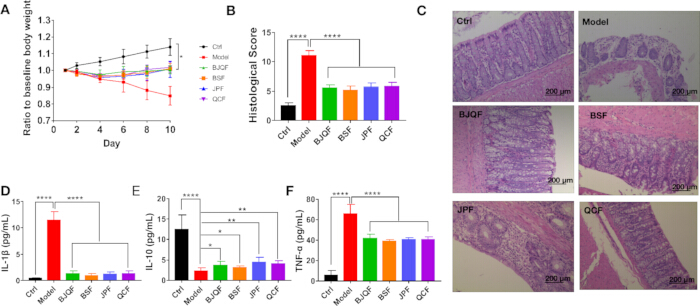
Figure 1: Therapeutic effects of BJQF and its decomposed sub-formulas on DSS-induced colitis in rats. (A) Dynamic changes in the ratio of body weight to baseline in each group over the 10-day experimental period; (B) Quantitative histological scores of colon tissues across all experimental groups; (C) Representative hematoxylin-eosin (HE) stained colon tissue sections from each group (scale bar = 200 µm); (D–F) Serum concentrations of inflammatory cytokines in each group: (D) IL-1β, (E) IL-10, (F) TNF-α. Data are expressed as mean ± SD. *p < 0.05, **p < 0.01, ****p < 0.0001. Please click here to view a larger version of this figure.
TCM treatment remodels gut microbial community structure in UC rats
After quality filtering, an average of 68,450 ± 28,297 high-quality clean reads were obtained per sample. TCM treatment altered bacterial α diversity (Chao1 index, Figure 2A). BJQF significantly reduced elevated α diversity in UC rats (p(FDR) < 0.05). Among sub-formulas, JPF significantly reduced diversity (p(FDR) < 0.05), while BSF and QCF showed no significant effect. For β diversity, unweighted PCoA of UniFrac distances showed clear separation across groups, indicating that TCM reshaped overall microbial composition (Figure 2B). Hierarchical clustering further demonstrated distinct clustering between model and treatment groups (Figure 2C).
A total of 13 phyla, 22 classes, 36 orders, 65 families, and 158 genera were detected. At the phylum level, Firmicutes, Bacteroidetes, Actinobacteria, Verrucomicrobia, and Proteobacteria dominated (>99% total abundance, Figure 2D). Firmicutes was dominant in controls (86.98%), significantly decreased in UC (p(FDR) < 0.05), and restored by TCM (p(FDR) < 0.05), with BJQF showing the strongest effect. Bacteroidetes and Proteobacteria were significantly increased in UC and reversed by all TCM formulas. At the genus level (abundance >1%, Figure 2E), Lactobacillus was dominant in controls (59.77%), markedly reduced in UC (3.57%, p(FDR) < 0.05), and restored by TCM (p(FDR) < 0.05). BJQF and JPF showed stronger effects on Lactobacillus restoration. UC also increased Bacteroides, Ruminococcus_2, Bifidobacterium, and Blautia, and decreased Enterorhabdus and Corynebacterium_1 — most of which were reversed by TCM. Overall, all formulas ameliorated UC-associated microbial dysbiosis, with BJQF and JPF showing the most pronounced effects.
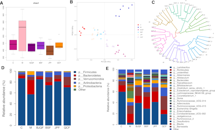
Figure 2: The composition of the gut microbiota in groups. (A) Index of bacterial α diversity assessed using the Chao 1 (p(FDR) < 0.05 vs Model); (B) PCoA of UniFrac distances based on OTU composition and abundances in different groups; (C) Hierarchical clustering analysis based on the distance matrix; (D) Relative abundance of phylum level (p(FDR) < 0.05 vs Model); and (E) Relative abundance of genus level (p(FDR) < 0.05 vs Model). Please click here to view a larger version of this figure.
LEfSe identifies UC-associated and TCM-associated differential taxa
LEfSe analysis was performed to identify taxa differentially enriched across groups (LDA > 3, p < 0.05, Figure 3). UC-enriched taxa included Bacteroidaceae, Bacteroides, Clostridiales, Clostridia, Bacteroidia, Bacteroidales, Bacteroidetes, Faecalibaculum, and Ruminococcaceae, mainly from Bacteroidetes and Firmicutes. After TCM treatment, most BJQF-enriched taxa belonged to Firmicutes, with only Bifidobacteriaceae, Bifidobacteriales, and Bifidobacterium from Actinobacteria. BSF-enriched taxa were also mainly Firmicutes. JPF-enriched taxa such as Turicibacter and Dorea belonged to Firmicutes. QCF showed the strongest enrichment of Verrucomicrobia (including Akkermansia), indicating high sensitivity of this phylum to QCF intervention. In general, Firmicutes was the most responsive phylum to BJQF, BSF, and JPF.
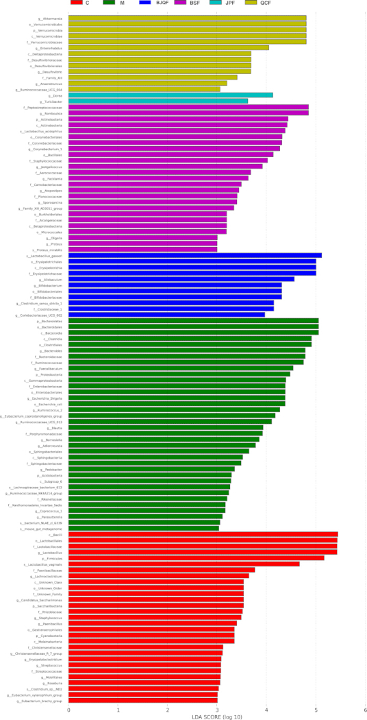
Figure 3: LEfSe identified the most differentially abundant OTUs in different groups. Significant differences in LDA scores (p < 0.05) were produced among classes (Kruskal–Wallis test) and between subclasses (Wilcoxon’s test). Differential taxa were determined based on an LDA threshold score of > 3 and a statistical significance level of 0.05. (p: phylum c: class o: order f: family g: genus). Please click here to view a larger version of this figure.
TCM intervention normalizes fecal bile acid profiles in UC rats
Given BA malabsorption in IBD, the authors used LC-MS/MS to quantify fecal BAs. PCA showed clear separation between control and model groups, while all treatment groups clustered closer to controls, indicating partial normalization of BA metabolism (Figure 4A). The most altered BAs in the model group were elevated LCA-3S and reduced GDCA (Figure 4B). After treatment, the most upregulated BAs were ILCA (BJQF), GCDCA (BSF), DLCA (JPF), and GCDCA (QCF); the most downregulated BAs were TLCA-3S (BJQF), LCA-3S (BSF), LCA-3S (JPF), and TLCA-3S (QCF) (Figure 4C–F). Violin plots visualized common differential BAs across groups (Figure 5). Notably, all TCM formulas increased 23-DCA, 3-oxo-DCA, and DLCA, and decreased HCA, LCA-3S, TCA, and TLCA-3S.
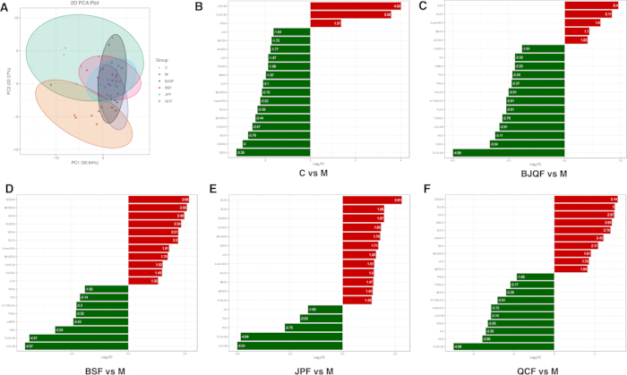
Figure 4: TCM formulas regulated DSS-induced bile acid disorder in the UC model. (A) PCA plot of bile acid of Control, Model, BJQF, BSF, JPF, and QCF groups. (B) Bar diagram of fold change in Model versus Control. (C) Bar diagram of fold change in BJQF versus Model. (D) Bar diagram of fold change in BSF versus Model. (E) Bar diagram of fold change in JPF versus Model, and (F) Bar diagram of fold change in QCF versus Model. Please click here to view a larger version of this figure.
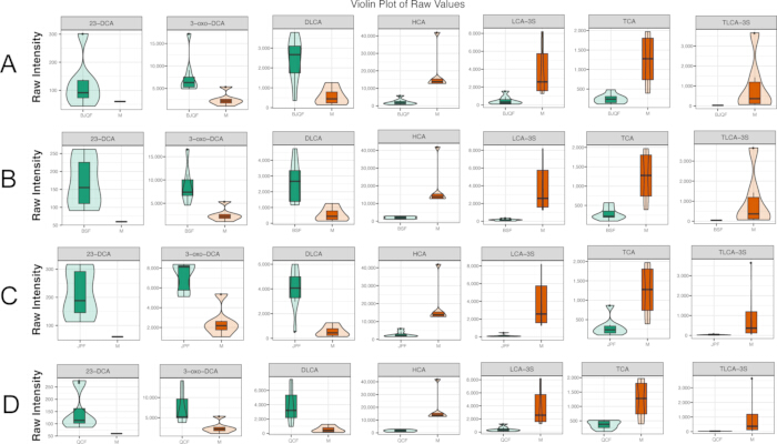
Figure 5: Violin Plot of differential BAs in each comparison group. (A) Differential BAs in BJQF versus Model. (B) Differential BAs in BSF versus Model. (C) Differential BAs in JPF versus Model and (D) Differential BAs in QCF versus Model. Please click here to view a larger version of this figure.
Correlation between fecal bile acids and gut microbial abundance
Spearman correlation analysis was performed between differential phyla and differential BAs (Figure 6A–F, Figure 7A–D). Chord charts show correlations with |r| ≥ 0.8 (Figure 6B, 6D, 6F). In the model vs. control comparison, 23-DCA was positively correlated with Saccharibacteria and Firmicutes, and negatively correlated with Proteobacteria and Bacteroidetes (Figure 6B). In the BJQF vs. model comparison, Proteobacteria was associated with TLCA-3S, LCA-3S, TCA, α-MCA, 6,7-DKLCA, HCA, CDCA, and THCA (Figure 6D). In the BSF vs. model comparison, Proteobacteria were positively correlated with TDCA, 6,7-DKLCA, and LCA-3S; Saccharibacteria were positively correlated with GUDCA and 23-DCA (Figure 6F).
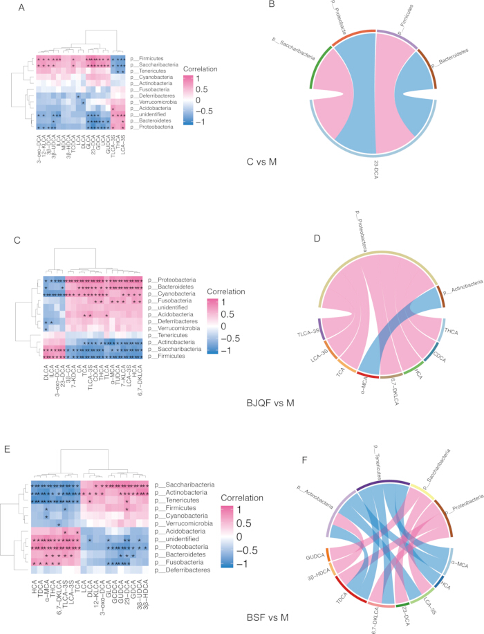
Figure 6: Correlations between fecal differential bile acids (BAs) and the abundance of differential bacteria at the phylum level. (A, C, E) Spearman correlation analyses for the Control vs. Model, BJQF vs. Model, and BSF vs. Model groups, respectively. (B, D, F) Corresponding chord charts illustrating strong correlations (|r| ≥ 0.8) for the Control vs. Model, BJQF vs. Model, and BSF vs. Model groups, respectively. Please click here to view a larger version of this figure.
Chord charts show correlations with |r| ≥ 0.8 (Figure 7B, 7D). In the JPF vs. model comparison, Saccharibacteria was negatively associated with TLCA-3S, LCA-3S, and HCA, and positively correlated with 23-DCA (Figure 7B). In the QCF vs. model comparison, Saccharibacteria was negatively correlated with HCA, CA, LCA-3S, THCA, and TLCA-3S, and positively correlated with 23-DCA (Figure 7D). These results indicate that different TCM formulas modulate the bacteria–BA interaction network through distinct patterns.
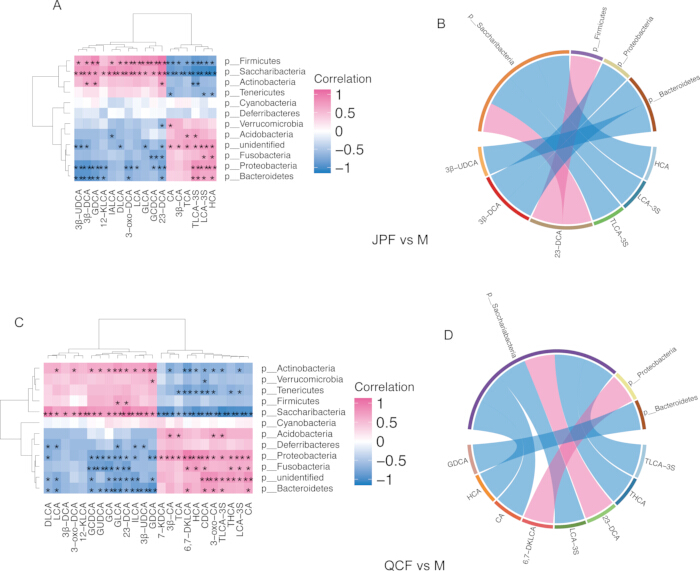
Figure 7: Correlations between fecal differential bile acids (BAs) and the abundance of differential bacteria. (A, C) Spearman correlation analyses for the JPF vs. Model and QCF vs. Model groups, respectively. (B, D) Corresponding chord charts illustrating strong correlations (|r| ≥ 0.8) for the JPF vs. Model and QCF vs. Model groups, respectively. Red indicates positive correlations, whereas blue indicates negative correlations. p < 0.05, p < 0.01. Please click here to view a larger version of this figure.
DATA AVAILABILITY:
All the raw data have been uploaded to the NCBI Sequence Reads Archive (SRA) database (Accession Number: PRJNA749732). https://www.ncbi.nlm.nih.gov/bioproject/PRJNA749732
Discussion
This study investigates the therapeutic mechanism of BJQF and its decomposed sub-formulas in alleviating DSS-induced UC via the gut microbiota–bile acid metabolic axis. This multi-omics analysis suggests that BJQF exerts anti-colitis effects by restoring UC-associated microbial dysbiosis and normalizing the fecal BA pool. Its sub-formulas exhibit distinct module-specific regulatory effects, with JPF showing efficacy most similar to the full formula, revealing the core role of the Jianpi strategy. The authors also identify 23-DCA as a key BA node linking microbial remodeling and UC amelioration, and TLCA-3S/LCA-3S as potential efficacy biomarkers.
BJQF-mediated restoration of microbial dysbiosis is potentially associated with normalization of the BA pool and reduced inflammation. UC is driven by immune-microbiota-metabolism network dysregulation, and core phyla imbalance is a typical feature in both patients and animal models10. These results confirm that DSS-induced UC rats show reduced Firmicutes and Lactobacillus, and increased Bacteroidetes and Proteobacteria — consistent with clinical UC profiles11. BJQF and JPF most effectively reversed these abnormalities, while BSF and QCF showed moderate effects. Given that Firmicutes taxa such as Lactobacillus and Clostridium are well documented to harbor the core enzymatic machinery for secondary BA biosynthesis12, this suggests a potential associative link: BJQF, especially its Jianpi module, restores Firmicutes with BA biosynthetic capacity, which may enhance secondary BA biotransformation and correct metabolic disorder. At the genus level, BJQF and JPF also reversed UC-induced changes in Bacteroides, Ruminococcus_2, Enterorhabdus, and Corynebacterium_1, further repairing dysbiosis and BA synthesis-related functions.
Restoration of microbial dysbiosis may contribute to BA pool normalization, a key metabolic mechanism of anti-colitis effects. BA metabolism is highly dependent on microbial activity, and secondary BAs are critical for immune homeostasis and barrier integrity13. The LC-MS/MS data show that UC rats exhibit reduced protective secondary BAs (23-DCA, 3-oxo-DCA, DLCA) and accumulated sulfated BAs (TLCA-3S, LCA-3S) — consistent with previous reports of UC-associated BA dysregulation14. Sulfated BAs have low intestinal absorbability and cannot enter enterohepatic circulation15 as their accumulation reflects impaired BA metabolism and is linked to pro-inflammatory pathway activation16. All formulas reversed this disorder, with BJQF and JPF showing the strongest effects.
Correlation analysis confirms 23-DCA as the key BA node associated with core phyla changes: positively correlated with Firmicutes and negatively with Proteobacteria. As a secondary BA with anti-inflammatory and barrier-protective effects17, 23-DCA upregulation by BJQF/JPF is potentially attributed to Firmicutes enrichment and enhanced biosynthesis, and correlates with reduced cytokines and improved histology. Based on these correlational findings, the authors propose a hypothetical mechanistic model: BJQF restores Firmicutes abundance > enhances microbial secondary BA biosynthesis > upregulates protective 23-DCA > inhibits intestinal inflammation. This model remains to be validated by functional experiments.
This study bridges TCM theory and modern molecular biology by mapping BJQF functional modules to specific microbial and metabolic changes. The Jianpi concept is the core link. Consistent with TCM theory of "mutual reinforcement between spleen and kidney"18, BSF showed moderate Firmicutes enrichment and 23-DCA upregulation. QCF mainly enriched Verrucomicrobia (Akkermansia) and regulated xenobiotic metabolism, reflecting the "clearing dampness-heat" effect. The full formula exerts synergistic effects superior to any single module, verifying the rationality of TCM compound design based on syndrome differentiation.
Correlation analysis further reveals distinct bacteria–BA interaction networks shaped by each formula. These differences reflect module-specific regulatory characteristics and provide a basis for individualized clinical application: JPF for spleen-deficiency dominant UC with Firmicutes-centered dysbiosis, QCF for dampness-heat dominant UC with mucosal barrier damage, and full BJQF for the classic combined syndrome. Consistent with the findings, recent studies also support the gut microbiota–bile acid axis as a core therapeutic target for UC and TCM intervention19,20,21,22. Microbial dysbiosis-driven secondary BA deficiency is a core feature of active UC, and restoring BA homeostasis can effectively alleviate inflammation and mucosal damage23.
This study has several limitations. First, all associations between microbial remodeling and BA normalization are based on correlational multi-omics analysis; causal relationships have not been validated by functional experiments such as fecal microbiota transplantation. Second, the sample size is relatively moderate, which may limit statistical power and generalizability. Third, only fecal BAs were quantified; serum and mucosal BA profiles, which reflect enterohepatic circulation, were not measured. Fourth, the specific active phytochemicals responsible for regulating microbiota and BA have not been identified. Future work will validate causal relationships via FMT and germ-free animal experiments, identify key active components of BJQF, and conduct clinical pilot studies to verify translational value.
Disclosures
The authors have nothing to disclose.
AUTHORS’ CONTRIBUTION:
Hongyu Chen contributed to the conceptualization, methodology, validation, formal analysis, investigation, visualization, and writing of the original draft of the manuscript. Song Zhao contributed to the methodology, project administration, investigation, visualization, funding acquisition, and writing of the original draft. Luzhou Xu contributed to the conceptualization, resource provision, manuscript review and editing, project administration, and overall supervision of the study.
Acknowledgements
This work was supported by the National Natural Science Foundation of China (81774243).
Materials
| Name | Company | Catalog Number | Comments |
|---|---|---|---|
| 0.2 mL PCR Tubes | Axygen | PCR-02-C | For 16S rRNA gene PCR amplification reactions |
| 1.5 mL Sterile Centrifuge Tubes | Axygen | MCT-150-C | Sterile containers for sample collection, storage and centrifugation |
| 10% Neutral Buffered Formalin | Solarbio Science & Technology | 1008128 | For fixation of colon tissue specimens |
| 6500 QTRAP MS/MS System | Sciex | 5009065 | For MRM-based quantitative profiling of fecal bile acids |
| -80 ºC Ultra-low Temperature Freezer | Thermo Fisher Scientific | 702-UR | For cryopreservation of fecal samples and biological specimens |
| Agarose | Sigma-Aldrich | A9539 | For DNA integrity detection via gel electrophoresis |
| Alpiniae Oxyphyllae Fructus | Nanjing TCM Co., Ltd. | Batch 20230321 | Raw herbal material, 15 g per dose, used in BJQF and BSF |
| Astragali Radix | Nanjing TCM Co., Ltd. | Batch 20230312 | Raw herbal material, 15 g per dose, used in BJQF and JPF |
| Aucklandiae Radix | Nanjing TCM Co., Ltd. | Batch 20230217 | Raw herbal material, 6 g per dose, used in BJQF and QCF |
| Bile Acid Standard Mix | Sigma-Aldrich | B4893 | For qualitative and quantitative calibration of fecal bile acids |
| Coptidis Rhizoma | Nanjing TCM Co., Ltd. | Batch 20230118 | Raw herbal material, 4 g per dose, used in BJQF and QCF |
| Dextran sulfate sodium (DSS, 36–50 kDa) | MP Biomedicals | 160110 | For rat UC induction, prepared as 3.5% (wt/vol) drinking solution for 10 consecutive days |
| Glycyrrhizae Radix | Nanjing TCM Co., Ltd. | Batch 20230411 | Raw herbal material, 5 g per dose, used in BJQF and JPF |
| GraphPad Prism | GraphPad Software | v7.00 | For statistical analysis, significance testing and scientific graph plotting |
| Hematoxylin-Eosin (H&E) Staining Kit | Beyotime Biotechnology | C0105S | For colon tissue section staining, histopathological observation and inflammation scoring |
| High-speed Refrigerated Centrifuge | Eppendorf | 5810R | For sample centrifugation in nucleic acid extraction and metabolomics preparation |
| HPLC-grade Methanol | Merck KGaA | 1.06007.4008 | For LC-MS/MS sample preparation and mobile phase preparation |
| Illumina MiSeq Sequencing Platform | Illumina Inc. | SY-410-1003 | For 16S rRNA gene high-throughput sequencing and gut microbiota profiling |
| KEGG Database | Kanehisa Laboratories | Release 107.0 | For microbial functional pathway annotation |
| Male SPF-grade SD rats | Shanghai Xipu-Bikai Experimental Animal Co., Ltd. | / | 44 rats weighing 200 ± 20 g, for UC model construction and follow-up experiments |
| Multiskan™ GO Microplate Reader | Thermo Fisher Scientific Oy | 51119080 | For ELISA absorbance reading and cytokine quantification |
| NanoDrop 2000 Spectrophotometer | Thermo Fisher Scientific | ND-2000 | For quantification and purity assessment of fecal bacterial genomic DNA |
| Paraffin Embedding Cassettes | Leica Biosystems | 3801520 | For colon tissue dehydration and paraffin embedding |
| PowerFecal® DNA Isolation Kit | MoBio (Qiagen) | 12830-50 | For extraction and purification of fecal bacterial genomic DNA |
| Psoraleae Fructus | Nanjing TCM Co., Ltd. | Batch 20230129 | Raw herbal material, 15 g per dose, used in BJQF and BSF |
| R Statistical Environment | R Foundation for Statistical Computing | v4.2.0 | For bioinformatics analysis and statistical computing |
| Rat IL-10 ELISA Kit | Lun Chang Shuo Biotech | YD-30194 | For quantitative detection of serum IL-10 concentration |
| Rat IL-1β ELISA Kit | Lun Chang Shuo Biotech | YD-30206 | For quantitative detection of serum IL-1β concentration |
| Rat TNF-α ELISA Kit | Lun Chang Shuo Biotech | YD-31063 | For quantitative detection of serum TNF-α concentration |
| Sanguisorbae Radix | Nanjing TCM Co., Ltd. | Batch 20230309 | Raw herbal material, 10 g per dose, used in BJQF and QCF |
| Scutellariae Radix | Nanjing TCM Co., Ltd. | Batch 20230407 | Raw herbal material, 10 g per dose, used in BJQF and QCF |
| Shim-pack UFLC UPLC System | Shimadzu Corporation | CBM-20A | For chromatographic separation of fecal bile acid samples |
| Stir-fried Atractylodis Macrocephalae Rhizoma | Nanjing TCM Co., Ltd. | Batch 20230225 | Raw herbal material, 10 g per dose, used in BJQF and JPF |
References
- Gilbert JA, et al. Current understanding of the human microbiome. Nat Med. 2018;24(4):392-400.
- Fiorucci S, et al. Bile acid signaling in inflammatory bowel diseases. Dig Dis Sci. 2021;66(3):674-693.
- Sinha SR, et al. Dysbiosis-induced secondary bile acid deficiency promotes intestinal inflammation. Cell Host Microbe. 2020;27(4):659-670.e5.
- Zhan K, et al. Gut microbiota-bile acid crosstalk in diarrhea-irritable bowel syndrome. Biomed Res Int. 2020;2020:3828249.
- Zhang C, Jiang M, Lu A. Considerations of traditional Chinese medicine as adjunct therapy in the management of ulcerative colitis. Clin Rev Allergy Immunol. 2013;44(3):274-283.
- Zhang S, et al. Consensus on Chinese medical diagnosis and treatment of ulcerative colitis (2009). Chin J Integr Tradit West Med. 2010;30(5):527-532.
- Chen YL, et al. Systems pharmacology approach reveals protective mechanisms of Jian-Pi Qing-Chang decoction on ulcerative colitis. World J Gastroenterol. 2019;25(21):2603-2622.
- Hu J, et al. Qingchang Huashi Formula attenuates DSS-induced colitis in mice by restoring gut microbiota-metabolism homeostasis and goblet cell function. J Ethnopharmacol. 2021;266:113394.
- Cooper HS, et al. Clinicopathologic study of dextran sulfate sodium experimental murine colitis. Lab Invest. 1993;69(2):238-249.
- Zuo T, Ng SC. The gut microbiota in the pathogenesis and therapeutics of inflammatory bowel disease. Front Microbiol. 2018;9:2247.
- Kobayashi T, et al. Ulcerative colitis. Nat Rev Dis Primers. 2020;6(1):74.
- Wahlström A, Sayin SI, Marschall HU, Bäckhed F. Intestinal crosstalk between bile acids and microbiota and its impact on host metabolism. Cell Metab. 2016;24(1):41-50.
- Joyce SA, Gahan CGM. Bile acid-microbiota crosstalk in inflammatory bowel disease. Nat Rev Gastroenterol Hepatol. 2020;17(11):694-707.
- Duboc H, et al. Connecting dysbiosis, bile-acid dysmetabolism and gut inflammation in inflammatory bowel diseases. Gut. 2013;62(4):531-539.
- Alnouti Y. Bile acid sulfation: A pathway of bile acid elimination and detoxification. Toxicol Sci. 2009;108(2):225-246.
- Ungaro R, et al. Ulcerative colitis. Lancet. 2017;389(10080):1756-1770.
- Tang Y, Li Y, Zhang L, et al. 23-Deoxycholic acid ameliorates colitis by regulating gut microbiota and activating FXR signaling. J Ethnopharmacol. 2022;298:115463.
- Lu YH, Cong LL, Zhang Y. TCM syndrome classification and its correlation with gut microbiota in ulcerative colitis. Chin J Integr Tradit West Med. 2020;40(7):867-871.
- Zhang Y, Li X, Wang H. Traditional Chinese medicine for inflammatory bowel disease: From empirical therapy to mechanism-based treatment. J Ethnopharmacol. 2022;296:115347.
- Li J, Wang Y, Zhang L, et al. Targeting the gut microbiota-bile acid axis: a novel strategy for TCM compound prescriptions in treating chronic inflammatory diseases. Pharmacol Res. 2023;192:106589.
- Abdo W, et al. Gut Microbiota and Bile Acid Metabolism in Inflammatory Bowel Disease: Pathophysiological Links and Therapeutic Implications. Arch Med Res. 2023;54(8):789-801.
- Liu M, et al. Traditional Chinese Medicine Targeting Gut Microbiota-Bile Acid Axis for Ulcerative Colitis: A Systematic Review. Arch Med Res. 2022;53(6):542-553.
- Chen L, et al. Secondary Bile Acids as Key Mediators of Intestinal Mucosal Barrier Protection in Inflammatory Bowel Disease. Arch Med Res. 2023;54(2):189-200.