This study investigates the impact of Glioma-pretrained federated learning on external generalization in multi-modal MRI meningioma segmentation
A subscription to JoVE is required to view this content. Sign in or start your free trial.
Research Article
Improving Cross-Center Generalization for Multi-modal MRI Meningioma Segmentation via Glioma-Pretrained Federated Learning
In This Article
Summary
Abstract
Automated meningioma segmentation on multi-modal MRI remains challenging when models are transferred across institutions, because scanner protocols, image characteristics, and annotation styles may differ between centers. Federated learning (FL) provides a privacy-preserving strategy for multi-center model development, but standard aggregation may not fully overcome cross-center domain shift. This study aimed to quantify the external generalization gap in MRI meningioma segmentation and evaluate whether Glioma-pretrained FL could improve robustness without centralized data pooling. A UMamba 2D architecture was used for binary meningioma segmentation using T1, T1c, and T2 MRI as model inputs. The protocol included 450 BraTS2023-Men cases as the source-domain meningioma dataset and 174 independent clinical cases from our institute as the external validation cohort. Three final meningioma segmentation strategies were quantitatively evaluated under the same external validation setting: centralized training on BraTS2023-Men, meningioma-pretrained FL across three simulated clients, and Glioma-pretrained FL initialized from a BraTS2023-Gli source model before federated optimization. Centralized training on Glioma was used only to generate the glioma-pretrained initialization and was not reported as an independently evaluated final meningioma segmentation strategy. Model performance was assessed using the Dice similarity coefficient (DSC) and Intersection over Union (IoU). Centralized training on BraTS2023-Men showed a clear external generalization drop, with DSC decreasing from 0.8958 on the internal BraTS2023-Men test set to 0.7452 on the SPHS cohort. Meningioma-pretrained FL yielded lower external performance (DSC = 0.7122), whereas Glioma-pretrained FL achieved comparable performance to centralized training on BraTS2023-Men and improved over meningioma-pretrained FL (DSC = 0.7503; IoU = 0.6301; Holm-adjusted p < 0.001). These results suggest that glioma-pretrained initialization provides a more robust starting point for federated meningioma segmentation and may improve external generalization while preserving institutional data privacy.
Introduction
Meningiomas are the most prevalent primary intracranial neoplasm in adults, accounting for approximately 38% of all primary brain and central nervous system tumors in the United States1. Originating from meningothelial (arachnoid cap) cells, they encompass World Health Organization (WHO) grades I–III, spanning a wide clinical spectrum: grade-I tumors (approximately 80%) are generally associated with favorable outcomes following gross total resection, whereas grade-II (atypical) and grade-III (anaplastic) lesions exhibit substantially higher recurrence rates of 20–40% and exceeding 70% at ten years, respectively2,3. Clinical decision-making—including microsurgical resection, stereotactic radiosurgery (SRS), fractionated radiotherapy, and active surveillance—depends on accurate and reproducible volumetric delineation of tumor extent across longitudinal imaging studies. Robust segmentation of meningiomas from magnetic resonance imaging (MRI) is therefore a prerequisite for consistent treatment planning and outcome assessment.
Multi-sequence MRI forms the foundation of meningioma characterization in routine clinical practice, with each sequence encoding complementary aspects of tumor biology and surrounding tissue response. Contrast-enhanced T1-weighted imaging (T1c) highlights regions of blood–brain barrier disruption through gadolinium-induced T₁ relaxation shortening, enabling clear visualization of enhancing tumor parenchyma and definition of the primary lesion boundary. Non-contrast T1-weighted imaging (T1) provides structural context for evaluating the arachnoid interface, dural attachment, and adjacent osseous involvement. T2-weighted imaging (T2) captures intra-tumoral heterogeneity and peritumoral edema, both of which are essential for understanding mass effect and for delineating treatment volumes in radiosurgical planning3,4. Manual contouring remains the standard approach for meningioma delineation. During this process, clinicians typically consider complementary contrast information across multiple MRI sequences to define tumor extent. However, this procedure is time-consuming and is associated with substantial inter-observer variability, particularly in anatomically ambiguous regions such as the dural tail, where reported agreement metrics fall below 0.805,6. These limitations motivate the development of automated segmentation approaches that deliver consistent, scalable performance.
Deep learning has substantially advanced medical image segmentation, supported by benchmark datasets such as BraTS7 and by the emergence of multiple architectural paradigms, including convolutional models such as U-Net8 and nnU-Net9, Transformer-based models such as UNETR and Swin-UNETR10,11, and state space model (SSM)-based approaches such as UMamba12. Recent work has further explored explainable attention-guided Swin Transformer networks and global-local Vision Transformer/axial state-space designs for 3D brain tumor segmentation13,14, highlighting the continued development of transformer- and state-space-based segmentation pipelines. Despite strong benchmark performance, meningioma-specific studies have historically been limited by relatively small cohort sizes (typically 50–200 cases)15,16. The release of the BraTS 2023 meningioma dataset17, with over 1,000 cases, partially alleviates this limitation; however, most models are still trained and evaluated within curated datasets, and their performance often degrades when applied to independent clinical data18. This highlights a core issue: achieving robust cross-center generalization requires exposure to diverse multi-center data.
In practice, aggregating large-scale multi-center datasets is constrained by data privacy regulations and institutional policies, making centralized training difficult or infeasible. Federated learning (FL) addresses this constraint by enabling collaborative model training without sharing raw data, where each institution trains locally and contributes model updates to a global model via aggregation, as in Federated Averaging (FedAvg)19. Related work has also extended privacy-preserving FL to human-intention modeling in pediatric cerebral palsy using extended reality20, and lightweight deep learning with virtual-reality visualization has been proposed for offline tumor segmentation in rural environments21, reflecting the broader need for deployable and privacy-aware medical AI workflows. While FL provides a practical framework for multi-center learning, it introduces a new challenge: each participating site typically has a limited local dataset, and independent local training before aggregation can lead to suboptimal local updates and degraded global performance, particularly when local data distributions diverge-a problem known as client drift22. A natural complementary strategy is to initialize the model using knowledge learned from a related source dataset23. In brain tumor imaging, glioma represents a closely related source task with partially shared imaging characteristics and tumor-context features, making it a practical source domain for pretraining. Although meningiomas and gliomas differ in biological characteristics and imaging appearance, they share common imaging modalities and anatomical context, allowing transferable low- and mid-level representations to be learned3. Prior work has shown that such cross-tumor pretraining can improve performance in data-limited settings23. Despite these developments, it remains unclear how FL performs for meningioma segmentation under realistic multi-center conditions, and whether glioma pretraining can improve federated training when each site has limited data. The relative benefits of FL alone versus Glioma-pretrained FL have not been evaluated under external clinical validation.
To address this gap, this study conducts a controlled comparison using an independent external cohort from SPHS. Specifically, we investigate three questions: (i) the magnitude of the cross-center generalization gap between models trained on benchmark data and external clinical data; (ii) whether FL can reduce this gap under limited local data conditions; and (iii) whether Glioma-pretrained FL (glioma -> meningioma) provides additional performance gains over both centralized training and standard federated approaches.
Access restricted. Please log in or start a trial to view this content.
Protocol
This study was a retrospective clinical study. The study protocol was reviewed and approved by the Ethics Committee of Second People’s Hospital of Shenzhen for the collection and use of the SPHS clinical dataset, with the approval number 20190910. Written informed consent was obtained from all patients included in the SPHS cohort prior to enrollment in this study.
Patient data
This study used three datasets: BraTS2023-Men, BraTS2023-Gli, and an external clinical cohort from SPHS (SPHS-Men). Their characteristics are summarized below.
BraTS2023-Men
BraTS2023-Men is the meningioma track of the BraTS 2023 benchmark24,25. For this study, 450 BraTS2023-Men cases were used as the source cohort for model development and internal evaluation. All images were preprocessed uniformly, including skull stripping, co-registration across modalities, and isotropic resampling to 1×1×1 mm3. Each case contains four MRI sequences (T1, T1c, T2, FLAIR) with voxel-wise annotations in three categories: non-enhancing tumor core (label 1), peritumoral edema (label 2), and enhancing tumor (label 3). For consistency with the clinical tumor definition used in this study, labels 1 and 3 were merged into a single tumor class, and label 2 (edema) was excluded, resulting in a binary segmentation target.
BraTS2023-Gli
BraTS2023-Gli is the glioma track of the BraTS benchmark and inherits the established BraTS multi-modal MRI conventions and annotation framework7,24,26,27. In this study, Centralized training on Glioma was performed using 450 BraTS2023-Gli cases to provide a source model for Glioma-pretrained FL, using the same binary tumor-versus-background formulation later used for the meningioma task. The goal of including this cohort was not to increase sample size relative to BraTS2023-Men, but to test whether a task-related brain-tumor source domain could provide a more transferable initialization for subsequent federated adaptation.
SPHS-Men
SPHS-Men is an external clinical dataset collected from the Second People’s Hospital of Shenzhen under institutional review board approval. A total of 174 cases were included for external evaluation. Unlike BraTS, the data were acquired using routine clinical protocols without standardized preprocessing, leading to variability in voxel spacing, slice thickness, field of view, and scanner-dependent reconstruction parameters. Annotations were originally provided with two labels: tumor mass and edema/peritumoral change. To align with the BraTS-derived tumor definition used in this study, only tumor mass was retained, and edema was excluded. All SPHS cases were subsequently processed using a harmonization-oriented pipeline before external evaluation, as described in MRI acquisition and preprocessing. After preprocessing, each voxel at spatial coordinate r is represented by a three-channel input consisting of T1c, T1n, and T2 intensities,

and the segmentation task is defined as binary classification Y(r) ∈ 0,1 indicating tumor versus background. Representative examples and qualitative differences across datasets are shown in Figure 1.
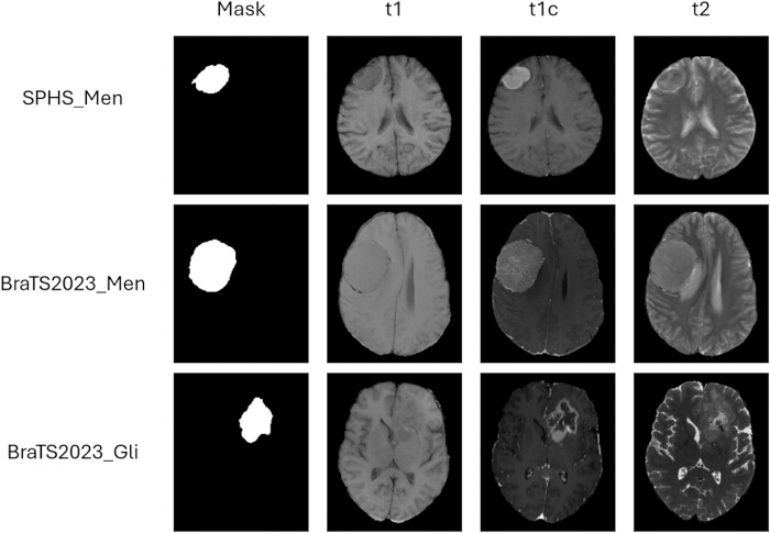
Figure 1. Representative cross-dataset comparison. Top row: SPHS-Men; middle: BraTS2023-Men; bottom: BraTS2023-Gli. Columns show tumor mask, T1, T1c, and T2. Qualitative differences in lesion size, enhancement pattern, and boundary definition reflect the compounded imaging and annotation shifts encountered in this study. Please click here to view a larger version of this figure.
MRI acquisition and preprocessing
All BraTS data were distributed with standardized preprocessing already applied, including skull stripping, co-registration, and isotropic resampling. For the SPHS cohort, raw DICOM images were first converted to NIfTI format28. The multimodal images were then rigidly registered to the native T1 space using mutual information-based alignment29. Because skull stripping is a necessary preprocessing step for brain MRI segmentation, brain extraction was explicitly performed before model training. Specifically, DeepBet was applied to the T1-weighted image to generate a brain mask30, and the resulting mask was propagated to the co-registered T1c and T2 volumes to remove non-brain tissue consistently across all modalities. The skull-stripped images were subsequently resampled to an isotropic resolution of 1 x 1 x 1 mm3. Binary tumor masks were finally refined using morphological closing and opening operations to suppress interpolation artifacts introduced during resampling. These harmonization steps were used to improve cross-dataset input consistency for external testing, but they were not intended to eliminate the underlying scanner-, protocol-, or annotation-level differences intrinsic to the SPHS cohort.
Study design
Model architecture
As shown in Figure 2, the UMamba 2D architecture integrates residual convolutional neural network modules with Mamba selective state-space model (SSM) blocks12,31. This hybrid design enables the simultaneous extraction of local texture features via convolutions and long-range global structural context via SSMs, with a linear computational cost with respect to the number of spatial positions.
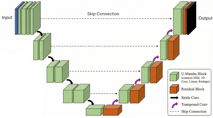
Figure 2. The UMamba 2D architecture. The encoder integrates residual convolutional blocks for local texture and boundary feature extraction with Mamba selective SSM modules for long-range context modeling. These components are connected via skip connections to a transposed-convolution decoder. The network input consists of a three-channel multi-modal MRI slice comprising T1, T1c, and T2 modalities. Please click here to view a larger version of this figure.
Encoder
Multi-modal MRI slices with C = 3 input channels are processed by a hierarchical encoder. At each stage s ∈ 1,…, S, the input feature map is first processed by a residual convolutional block to extract localized spatial patterns. The feature map is then flattened into a one-dimensional sequence and processed by a selective SSM module to capture long-range contextual dependencies. Spatial downsampling is performed using stride-2 convolutions, progressively generating hierarchical feature representations F_s ∈ R^C_s × H/2^s × W/2^s with increasing channel dimensions C_s ∈ 32, 64, 128, 256, 512.
Mamba selective SSM
The selective SSM operates on a one-dimensional sequence x ∈ R^L × d. Because spatial adjacency is disrupted during standard one-dimensional flattening of two-dimensional feature maps, a multi-directional scanning strategy is used: feature maps are scanned along four directions (row forward, row backward, column forward, and column backward) to preserve spatial context across both row and column directions. The continuous-time SSM is defined as:


where A ∈ R^N × N denotes the learnable state transition matrix initialized via the HiPPO (Hidden Polynomial Projection Operator) framework31. The projection matrices B(t) and C(t) are input-dependent, establishing a selective mechanism that enables the model to dynamically filter irrelevant tokens and retain important contextual information. The continuous system is discretized using a time step Δ via a zero-order hold (ZOH) transformation:




At each discrete step k, the parameters B, C, and Δ are computed as linear projections of the input token xk, enabling input-dependent feature propagation. The HiPPO initialization ensures that the eigenvalues satisfy |λj(barA)| < 1, which keeps the hidden state representations ‖hk‖ bounded across arbitrarily long spatial sequences.
Decoder and skip connections
The decoder reconstructs the original spatial resolution from the encoded representations. At each stage, transposed convolutions upsample the lower-resolution feature maps. Skip connections concatenate the multi-scale encoder features F_s with the corresponding upsampled decoder features, preserving high-frequency boundary information that is essential for precise voxel-level tumor delineation.
Training loss
Model optimization is guided by a composite loss function:

where p_θ(r) ∈ [0,1] is the predicted tumor probability at voxel r, Y(r) is the corresponding binary ground-truth label, and λ = 1.0 balances the two terms. The Dice loss directly optimizes the target evaluation metric and addresses the extreme foreground–background imbalance inherent to meningioma segmentation, where tumor voxels typically constitute less than 0.5% of the total intracranial volume. The binary cross-entropy (BCE) term provides per-voxel supervision, stabilizing training in this imbalanced setting.
Training configuration
All segmentation models were optimized using AdamW with an initial learning rate of η₀ = 3 × 10⁻4, cosine annealing schedule, and weight decay of 5 × 10⁻4. The centralized baseline models were trained for 200 epochs on the 450-case BraTS2023 cohorts with a batch size of 16. To avoid ambiguity in the federated setting, we use the following terminology throughout this manuscript: a communication round denotes one complete FL cycle consisting of server broadcast, local client optimization, and server aggregation; a mini-batch iteration denotes one parameter update on one mini-batch; and the local optimization performed by each client in one round consisted of 250 mini-batch iterations. In other words, each client performed one local training stage per round, and that local stage contained 250 parameter-update iterations. Thus, the term 'round' (r = 3) in this study refers only to FL communication, not to an epoch across the entire centralized dataset. An online data augmentation pipeline was applied uniformly across all training configurations, including random horizontal and vertical flipping (p=0.5 per axis), random in-plane rotation (±15^circ), random intensity scaling (factor uniformly sampled from [0.85, 1.15]), and additive Gaussian noise (sigma ∼ U[0, 0.05]) computed per two-dimensional slice. All augmentation was disabled during inference and validation. The framework was implemented in PyTorch 2.0.1 with MONAI 1.3 for data loading, and all computations were performed on NVIDIA GeForce RTX 3090 (24 GB GDDR6X) graphics processing units (GPUs). Reproducibility was ensured by fixing all random seeds across PyTorch, NumPy, and the Python random module to identical values for all strategies.
Experimental strategies
Three primary training strategies were compared to evaluate cross-center generalization (Figure 3). The labels meningioma-pretrained FL and Glioma-pretrained FL are descriptive names introduced in this manuscript to distinguish the source of the initial global model before federated adaptation.
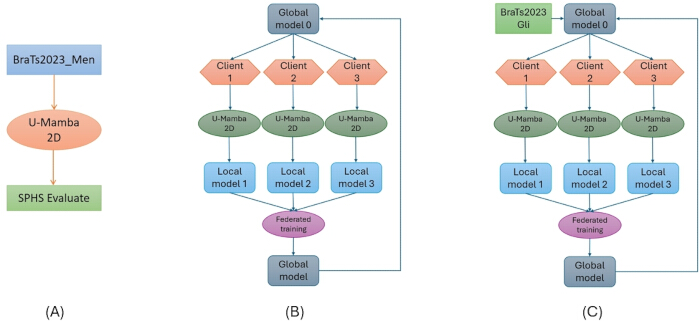
Figure 3. Experimental design overview. (A) Centralized training using 450 BraTS2023-Men cases followed by external evaluation on SPHS. (B) Meningioma-pretrained FL across three simulated clients. (C) Glioma-pretrained FL initialized from a centralized glioma model before federated adaptation. Please click here to view a larger version of this figure.
Strategy 1 (Centralized training)
A single UMamba 2D model was trained on all 450 BraTS2023-Men cases. This strategy represents the upper bound on in-domain performance and serves as the baseline for assessing cross-center degradation.
Strategy 2 (FL with MEN-domain initialization)
The 450 BraTS2023-Men cases were randomly assigned to three clients (|Di| = 150) for equal partitioning. The three BraTS2023-Men clients were therefore simulated from the same public cohort rather than constructed using institution-specific metadata or scanner-origin labels. To initialize federated training in the meningioma domain, the initial global model was first trained on a fixed BraTS2023-Men client split (client 1 in this implementation) and then used to initiate the federated procedure. This seed client was held constant across experiments for reproducibility and should be interpreted as a within-domain warm start rather than an optimized or privileged source client. Because the three clients were simulated by partitioning the same BraTS2023-Men cohort, this initialization choice reflects an implementation-level starting point rather than a biologically distinct institutional prior. The purpose of this design was not to strictly replicate a real multi-institution launch process, but to provide a controlled comparison between centralized training on all available MEN data and federated collaboration starting from a partial meningioma-pretrained FL. The global federated optimization objective is defined as:

where Di denotes the local training set of clients i, and ell is the Dice + cross-entropy loss. In communication round 1, aggregation weights were proportional to sample count (wi(1) = 1/3). To down-weight underperforming clients in subsequent communication rounds, rounds 2 and 3 used validation Dice-weighted aggregation:

where DSCi(r) is the case-averaged DSC on the local validation partition of client i after local training in communication round r, and ε = 10⁻6 is a small constant to prevent division by zero.
Strategy 3 (FL with glioma-pretrained initialization)
To obtain a task-related cross-domain warm start, Centralized training on Glioma was first performed using 450 BraTS2023-Gli cases with the same three MRI modalities (T1, T1c, and T2) and the same binary tumor-versus-background formulation adopted in the meningioma experiments.
Specifically, glioma labels were harmonized by combining the non-enhancing tumor core and enhancing tumor into a single foreground class, while peritumoral edema was excluded, so that the source and target tasks shared the same binary output definition. Because both source and target tasks were formulated as binary segmentation, the pretrained weights were transferred directly to initialize the full UMamba 2D model, including the segmentation head, rather than discarding and reinitializing the final layer. The same FedAvg protocol was then applied, with the initial global model initialized by the converged Centralized training on Glioma weights (θ⁽°⁾ = θ_Gli^*). The subsequent federated adaptation is given by:

where Δθ(r) represents the accumulated domain-specific parameter updates across r communication rounds. This strategy tests whether a Glioma-pretrained FL source model provides a better starting point for federated adaptation on meningioma clients.
Evaluation metrics
All strategies were evaluated on the SPHS cohort using a case-averaged protocol. The DSC and IoU were computed independently per case and then averaged, ensuring equal statistical weight across all subjects regardless of tumor volume:
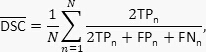
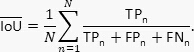
Case-averaged error compositions—comprising mean true positive (TP), false positive (FP), false negative (FN), and true negative (TN) voxel counts per case—were also reported. This breakdown distinguishes over-segmentation from under-segmentation errors, providing insight into directional failure modes.
Access restricted. Please log in or start a trial to view this content.
Results
The centralized UMamba 2D model (Strategy 1) achieved competitive in-domain performance on the BraTS2023-Men internal test set, with a case-averaged DSC of 0.8958, IoU of 0.8356, FN of 1,902, FP of 667, TN of 8,902,814, and TP of 22,616. When evaluated on the SPHS external validation cohort, the centralized model yielded a DSC of 0.7452 ± 0.1992 and an IoU of 0.6241 ± 0.1941, corresponding to a cross-center generalization gap of ΔDSC = 0.1506 (Table 1, ID 1). As an additional self-configuring baseline, n...
Access restricted. Please log in or start a trial to view this content.
Discussion
The central finding of this study is that initialization quality—rather than the federated aggregation protocol itself—is the decisive factor in closing the cross-center generalization gap for privacy-preserving meningioma segmentation. While the centralized UMamba 2D model achieved strong in-domain performance on the BraTS2023-Men cohort (DSC = 0.8958), it suffered a substantial degradation when evaluated on the independent SPHS clinical dataset (Δ DSC = 0.1506). This performance drop underscores the vu...
Access restricted. Please log in or start a trial to view this content.
Disclosures
The authors have no conflicts of interest to declare.
Acknowledgements
This work was supported by the National Natural Science Foundation of China (Grant 12405382), the Shenzhen Science and Technology Innovation Commission (Grant JCYJ20220530143602006), the Shenzhen Basic Research Project (Natural Science Foundation) (Grant JCYJ20220530150416036), and the Inner Mongolia Science and Technology Department Social Welfare Project (Project No. 2026YFSH0155).
Access restricted. Please log in or start a trial to view this content.
Materials
| Name | Company | Catalog Number | Comments |
|---|---|---|---|
| BraTS2023-Gli dataset | ASNR-MICCAI BraTS Challenge / Synapse (Sage Bionetworks) | N/A | Public glioma MRI dataset used for glioma-pretrained initialization. In this study, 450 cases were used under the binary tumor-versus-background formulation. https://www.synapse.org/Synapse:syn51156910/wiki/622351 |
| BraTS2023-Men dataset | ASNR-MICCAI BraTS Challenge / Synapse (Sage Bionetworks) | N/A | Public meningioma MRI dataset used as the source cohort for model development and internal evaluation. In this study, 450 cases were used. https://arxiv.org/abs/2305.07642 |
| DeepBet | Academic software by Fisch et al. | N/A | Brain extraction tool applied to the T1-weighted image to generate a brain mask for the SPHS cohort. https://arxiv.org/abs/2308.07003 |
| MONAI | Project MONAI | Version 1.3 | Medical imaging AI framework used for data loading and deep learning workflow implementation. https://docs.nvidia.com/monai/index.html |
| NVIDIA GeForce RTX 3090 GPU | NVIDIA | GeForce RTX 3090 | Graphics processing unit with 24 GB GDDR6X memory used for model training and computation. https://www.nvidia.com/en-me/geforce/graphics-cards/30-series/rtx-3090-3090ti/ |
| PyTorch | PyTorch Foundation, Linux Foundation | Version 2.0.1 | Deep learning framework used to implement and train the UMamba 2D segmentation models. https://pytorch.org/foundation/ |
| Python | Python Software Foundation | Version 3.10 | Programming environment used for model training, preprocessing, evaluation, and statistical analysis. Replace with the exact Python version if known. https://www.python.org/psf-landing/ |
| SPHS-Men clinical cohort | Second People’s Hospital of Shenzhen | N/A | External clinical multi-modal MRI cohort used for independent evaluation. A total of 174 cases were included. |
| UMamba 2D model implementation | Bowang-lab / U-Mamba authors | U-Mamba 2D configuration | State space model-based segmentation architecture used for binary meningioma segmentation. https://u-mamba.github.io/ |
References
- Ostrom, Q. T., et al. CBTRUS statistical report: primary brain and other central nervous system tumors diagnosed in the United States in 2014-2018. Neuro Oncol. 23 (Suppl 3), (2021).
- Goldbrunner, R., et al. EANO guideline on the diagnosis and management of meningiomas. Neuro Oncol. 23 (11), 1821-1834 (2021).
- Huang, R. Y., et al. Imaging and diagnostic advances for intracranial meningiomas. Neuro Oncol. 21 (Suppl 1), (2019).
- Combs, S. E., et al. ESTRO ACROP guideline for target volume delineation of skull base tumors. Radiother Oncol. 156, 80-94 (2021).
- Growcott, S., et al. Inter-observer variability in target volume delineations of benign and metastatic brain tumours for stereotactic radiosurgery. Clin Oncol (R Coll Radiol). 32 (1), 13-25 (2020).
- Kriwanek, F., et al. Impact of SSTR PET on inter-observer variability of target delineation of meningioma. Cancers (Basel). 14 (18), 4435 (2022).
- Menze, B. H., et al. The multimodal brain tumor image segmentation benchmark (BRATS). IEEE Trans Med Imaging. 34 (10), 1993-2024 (2015).
- Ronneberger, O., Fischer, P., Brox, T. U-Net: convolutional networks for biomedical image segmentation. , (2015).
- Isensee, F., et al. nnU-Net: a self-configuring method for deep learning-based biomedical image segmentation. Nat Methods. 18 (2), 203-211 (2021).
- Hatamizadeh, A., et al. UNETR: transformers for 3D medical image segmentation. , (2022).
- Hatamizadeh, A., et al. Swin UNETR: Swin transformers for semantic segmentation of brain tumors in MRI. , (2021).
- Ma, J., Li, F., Wang, B. U-Mamba: enhancing long-range dependency for biomedical image segmentation. arXiv. , (2024).
- Ranjbarzadeh, R., Keles, A., Anari, S., Zarbakhsh, P., Bendechache, M., Nagar, A. K., Jat, D. S., Mishra, D. K., Joshi, A. Explainable attention-guided Swin Transformer networks for brain tumor segmentation from 3D MRI. Intelligent Sustainable Systems: Selected Papers of WorldS4 2025. , 110-128 (2026).
- Ranjbarzadeh, R., Keles, A., Anari, S., Cunneen, M., Bendechache, M. A global-local 3D brain tumor segmentation model using Vision Transformers and axial statespace modeling. , (2026).
- Laukamp, K. R., et al. Fully automated detection and segmentation of meningiomas using deep learning on routine multiparametric MRI. Eur Radiol. 29 (1), 124-132 (2019).
- Boaro, A., et al. Deep neural networks allow expert-level brain meningioma segmentation. Sci Rep. 12 (1), 15462 (2022).
- LaBella, D., et al. A multi-institutional meningioma MRI dataset for automated multi-sequence image segmentation. Sci Data. 11 (1), 496 (2024).
- Zhang, L., et al. Generalizing deep learning for medical image segmentation to unseen domains via deep stacked transformation. IEEE Trans Med Imaging. 39 (7), 2531-2540 (2020).
- McMahan, B., Moore, E., Ramage, D., Hampson, S., y Arcas, B. A. Communication-efficient learning of deep networks from decentralized data. , 1273-1282 (2017).
- Anari, S., Ranjbarzadeh, R., Cunneen, M., Bendechache, M. Privacy-preserving federated learning for human intention modeling in pediatric cerebral palsy using extended reality. , 1565-1570 (2025).
- Ranjbarzadeh, R., Anari, S., Cunneen, M., Bendechache, M. Lightweight deep learning with virtual reality visualization for offline tumor segmentation in rural environments. , 1577-1582 (2025).
- Karimireddy, S. P., et al. SCAFFOLD: stochastic controlled averaging for federated learning. , (2020).
- Tabassum, M., Di Ieva, A., Liu, S. Meta transfer learning for brain tumor segmentation using nnUNet in meningioma and metastasis cases. Sci Rep. 15 (1), 37599 (2025).
- Karargyris, A., et al. Federated benchmarking of medical artificial intelligence with MedPerf. Nat Mach Intell. 5, 799-810 (2023).
- LaBella, D., et al. The ASNR-MICCAI Brain Tumor Segmentation (BraTS) Challenge 2023: intracranial meningioma. arXiv. , (2023).
- Baid, U., et al. The RSNA-ASNR-MICCAI BraTS 2021 benchmark on brain tumor segmentation and radiogenomic classification. arXiv. , (2021).
- Bakas, S., et al. Advancing the cancer genome atlas glioma MRI collections with expert segmentation labels and radiomic features. Sci Data. 4, 170117 (2017).
- Li, X., Morgan, P. S., Ashburner, J., Smith, J., Rorden, C. The first step for neuroimaging data analysis: DICOM to NIfTI conversion. J Neurosci Methods. 264, 47-56 (2016).
- Klein, S., Staring, M., Murphy, K., Viergever, M. A., Pluim, J. P. W. Elastix: a toolbox for intensity-based medical image registration. IEEE Trans Med Imaging. 29 (1), 196-205 (2010).
- Fisch, L., et al. DeepBet: fast brain extraction of T1-weighted MRI using convolutional neural networks. Comput Biol Med. 179, 108845 (2024).
- Gu, A., Dao, T. Mamba: linear-time sequence modeling with selective state spaces. arXiv. , (2023).
- Sheller, M. J., et al. Federated learning in neuro-oncology for multi-institutional collaborations without sharing patient data. Neuro Oncol. 21 (Suppl 6), (2019).
- Karimi, D., Warfield, S. K., Gholipour, A. Transfer learning in medical image segmentation: new insights from analysis of model parameters and learned representations. Artif Intell Med. 116, 102078 (2021).
- Bonawitz, K., et al. Practical secure aggregation for privacy-preserving machine learning. , 1175-1191 (2017).
- McMahan, H. B., Ramage, D., Talwar, K., Zhang, L. Learning differentially private recurrent language models. arXiv. , (2017).
- Zhu, L., Liu, Z., Han, S. Deep leakage from gradients. , (2019).
- Li, T., Sahu, A. K., Talwalkar, A., Smith, V. Federated optimization in heterogeneous networks. Proc Mach Learn Syst. 2, 429-450 (2020).
Access restricted. Please log in or start a trial to view this content.