This protocol eliminates the Cas9 protein after genome editing to facilitate robust rescue experiments in stably transduced knockout cell lines.
Method Article
Targeting Cas9 to Perform Rescue Experiments in Stably Transduced Knockout HEK293 Cells
In This Article
Summary
Abstract
CRISPR-Cas9 gene editing technology has revolutionized molecular biology. Often, this technology is employed to delete a gene encoding a protein of interest. The resulting phenotype provides valuable insight into the protein's function. The functional importance of the target protein can be confirmed by reintroducing the protein to restore the lost function (rescue). This is typically accomplished by introducing the protein-coding cDNA in trans using an expression vector. However, in knockout cell lines that stably express the CRISPR-Cas9 system, the newly introduced expression plasmid may also be cleaved by Cas9. The protocol presented here provides a strategy to circumvent this potential barrier to rescue experiments. This approach is demonstrated using HEK293 cells in which the gene encoding the E3 ubiquitin ligase scaffold protein CUL4B was disrupted by CRISPR-Cas9. Transduction of these cells with a guide RNA (gRNA) targeting the integrated Cas9 transgene resulted in the loss of detectable Cas9 protein. Cas9 ablation enabled restoration of CUL4B expression and function following introduction of a CUL4B expression plasmid. These results provide proof of concept for a broadly applicable approach to studying protein function through rescue experiments.
Introduction
CRISPR-Cas9 is a powerful genome-editing tool derived from a naturally occurring adaptive immune system used by prokaryotes to defend against bacteriophage infection. Owing to its low cost, high success rate, and ease of use, CRISPR-Cas9 technology has surpassed other genome-editing tools, such as zinc finger nucleases (ZFNs) and transcription activator-like effector nucleases (TALENs), for precise genetic modification, particularly in cell culture. The two-component system comprises a Cas9 nuclease and a guide RNA (gRNA). Through Watson-Crick base pairing, the gRNA directs Cas9 to a genomic target adjacent to a protospacer adjacent motif (PAM). The PAM sequence serves as a critical recognition site that licenses nuclease activity. At that site, Cas9 generates a double-strand break (DSB). In the absence of a homologous repair template, the cell repairs the DSB through non-homologous end joining (NHEJ). This repair mechanism can introduce insertions or deletions (indels), leading to frameshift mutations and functional knockout of the target gene. Target gene disruption is therefore achieved by introducing Cas9 together with an engineered gRNA. The 20-nucleotide gRNA sequence is complementary to a genomic site immediately adjacent to the PAM. The resulting functional loss of the target gene reveals that gene’s specific roles in cellular processes1.
A rescue experiment, or complementation assay, involves restoration of the missing protein to demonstrate reversal of the observed phenotype. This approach serves as a rigorous control to rule out unintended experimental effects, such as those resulting from 'off-target' CRISPR-Cas9 deletions. The most common method for reintroducing a protein of interest is through transfection or transduction of an exogenous expression vector. However, this introduced DNA is susceptible to the same CRISPR-Cas9 targeting as the endogenous locus. This risk is particularly high in stably transduced cell lines, where constitutive Cas9 expression is coupled with antibiotic selection to ensure a homogeneous and consistent knockout population; a strategy often required in cell lines that are difficult to transfect or transduce, such as those derived from immune, hematopoietic, or neuronal lineages.
One method to circumvent vector targeting is to introduce exogenous mRNA encoding the protein of interest, thereby bypassing Cas9-mediated cleavage and the need for nuclear transcription. However, compared to plasmid DNA, synthetic mRNA is more costly to produce, inherently less stable, and yields highly transient expression. Furthermore, it requires specific chemical modifications to avoid triggering cytosolic innate immune responses2.
To avoid Cas9/gRNA complex targeting of expression plasmids, wobble base engineering of the coding sequence can be employed. By exploiting the degeneracy of the genetic code, synonymous mutations can be introduced to eliminate the required PAM sequence and/or disrupt gRNA complementarity without changing the amino acid sequence of the encoded protein. However, the employment of synonymous mutations is not without risk. These alterations can introduce suboptimal codon usage, which may disrupt translation kinetics and lead to protein misfolding, reduced mRNA stability, or deleterious changes in RNA secondary structure3,4. Although less frequent, other potential consequences include the introduction of cryptic polyadenylation signals, the disruption of post-transcriptional regulatory motifs, and increased CpG density5,6. Furthermore, engineered sequences may inadvertently trigger cellular stress responses if the resulting mRNA structure interferes with ribosomal progression7,8.
Beyond the challenges associated with the use of synthetic mRNAs or wobble-base engineering, the constitutive expression of Cas9 post-editing introduces several confounding variables in downstream assays. Because Cas9 specificity can be imperfect, persistent expression increases the likelihood of off-target cleavage, leading to the cumulative acquisition of unintended mutations over time9,10,11. Furthermore, chronic genomic surveillance and repetitive DSBs can trigger a persistent DNA damage response, ultimately driving cells toward cell cycle arrest, senescence, or apoptosis12,13. This constitutive production also imposes a metabolic burden, thereby potentiating clonal drift in which low-expressing cells outcompete high-expressing cells, resulting in inconsistent transgene levels across successive passages. Finally, as a large prokaryotic protein, Cas9 may trigger stress via innate immune signaling or overwhelm the host’s proteostasis machinery, inducing the unfolded protein response14,15,16.
To mitigate these complications, several transient Cas9 inhibition strategies have been developed, including phage-derived Anti-CRISPR proteins (Acrs)17,18, small-molecule inhibitors or degron-fused systems like PROTACs19,20, and optogenetic control mechanisms19,21. However, these approaches introduce practical hurdles that limit their utility for rescue assays. Acrs inhibit through molecular mimicry and therefore require stoichiometric optimization for precision. Acrs may also exacerbate the same proteostatic and immunogenic stresses associated with Cas9 overexpression. Chemical modulation introduces pharmacokinetic complexities, potential cytotoxicity, and PROTAC-associated 'hook effects' where excessive ligand concentrations paradoxically prevent productive ternary complex formation22. Optogenetic platforms are limited by cellular phototoxicity and the requirement for specialized hardware to maintain consistent light delivery19,21. Importantly, because these methods provide only temporary or reversible inhibition, they are often insufficient for applications in which Cas9 is constitutively expressed.
In contrast, self-inactivating CRISPR systems offer a permanent solution by autonomously and permanently disrupting the Cas9 transgene immediately following successful target gene editing. By eliminating residual nuclease activity, this approach permanently shields subsequent rescue vectors from cleavage while simultaneously reducing long-term proteostatic stress and off-target genomic drift. Although self-directed gRNAs can be delivered via transient ribonucleoprotein or lipid-based transfections23,24, these methods are prone to dilution and stochastic variation, resulting in a "mosaic" population in which some cells escape inactivation25,26,27.
To overcome these limitations, the protocol described herein leverages a lentiviral delivery system for Cas9 self-inactivation, offering distinct practical advantages for stable cell line modification. Lentiviral delivery ensures the linked inheritance of the Cas9 transgene and its self-directed regulator, effectively preventing the 'dilution escape' typical of rapidly dividing populations. Furthermore, the expanded cargo capacity of lentiviral vectors permits the co-expression of selection markers to ensure a homogeneous, Cas9-deleted population, while their broad tropism, conferred by VSV-G pseudotyping, facilitates high-efficiency delivery into hard-to-transfect cell types25,28,29.
As a proof-of-concept demonstration of this method, this protocol details the self-inactivation of Cas9 to facilitate phenotypic rescue of the E3 ubiquitin ligase scaffold protein CUL4B in CUL4B-knockout HEK293 cells30.
Protocol
This study used established immortalized human cell lines (HEK293 and HEK293FT) and did not involve human participants, primary human tissues, or animal experiments. Therefore, institutional ethics approval was not required.
1. Generation of Cas9-specific gRNA lentiviral construct
- Insert the Cas9-specific gRNA sequence (Table 1) into the pLentiGuide-Puro lentiviral vector.
- Replace the antibiotic resistance gene with one that differs from the resistance marker used to select the stably transduced CRISPR-Cas9 cell line.
- Verify successful insertion and sequence fidelity of both the Cas9-specific gRNA and the replacement antibiotic resistance gene by Sanger sequencing using vector-specific primers.
NOTE: In this protocol, the puromycin resistance (PuroR) gene was replaced with the hygromycin B resistance (HygroR) gene to generate the vector Cas9 gRNA1_pLentiGuide-Hygro. Alternative antibiotic resistance genes include blasticidin S (BlastR), G418/Geneticin (NeoR), and zeocin (ZeoR).
2. Production of lentivirus encoding Cas9-specific gRNA
- Seed 2.5 × 10⁶ HEK293FT cells in a 10 cm tissue culture-treated dish.
NOTE: Maintain the cells in Dulbecco's Modified Eagle Medium (DMEM; high glucose) supplemented with 10% fetal bovine serum (FBS) (hereafter referred to as cDMEM) and incubate them at 37 °C in a humidified incubator containing 5% CO2. HEK293FT cells are used because they generate higher lentiviral titers than other HEK293-derived cell lines31. - Incubate the cells for 12 h until they reach at least 60% confluence before transfection.
CAUTION: Perform all procedures involving third-generation lentiviral vectors under Biosafety Level 2 (BSL-2) conditions using appropriate personal protective equipment (PPE) and approved decontamination procedures (e.g., a Class II biological safety cabinet, secondary containment, and 70% ethanol), as described by Pauwels et al32. - Prepare a standard calcium phosphate (CaPhos) transfection mixture. Dilute 20 µg of total plasmid DNA in sterile molecular biology-grade water to a final volume of 450 µL. Add 50 µL of 2.5 M CaCl2, followed by 500 µL of 2× HEPES-buffered saline (HBS; pH 7.05), to obtain a final volume of 1 mL in a 1.5 mL microcentrifuge tube.
NOTE: Use sequence-verified, predominantly supercoiled plasmid DNA that meets the following quality criteria: A260/A280 = 1.8–2.0, A260/A230 = 2.0–2.2, and endotoxin <0.1 EU/µg. Prepare the transfection mixture using an approximate equimolar ratio consisting of 5 µg Cas9 gRNA1_pLentiGuide-Hygro, 5.6 µg pLP1 (Gag/Pol), 3.8 µg pLP2 (Rev), and 5.6 µg pLP/VSVG (vesicular stomatitis virus glycoprotein). - Mix the CaPhos transfection solution thoroughly by gentle pipetting.
- Add 1 mL of the CaPhos transfection mixture dropwise to the HEK293FT cell culture.
- Gently rock the culture dish to distribute the transfection complexes evenly.
- Incubate the cells for 12 h.
NOTE: Calcium phosphate transfection is generally unaffected by standard maintenance antibiotics33. However, an antibiotic-free medium is recommended for more sensitive transfection methods, such as lipofection, because polyanionic antibiotics can interfere with DNA-lipid complex formation34. - Aspirate and discard the culture medium.
- Wash the cells once with 10 mL of room-temperature sterile 1× phosphate-buffered saline (PBS).
- Aspirate and discard the PBS.
- Add 10 mL of fresh room-temperature cDMEM.
- Return the cells to the incubator and allow lentiviral particles to accumulate in the culture medium for 36 h.
NOTE: Under these conditions, unconcentrated lentiviral supernatant is estimated to yield approximately 1.0 × 10⁶ transducing units (TU)/mL after 48 h, although the actual titer should be determined experimentally as described by Kutner et al.35 or Barde et al.36. Store lentiviral supernatant at −80 °C for up to 2 years. Avoid repeated freeze-thaw cycles, as they can reduce viral titer by approximately 55%37,38,39,40.
3. Harvesting lentivirus encoding Cas9-specific gRNA and transduction of target cells
- Seed 1.0 × 10⁶ adherent target cells in a 10 cm tissue culture-treated dish.
- Incubate the target cells for 12 h before lentiviral transduction.
NOTE: The target cells are HEK293 cells stably expressing Streptococcus pyogenes Cas9, a CUL4B-targeting gRNA (Table 1), and a puromycin resistance (PuroR) gene. Maintain the cells in cDMEM containing 10 µg/mL puromycin and verify CUL4B disruption by western blot analysis before Cas9 self-inactivation. Perform all subsequent experiments using early-passage HEK293ΔCUL4B cells. - Harvest the lentivirus-containing medium by carefully transferring the supernatant from the HEK293FT producer cells into a sterile 15 mL conical tube, taking care not to disturb the cell monolayer.
NOTE: Discard the HEK293FT producer cells according to established biosafety guidelines32. - Centrifuge the collected supernatant at 1,000 × g for 10 min at room temperature (RT) to pellet any residual HEK293FT producer cells.
- Aspirate and discard the culture medium from the previously seeded HEK293ΔCUL4B cells.
- Transfer 7 mL of the clarified lentiviral supernatant to the 10 cm dish containing the HEK293ΔCUL4B cells. Avoid disturbing the pellet at the bottom of the centrifuge tube and leave at least 1 mL of residual supernatant behind to minimize carryover of producer cells.
NOTE: Filter the supernatant through a 0.45 µm filter, if desired, to further eliminate residual producer cells. Discard the remaining supernatant in accordance with biosafety guidelines32. At an estimated titer of 1.0 × 10⁶ TU/mL, a 7 mL inoculum is expected to produce a multiplicity of infection (MOI) between 3.5 and 5.541. - Incubate the transduced HEK293ΔCUL4B cells for 24 h to permit complete lentiviral transduction and integration.
- Aspirate and discard the viral supernatant.
- Wash the cells once with 10 mL of room-temperature sterile 1× PBS.
- Aspirate and discard the PBS.
- Add 10 mL of fresh room-temperature cDMEM.
- Return the cells to the incubator and culture them for four days to allow transgene expression and Cas9 disruption.
NOTE: If the lentiviral Cas9 gRNA vector does not encode a fluorescent reporter, transduce a parallel sentinel culture with a similarly generated lentiviral vector expressing a fluorescent reporter. Use the sentinel culture to estimate transduction efficiency and the timing of target gene depletion by qualitative fluorescence microscopy or quantitative flow cytometry. In this study, GFP expression was readily detected by fluorescence microscopy four days after transduction.
4. Antibiotic selection of Cas9-specific gRNA transductants
NOTE: Four days after transduction, the target cells should be at or near confluence and require transfer to a larger culture vessel. Perform cell transfer simultaneously with the initiation of antibiotic selection.
- Aspirate and discard the culture medium from the HEK293ΔCUL4B cells.
- Wash the cells once with 10 mL of room-temperature sterile 1× phosphate-buffered saline (PBS).
- Aspirate and discard the PBS.
- Add 2 mL of sterile, room-temperature trypsin-EDTA solution to the culture dish.
- Incubate the cells for 5 min to promote detachment.
- Add 8 mL of room-temperature cDMEM to neutralize the trypsin.
- Transfer the cell suspension to a sterile 15 mL conical tube.
- Triturate the cell suspension 15–20 times to dissociate cellular aggregates thoroughly.
- Centrifuge the cells at 200 × g for 10 min at room temperature.
- While the cells are centrifuging, prepare a T150 tissue culture-treated flask containing 20 mL of cDMEM supplemented with 250 µg/mL hygromycin B.
NOTE: The hygromycin B concentration required for selection may vary among cell types. Perform a kill curve using 50–500 µg/mL hygromycin B to determine the minimum concentration required to eliminate all untransduced cells within 7–10 days. If an alternative antibiotic resistance marker is used in the Cas9 gRNA vector, perform a kill curve using 2–10 µg/mL blasticidin S, 400–800 µg/mL G418/Geneticin, or 50–400 µg/mL zeocin. - Aspirate and discard the supernatant without disturbing the cell pellet.
- Resuspend the cell pellet in 10 mL of cDMEM supplemented with 250 µg/mL hygromycin B.
- Transfer the 10 mL cell suspension into the prepared T150 flask containing 20 mL of selection medium.
- Gently rock the flask to distribute the cells evenly.
- Incubate the cells under selection for three days.
- On day 3 of selection, aspirate the selection medium and replace it with 30 mL of fresh cDMEM supplemented with 250 µg/mL hygromycin B.
NOTE: Replacing the selection medium removes non-adherent dead cells, replenishes nutrients, and maintains antibiotic activity. - Continue incubation until the surviving cell monolayer reaches approximately 90% confluence, typically after an additional four days.
5. Validation of Cas9 clearance and in trans CUL4B rescue
NOTE: Once the cells reach approximately 90% confluence, confirm Cas9 ablation in the initial experiments, and immediately cryopreserve the remaining cell population42. This approach minimizes the development of secondary compensatory mechanisms.
- Seed early-passage HEK293ΔCUL4B cells stably expressing a Cas9-targeting gRNA (hereafter referred to as HEK293ΔCUL4BΔcas9) into four wells of a 6-well tissue culture-treated plate at a density of 2.5 × 10⁵ cells per well.
- Seed HEK293ΔCUL4B cells into four wells of a separate 6-well plate. Seed parental HEK293 cells into at least one additional well to serve as a control.
NOTE: Use the HEK293ΔCUL4B and parental HEK293 cultures as controls for Cas9 and CUL4B expression, respectively. - Incubate all cultures for 12 h to allow complete cell attachment and active proliferation.
- Prepare calcium phosphate (CaPhos) transfection mixtures containing 0, 0.5, 1.0, or 2.0 µg of pCMV-FLAG-CUL4B. Adjust each mixture to a constant total of 10 µg plasmid DNA by adding an empty pCMV vector.
- Dilute each DNA mixture in sterile molecular biology-grade water to a final volume of 180 µL, add 20 µL of 2.5 M CaCl2, and then add 200 µL of 2× HEPES-buffered saline (HBS; pH 7.05) to obtain a final volume of 400 µL in a 1.5 mL microcentrifuge tube.
NOTE: When using expression plasmids driven by strong promoters (e.g., CMV), perform a DNA titration to achieve protein expression levels comparable to endogenous expression. Use sequence-verified, predominantly supercoiled plasmid DNA with A260/A280 values of 1.8–2.0, A260/A230 values of 2.0–2.2, and an endotoxin level of <0.1 EU/µg. - Mix each CaPhos transfection solution thoroughly by gentle pipetting.
- Add 200 µL of each transfection mixture dropwise to the designated well containing HEK293ΔCUL4BΔcas9 cells. Change pipette tips between samples to prevent cross-contamination.
- Add the remaining 200 µL of each corresponding transfection mixture dropwise to the designated well containing HEK293ΔCUL4B cells.
- Incubate the transfected cultures for 12 h.
- Aspirate the culture medium from each well.
- Wash the cells once with 1 mL of room-temperature sterile 1× PBS.
- Aspirate and discard the PBS.
- Add 2 mL of fresh room-temperature cDMEM to each well.
- Incubate the cultures for an additional 24 h to allow CUL4B expression and downstream functional responses.
- Harvest the cells using standard trypsinization procedures.
- Transfer each cell suspension to an individual 1.5 mL microcentrifuge tube.
- Wash each cell suspension once with 1× PBS.
- Briefly pulse the samples to 14,000 × g at room-temperature to pellet the cells.
- Aspirate and discard the PBS supernatant.
CAUTION: β-mercaptoethanol is a volatile skin and respiratory irritant with a strong odor. Handle stock solutions in a certified chemical fume hood while wearing appropriate personal protective equipment, including chemical-resistant gloves, a laboratory coat, and safety glasses. Dispose of contaminated tips, tubes, and waste according to institutional hazardous waste guidelines. - Lyse each cell pellet directly in 200 µL Laemmli sample buffer supplemented with 5% (v/v) β-mercaptoethanol.
- Denature the proteins by heating the samples at 95 °C for 15 min.
- Resolve 20–40 µg of total protein per lane by standard sodium dodecyl sulfate-polyacrylamide gel electrophoresis (SDS-PAGE).
- Transfer the resolved proteins onto a polyvinylidene fluoride (PVDF) membrane.
- Block the membrane for 30 min at room temperature using blocking buffer.
NOTE: Prepare the blocking buffer using either 5% (w/v) non-fat dry milk or 5% (w/v) bovine serum albumin (BSA) dissolved in standard wash buffer (0.1% Tween 20 in either 1× Tris-buffered saline or 1× PBS). - Wash the membrane three times for 10 min each using standard wash buffer.
- Incubate the membrane with primary antibodies against CUL4B, Cas9, WDR5, CDC6, or α-TUBULIN for 12 h at 4 °C with gentle rocking.
NOTE: Dilute the primary antibodies 1:1,000 in standard wash buffer. - Remove the primary antibody solution and wash the membrane three times for 10 min each using standard wash buffer.
- Incubate the membrane with the appropriate horseradish peroxidase (HRP)-conjugated anti-mouse or anti-rabbit IgG secondary antibody for 12 h at 4 °C with gentle rocking.
NOTE: Dilute the secondary antibodies 1:3,000 in blocking buffer. - Remove the secondary antibody solution and wash the membrane three times for 10 min each using standard wash buffer.
- Apply the enhanced chemiluminescent (ECL) substrate uniformly across the membrane and incubate for 1 min.
- Capture the chemiluminescent signals using a digital imaging system.
NOTE: Confirm successful gene disruption, if desired, by Sanger sequencing of the targeted genomic region following PCR amplification of isolated genomic DNA43.
Results
To generate Cas9-ablated knockout lines for experimental reconstitution, lentivirus encoding a Cas9-targeting gRNA (Cas9 gRNA1_pLentiGuide-Hygro) was produced in HEK293FT cells using standard packaging plasmids (pLP1, pLP2, and pLP/VSVG). The resulting viral supernatant was harvested and used to transduce target cells. Survival during hygromycin B selection confirmed successful lentiviral transduction, and subsequent western blot analysis verified Cas9 ablation (Figure 1).
CUL4B protein was readily detected in parental HEK293 cells but was absent from empty vector-transfected HEK293ΔCUL4B and HEK293ΔCUL4BΔcas9 cells. Consistent with the loss of CUL4B, WDR5 and CDC6 exhibited the expected increases and decreases in steady-state expression, respectively, confirming disruption of CUL4B function. Cas9 protein was detected in HEK293ΔCUL4B cells but was undetectable in parental HEK293 and HEK293ΔCUL4BΔcas9 cells, confirming successful Cas9 ablation. As expected, transfection of the CUL4B expression plasmid into HEK293ΔCUL4B cells resulted in only minimal restoration of CUL4B protein expression. In contrast, robust CUL4B expression was observed following transfection of HEK293ΔCUL4BΔcas9 cells, accompanied by restoration of downstream protein expression, as evidenced by reduced WDR5 and increased CDC6 levels. Together, these findings demonstrate that Cas9 ablation permits efficient restoration of CUL4B expression and function by preventing Cas9-mediated cleavage of the rescue plasmid (Figure 2).
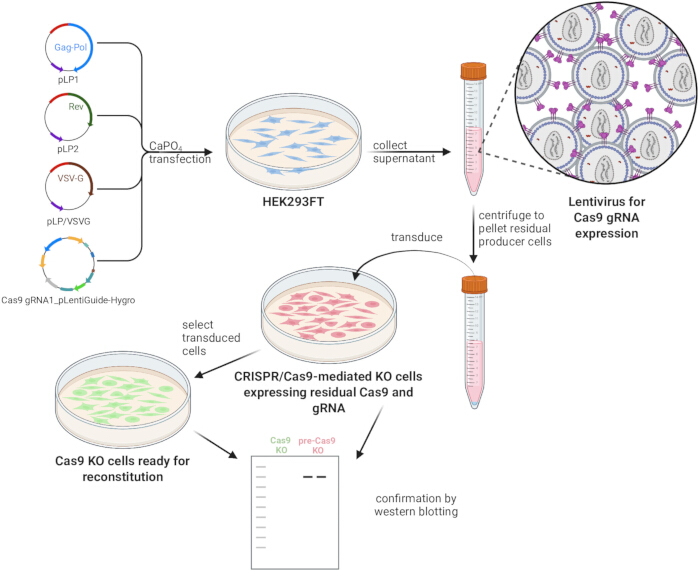
Figure 1: Experimental workflow for the strategic ablation of Cas9 and validation of CUL4B rescue. This workflow outlines the generation of Cas9-ablated CRISPR knockout cells by co-transfecting HEK293FT producer cells with pLP1, pLP2, pLP/VSVG, and a Cas9 gRNA1_pLentiGuide-Hygro construct via calcium phosphate transfection. The resulting lentiviral supernatant is collected, centrifuged to clear residual producer cells, and used to transduce target cells. Following selection, successful Cas9 ablation is confirmed via western blot analysis to establish a clean knockout cell line ready for experimental CUL4B reconstitution. Please click here to view a larger version of this figure.
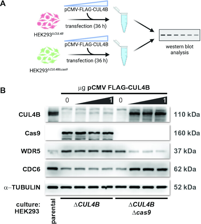
Figure 2: Rescue of CUL4B expression and function in CRISPR-Cas9-edited HEK293 cells. (A) Experimental schematic of the transfection workflow. HEK293ΔCUL4B and HEK293ΔCUL4BΔcas9 cells were transfected with either an empty vector or increasing amounts of pCMV-FLAG-CUL4B. Cells were harvested 36 h post-transfection, and lysate proteins were resolved by SDS-PAGE. (B) Western blot analysis of rescue efficiency and downstream functional controls. The indicated proteins were visualized by Western blot. WDR5 and CDC6 served as controls for CUL4B function. α-TUBULIN served as a loading control. Parental HEK293 cells served as the control for baseline expression. Blots are representative of replicate experiments (n = 2). Please click here to view a larger version of this figure.
| Target Gene | gRNA Sequence (5′→3′) | PAM (5′→3′) | Target Location | Target Description |
| CUL4B | ATCAAACCCTACAAACTCCA | AGG | Xq24 Exon 3 | N-terminal region of the human CUL4B ORF for frame-shift knockout |
| cas9 | CGTGATCACCGACGAGTACA | AGG | ORF (hSpCas9) | conserved internal region of the cas9 transgene to trigger self-inactivating indels |
Table 1: CRISPR-Cas9 guide RNA sequences and genomic target specifications.
Discussion
This protocol enabled robust restoration of target protein expression and function in CRISPR-Cas9-edited knockout cell lines in which Cas9 was ablated post-editing. A notable observation from this work is the substantial degree to which constitutive Cas9 expression suppresses plasmid-driven transgene expression. The strength of this protocol lies in its ability to rescue both the lost protein and its associated phenotype. This method also establishes a framework for phenotypic exploration of a wide array of protein alterations. For example, the introduction of a phosphomimetic (e.g., serine to aspartic acid) variant may provide valuable insight into the role of that post-translational modification without the confounding background of the native protein.
Consequently, this protocol is broadly applicable to standard adherent (e.g., HeLa, A549) and suspension (e.g., Jurkat, THP-1) cell lines. However, this sequential selection framework may prove impractical for primary or non-dividing cells (e.g., primary neurons or macrophages). In these sensitive systems, the rapid clearance of non-transduced bystander cells releases cytotoxic debris and pro-apoptotic stress signals into the shared microenvironment, driving indirect toxicity46,47. It may similarly fail in highly adaptable, aneuploid cancer lines (e.g., U87 glioblastoma or A375 melanoma) that are prone to escaping CRISPR-mediated phenotypes via rapid chromosomal rearrangements or the rapid selection of pre-existing resistant clones48,49. Ultimately, suitability depends on a cell line's capacity to tolerate the metabolic load of dual-vector expression, withstand these indirect selection pressures, and maintain stable growth kinetics throughout the multi-week protocol.
While this approach serves as a versatile proof of concept for most cell lines, its implementation requires a careful balance between delivery efficiency and genomic integrity. As outlined, using lentiviral delivery for 'self'-directed gRNA ensures a homogeneous Cas9-ablated population, especially in hard-to-transfect cells. However, the risk of insertional mutagenesis from semi-random integration remains a concern. These risks are manageable by optimizing the MOI. In permissive lines like HEK293, an MOI of 1–5 achieves high saturation while minimizing integration-driven mutagenesis and cytotoxicity41. In contrast, refractory immune or neuronal cells typically require MOIs of 10–50, often necessitating enhancers such as polybrene and/or spinoculation to achieve similar efficiencies50,51,52,53,54,55.
This technical trade-off extends to the choice between polyclonal- and monoclonal-selected populations. The HEK293ΔCUL4BΔcas9 cells generated here are polyclonal, a choice that accelerates experimental timelines by bypassing the weeks-long expansion required for single-cell subcloning. Furthermore, a polyclonal pool captures the average phenotype of the culture, effectively buffering against clonal artifacts or site-specific integration issues. While monoclonal lines are more likely to yield consistent results owing to genetic homogeneity, the results presented here suggest that polyclonal selection is a viable and often better alternative when speed and population representation are prioritized.
The temporal stability of the resulting phenotype may also be a critical variable. In the system presented here, the regulatory impact of CUL4B on WDR5 and CDC6 diminished over successive passages of the knockout cell lines, correlating with a compensatory upregulation of the paralog CUL4A (data not shown). This mirrors the findings of Nakagawa et al., who showed that CUL4A can partially restore CUL4B function in CUL4B-depleted cells44. Such homeostatic compensation is common in cellular biology, particularly when targeting a protein with high structural homology and functional redundancy to other cellular proteins.
To navigate these secondary compensatory mechanisms, two distinct strategic approaches can be employed, depending on the experimental goals and the cell line used. For researchers prioritizing efficiency or working with highly adaptive cell lines, a condensed experimental timeline is essential. This strategy involves immediate validation of target protein loss, ideally once the culture reaches 90% confluence post-selection, followed promptly by self-inactivating Cas9 targeting and functional assays. By adopting this accelerated workflow, one is more likely to capture the primary biological consequences of the knockout before the cellular homeostatic machinery has sufficient time to upregulate redundant paralogs or otherwise confound the resulting phenotype.
Alternatively, for studies requiring long-term stability or high-precision rescue experiments, particularly in transfection-refractory models such as immune and neuronal cells, a tetracycline-controlled (Tet-On/Off) system offers a viable solution56,57. While implementing this inducible framework necessitates additional rounds of lentiviral transduction and the use of distinct antibiotic resistance markers, it provides a mechanism for sustained restoration of the target protein to stabilize the cellular environment before triggering transient, doxycycline-mediated 'loss and rescue' events57.
Disclosures
The authors used Google Gemini to assist with refining technical terminology and editing the manuscript. The AI was used as a tool for linguistic improvement and did not contribute to the original synthesis of data, the conceptualization of the study, or the primary drafting of the text. The authors reviewed and edited the output and take full responsibility for the content of the final manuscript. The authors declare no conflict of interest.
Acknowledgements
This work was supported by startup funds provided by Le Moyne College, the Le Moyne College Research and Development Committee, the Le Moyne College Student Research Committee, the Walter L. '66 and MaryAnne Poland Jesuit Center for Research and Teaching Innovation Fund, and the Le Moyne College Department of Biological and Environmental Sciences. The authors acknowledge the use of BioRender.com in preparing the graphical illustrations. Figure 1 was created in BioRender (Sharifi, J., 2026; https://BioRender.com/72u0wl3), and the top panel of Figure 2 was created in BioRender (Sharifi, J., 2026; https://BioRender.com/lc6o2lu).
Materials
| Name | Company | Catalog Number | Comments |
|---|---|---|---|
| anti-Cas9 antibody | Cell Signaling | 65832 | This mouse monoclonal antibody provides specific detection of S. pyogenes Cas9 in human lysates, making it ideal for verifying self-directed gRNA-mediated Cas9 clearance. |
| anti-CDC6 antibody | Cell Signaling | 3387 | This rabbit monoclonal antibody provides specific detection of human CDC6, making it ideal for monitoring downstream functional impacts of CUL4B. |
| anti-CUL4B antibody | Sigma-Aldrich | C9995 | This affinity-purified rabbit polyclonal antibody provides specific detection of human CUL4B without cross-reacting with its closely related paralog CUL4A. |
| anti-mouse IgG, HRP-linked secondary antibody | Cell Signaling | 7076 | This horse anti-mouse IgG antibody is conjugated to horseradish peroxidase (HRP) and optimized for chemiluminescent detection of mouse monoclonal primary antibodies. |
| anti-rabbit IgG, HRP-linked secondary antibody | Cell Signaling | 7074 | This goat anti-rabbit IgG antibody is conjugated to horseradish peroxidase (HRP) and optimized for chemiluminescent detection of rabbit polyclonal and monoclonal primary antibodies. |
| anti-WDR5 antibody | Cell Signaling | 13105 | This rabbit monoclonal antibody provides specific detection of human WDR5, making it ideal for monitoring downstream functional impacts of CUL4B. |
| anti-α-TUBULIN antibody | Cell Signaling | 3873 | This mouse monoclonal antibody targets human alpha-tubulin and is ideal as a uniform loading control for normalization in western blot analysis. |
| Calcium Phosphate Transfection Kit | ThermoFisher Scientific | K278001 | This kit provides efficient transient transfection reagents optimized for the scalable, cost-effective production of lentiviral vectors in adherent HEK293FT producer cells. |
| Cas9 gRNA1_pLentiGuide-Hygro | GenScript Biotech Corporation | custom synthesis | cas9 gRNA sequence: CGTGATCACCGACGAGTACA, PAM sequence: AGG |
| Cas9 gRNA1_pLentiGuide-Puro | GenScript Biotech Corporation | custom synthesis | cas9 gRNA sequence: CGTGATCACCGACGAGTACA, PAM sequence: AGG |
| Dulbecco's Modified Eagle Medium | Gibco | D5796 | This high-glucose medium supplemented with L-glutamine and sodium pyruvate is optimized for sustaining growth, viability, and metabolic stability in fast-proliferating mammalian cell lines (e.g., HEK293FT) |
| Fetal Bovine Serum, Premium (Heat Inactivated) | Corning | 35-016-CV | This premium-grade, heat-inactivated serum provides essential growth factors and hormones while minimizing complement-mediated cytotoxicity for mammalian cell lines. |
| HEK293 | American Type Culture Collection | CRL-1573 | This well-characterized human embryonic kidney cell line is highly transfectable and serves as a robust, standard model for investigating gene knockout phenotypes and protein rescue frameworks. |
| HEK293FT cells | ThermoFisher Scientific | R70007 | This fast-growing, highly transfectable variant of the human embryonic kidney line stably expresses the SV40 large T antigen, making it optimized for maximizing viral titers during lentiviral vector production. |
| Hygromycin B | sigmaaldrich | H7772 | This aminonucleoside antibiotic is utilized as a selective agent in mammalian cell culture to isolate stable transductants expressing the hygromycin resistance marker. |
| pCLIP-All-hCMV-Puro plasmid (CUL4B gRNA) | Skyang Bio LLC | sgRNA3:1019408_b | CUL4B gRNA sequence: ATCAAACCCTACAAACTCCA, PAM sequence: AGG |
| pCMV-FLAGCUL4B | N/A | N/A | This mammalian expression plasmid utilizes a cytomegalovirus (CMV) promoter to drive constitutive expression of a FLAG-tagged Cullin 4B protein (CUL4B) and is a gift from Jianping Jin. |
| pCMV-M1 Mammalian Expression Vector | Addgene | 23007 | Empty expression vector control (a gift from Linda Wordeman) |
| pLentiGuide-Puro | Addgene | 52963 | This lentiviral transfer vector drives human U6-promoter expression of a customizable gRNA alongside an EF-alpha-promoter-driven puromycin resistance marker for CRISPR targeting in Cas9-expressing cells and is a gift from Feng Zhang. |
| pLX302 EGFP-V5 | Addgene | 141348 | GFP-encoding lentiviral vector for assessing transduction efficiency (a gift from Kevin Janes) |
| Puromycin | Sigma-Aldrich | P8833 | This aminonucleoside antibiotic is utilized as a selective agent in mammalian cell culture to isolate stable transductants expressing the puromycin resistance marker. |
| Sterile Phosphate Buffered Saline (PBS) | Corning | 21-040-CV | This standard balanced salt solution is utilized for washing cell monolayers to effectively remove trace culture media, serum proteins, or enzymes without disrupting osmotic pressure or cell viability. |
| Trypsin EDTA 1X | Corning | 25-051-CI | This standard enzymatic dissociation solution is utilized to detach adherent mammalian cells from culture vessels by cleaving peptide bonds while the EDTA chelates divalent cations to disrupt intercellular adhesion. |
| ViraPower Lentiviral Packaging Mix | ThermoFisher Scientific | K497500 | This optimized mixture of packaging plasmids (pLP1, pLP2, and pLP/VSVG) is utilized for co-transfection into producer cells to facilitate the high-titer assembly and replication-incompetent production of lentiviral expression vectors. |
References
- Doudna JA, Charpentier E. Genome editing: The new frontier of genome engineering with CRISPR-Cas9. Science. 2014;346(6213):1258096.
- Leonhardt C, et al. Single-cell mRNA transfection studies: Delivery, kinetics and statistics by numbers. Nanomedicine. 2014;10(4):679-688.
- Liu Y, Yang Q, Zhao F. Synonymous but not silent: The codon usage code for gene expression and protein folding. Annu Rev Biochem. 2021;90:375-401.
- Chamary JV, Hurst LD. Evidence for selection on synonymous mutations affecting stability of mRNA secondary structure in mammals. Genome Biol. 2005;6(9):R75.
- Mauro VP, Chappell SA. A critical analysis of codon optimization in human therapeutics. Trends Mol Med. 2014;20(11):604-613.
- Bauer AP, et al. The impact of intragenic CpG content on gene expression. Nucleic Acids Res. 2010;38(12):3891-3908.
- Kudla G, Murray AW, Tollervey D, Plotkin JB. Coding-sequence determinants of gene expression in Escherichia coli. Science. 2009;324(5924):255-258.
- Simms CL, Yan LL, Zaher HS. Ribosome collision is critical for quality control during no-go decay. Mol Cell. 2017;68(2):361-373.e5.
- Fu Y, et al. High-frequency off-target mutagenesis induced by CRISPR-Cas nucleases in human cells. Nat Biotechnol. 2013;31(9):822-826.
- Babu K, et al. Bridge helix of Cas9 modulates target DNA cleavage and mismatch tolerance. Biochemistry. 2019;58(14):1905-1917.
- Hsu PD, et al. DNA targeting specificity of RNA-guided Cas9 nucleases. Nat Biotechnol. 2013;31(9):827-832.
- Ihry RJ, et al. p53 inhibits CRISPR-Cas9 engineering in human pluripotent stem cells. Nat Med. 2018;24(7):939-946.
- Haapaniemi E, Botla S, Persson J, Schmierer B, Taipale J. CRISPR-Cas9 genome editing induces a p53-mediated DNA damage response. Nat Med. 2018;24(7):927-930.
- Yang S, Li S, Li XJ. Shortening the half-life of Cas9 maintains its gene editing ability and reduces neuronal toxicity. Cell Rep. 2018;25(10):2653-2659.e3.
- Kafri M, Metzl-Raz E, Jona G, Barkai N. The cost of protein production. Cell Rep. 2016;14(1):22-31.
- Mehta A, Merkel OM. Immunogenicity of Cas9 protein. J Pharm Sci. 2020;109(1):62-67.
- Liu Q, Zhang H, Huang X. Anti-CRISPR proteins targeting the CRISPR-Cas system enrich the toolkit for genetic engineering. FEBS J. 2020;287(4):626-644.
- Marino ND, Pinilla-Redondo R, Csörgő B, Bondy-Denomy J. Anti-CRISPR protein applications: Natural brakes for CRISPR-Cas technologies. Nat Methods. 2020;17(5):471-479.
- Gangopadhyay SA, et al. Precision control of CRISPR-Cas9 using small molecules and light. Biochemistry. 2019;58(4):234-244.
- Pandit B, et al. Bio-orthogonal chemistry-based strategy to turn off CRISPR-Cas9 activity in solution and live cells. NAR Mol Med. 2026;3(1):ugag008. Available from: https://doi.org/10.1093/narmme/ugag008
- Bubeck F, et al. Engineered anti-CRISPR proteins for optogenetic control of CRISPR-Cas9. Nat Methods. 2018;15(11):924-927.
- Kostic M, Jones LH. Critical assessment of targeted protein degradation as a research tool and pharmacological modality. Trends Pharmacol Sci. 2020;41(5):305-317.
- Kim S, Kim D, Cho SW, Kim J, Kim JS. Highly efficient RNA-guided genome editing in human cells via delivery of purified Cas9 ribonucleoproteins. Genome Res. 2014;24(6):1012-1019.
- Hendel A, et al. Chemically modified guide RNAs enhance CRISPR-Cas genome editing in human primary cells. Nat Biotechnol. 2015;33(9):985-989.
- Merienne N, et al. The self-inactivating KamiCas9 system for the editing of CNS disease genes. Cell Rep. 2017;20(12):2980-2991.
- Kelkar A, et al. Doxycycline-dependent self-inactivation of CRISPR-Cas9 to temporally regulate on- and off-target editing. Mol Ther. 2020;28(1):29-41.
- Dai Y, et al. Self-inactivating AAV-CRISPR at different ages enables sustained amelioration of Huntington's disease deficits in BAC226Q mice. Sci Adv. 2026;12(12):eaea8052.
- Sanjana NE, Shalem O, Zhang F. Improved vectors and genome-wide libraries for CRISPR screening. Nat Methods. 2014;11:783-784.
- Parr-Brownlie LC, et al. Lentiviral vectors as tools to understand central nervous system biology in mammalian model organisms. Front Mol Neurosci. 2015;8:14.
- Dobransky A, Root M, Hafner N, Marcum M, Sharifi HJ. CRL4-DCAF1 ubiquitin ligase-dependent functions of HIV viral protein R and viral protein X. Viruses. 2024;16(8)1313.
- Thermo Fisher Scientific. ViraPower Lentiviral Expression Systems User Guide. 2026. Available from: https://tools.thermofisher.com/content/sfs/manuals/virapower_lentiviral_system_man.pdf
- Pauwels K, et al. State-of-the-art lentiviral vectors for research use: Risk assessment and biosafety recommendations. Curr Gene Ther. 2009;9(6):459-474.
- Guo L, et al. Optimizing conditions for calcium phosphate-mediated transient transfection. Saudi J Biol Sci. 2017;24(3):622-629.
- Thermo Fisher Scientific. Cationic Lipid Transfection. 2026. Available from: https://www.thermofisher.com/us/en/home/references/gibco-cell-culture-basics/transfection-basics/methods/cationic-lipid-transfection.html
- Kutner RH, Zhang XY, Reiser J. Production, concentration and titration of pseudotyped HIV-1-based lentiviral vectors. Nat Protoc. 2009;4(4):495-505. Available from: https://doi.org/10.1038/nprot.2009.22
- Barde I, Salmon P, Trono D. Production and titration of lentiviral vectors. Curr Protoc Neurosci. 2010;53(1):4.21.1-4.21.23. Available from: https://currentprotocols.onlinelibrary.wiley.com/doi/abs/10.1002/0471142301.ns0421s53
- Kumru OS, et al. Physical characterization and stabilization of a lentiviral vector against adsorption and freeze-thaw. J Pharm Sci. 2018;107(11):2764-2774.
- American Type Culture Collection. Lentiviral Vector Reference Material Information Sheet. 2026. Available from: https://www.atcc.org/-/media/product-assets/documents/protocols/virology/lentiviral-vector-reference-material-information-sheet.pdf?rev=a934bd06c07f4969b0aafec16506cd44
- Higashikawa F, Chang L. Kinetic analyses of stability of simple and complex retroviral vectors. Virology. 2001;280(1):124-131.
- Kowolik CM, Yee JK. Preferential transduction of human hepatocytes with lentiviral vectors pseudotyped by Sendai virus F protein. Mol Ther. 2002;5(6):762-769.
- Pichavant C, Tremblay JP. Generation of lentiviral vectors for use in skeletal muscle research. In: DiMario JX, editor. Myogenesis: Methods and Protocols. Humana Press; Totowa, NJ; 2012. p. 285-295.
- Stacey GN, Masters JR. Cryopreservation and banking of mammalian cell lines. Nat Protoc. 2008;3(12):1981-1989.
- Bauer DE, Canver MC, Orkin SH. Generation of genomic deletions in mammalian cell lines via CRISPR/Cas9. J Vis Exp. 2015;(95):e52118.
- Nakagawa T, Xiong Y. X-linked mental retardation gene CUL4B targets ubiquitylation of H3K4 methyltransferase component WDR5 and regulates neuronal gene expression. Mol Cell. 2011;43(3):381-391.
- Zou Y, et al. CUL4B promotes replication licensing by up-regulating the CDK2-CDC6 cascade. J Cell Biol. 2013;200(6):743-756.
- Dachs GU, Hunt MA, Syddall S, Singleton DC, Patterson AV. Bystander or no bystander for gene-directed enzyme prodrug therapy. Molecules. 2009;14(11):4517-4545.
- Prise KM, O'Sullivan JM. Radiation-induced bystander signalling in cancer therapy. Nat Rev Cancer. 2009;9(5):351-360.
- Clark MJ, et al. U87MG decoded: The genomic sequence of a cytogenetically aberrant human cancer cell line. PLoS Genet. 2010;6(1):e1000832.
- Cormier JN, et al. Heterogeneous expression of melanoma-associated antigens and HLA-A2 in metastatic melanoma in vivo. Int J Cancer. 1998;75(4):517-524.
- Sigma-Aldrich. Successful Transduction Using Lentivirus. Darmstadt, Germany; 2023. Available from: https://www.sigmaaldrich.com/technical-documents/technical-article/genomics/advanced-gene-editing/successful-transduction-lentivirus
- Swainson L, Mongellaz C, Adjali O, Vicente R, Taylor N. Lentiviral transduction of immune cells. Methods Mol Biol. 2008;415:301-320.
- Davis HE, Morgan JR, Yarmush ML. Polybrene increases retrovirus gene transfer efficiency by enhancing receptor-independent virus adsorption on target cell membranes. Biophys Chem. 2002;97(2-3):159-172.
- Denning W, et al. Optimization of the transductional efficiency of lentiviral vectors: Effect of sera and polycations. Mol Biotechnol. 2013;53(3):308-314.
- O'Doherty U, Swiggard WJ, Malim MH. Human immunodeficiency virus type 1 spinoculation enhances infection through virus binding. J Virol. 2000;74(21):10074-10080.
- Sano S, et al. Lentiviral CRISPR/Cas9-mediated genome editing for the study of hematopoietic cells in disease models. J Vis Exp. 2019;(152):e59977.
- Gossen M, Bujard H. Tight control of gene expression in mammalian cells by tetracycline-responsive promoters. Proc Natl Acad Sci U S A. 1992;89(12):5547-5551.
- Gossen M, et al. Transcriptional activation by tetracyclines in mammalian cells. Science. 1995;268(5218):1766-1769.
Reprints and Permissions
This article has been published
Video Coming Soon