This review evaluates pearl powder, nacre, and nacre-derived water-soluble matrix (WSM) as osteogenic biomaterial candidates. It emphasizes stronger pearl-specific evidence for osteogenic activity than for high-dose cytotoxicity or autophagy-mediated injury, and identifies needs for standardized characterization, dose-response testing, stress assays, autophagic flux validation, and longer in vivo safety assessment.
Review Article
Nano-Pearl Powder and Nacre-Derived Water-Soluble Matrix in Osteogenesis: Evidence, Mechanistic Gaps, and Safety Considerations
In This Article
Summary
Abstract
Pearl powder, nacre, and nacre-derived water-soluble matrix (WSM) are natural mineral-organic materials with growing relevance for bone-regenerative research. Their biological activity appears to depend not only on calcium carbonate content but also on soluble matrix components that can influence osteoblast behavior, mineral deposition, and cellular stress responses. This narrative review compares pearl-specific osteogenic studies in cell, scaffold, and defect-repair models with selected comparator nanoparticle studies used to frame unresolved safety questions. The pearl-specific literature generally supports osteogenic activity, particularly in preosteoblast or osteoblast-lineage models and WSM-containing materials, but the evidence remains heterogeneous because species source, extraction method, particle size, release behavior, dose, and endpoint selection differ across studies. Evidence linking nano-pearl powder to autophagy-associated osteogenic differentiation is promising, especially in MC3T3-E1 models, but the existing pearl-specific literature provides limited direct evidence for high-dose toxicity, impaired autophagic flux, immunogenicity, heavy-metal-related risk, degradation-matched repair, or chronic biosafety. Future work should integrate material characterization, dose-response testing, oxidative and mitochondrial readouts, autophagic flux analysis, and osteogenic endpoints into a single experimental design. Such studies will be needed to define whether WSM-based or nano-pearl formulations can provide a reproducible and safe osteogenic window for bone-regenerative biomaterial development.
Introduction
Bone repair has two basic needs. A material has to fill the defect and also help osteoblast-lineage cells make extracellular matrix and mineral. Lamghari et al. showed that nacre stimulated bone marrow cells and supported bone formation. Rousseau showed that a nacre water-soluble matrix (WSM) fraction made MC3T3-E1 preosteoblast mineralization start earlier1,2. Conventional autografts, allografts, hydroxyapatite, and beta-tricalcium phosphate remain useful, but they can be limited by donor-site morbidity, supply, variable integration, brittleness, slow remodeling, or insufficient biological signaling. Pearl-derived materials are therefore being studied as active natural mineral-organic alternatives rather than as simple calcium carbonate fillers.
Pearl and nacre are related but not identical. Pearl powder is produced from the pearl body and contains calcium carbonate with a smaller organic fraction, whereas nacre is the mother-of-pearl shell layer with a highly organized aragonite-organic lamellar architecture. Nacre-derived WSM is a soluble fraction extracted from this matrix. These differences matter because whole-powder exposure, soluble-extract exposure, scaffold incorporation, and nanoscale-particle exposure may yield distinct release profiles, cell-contact patterns, and biological outcomes.
This review uses comparator nanoparticle studies only as hazard signals and hypothesis-generating evidence. Titanium dioxide, silver, hydroxyapatite, zirconia, and other non-pearl nanoparticles can raise reactive oxygen species (ROS), impair mitochondria, stress lysosomes, and alter autophagy in osteoblast-related models. These effects should guide pearl-specific safety testing, but they should not be interpreted as direct evidence that nano-pearl powder produces the same injury pattern.
Review and Perspective
Composition and bioactivity of pearl powder, nacre, and water-soluble matrix
Nacre is not simply calcium carbonate. The shell material carries soluble and insoluble organic matrices within the mineral phase. Soluble nacre proteins can modify calcium carbonate precipitation, and WSM proteomic studies have identified proteins with possible osteogenic activity, including mineral binding, matrix organization, and growth-factor-like signaling3,4,5. Claims about WSM bioactivity should therefore specify the species, extraction method, fraction, and model used rather than treating WSM as a single interchangeable material.
Preparation and characterization of nano-pearl powder
A minimum characterization checklist should be reported before biological testing: source species and tissue, sterilization method, particle-size distribution, morphology, crystal phase, zeta potential, dispersion stability in culture medium, aggregation state, endotoxin or bioburden status where relevant, WSM content, protein profile, calcium ion release, WSM release kinetics, and batch-to-batch reproducibility. Studies should also include controls that separate calcium carbonate effects from organic-matrix effects, such as calcium carbonate-only particles, protein-depleted or enzyme-treated WSM, particle-size-matched non-pearl controls, scaffold-only controls, and WSM-only controls, where applicable.
Recent scaffold work also shows that pearl-derived materials can be integrated with modern manufacturing strategies. Nacre WSM, nacre powder, and nacre-mimetic phases have been incorporated into porous, layered, and printed scaffolds6,7,8. The additive-manufacturing studies recommended during review further highlight controlled architecture, shape-memory behavior, photothermal functionality, and composite reinforcement as relevant design directions for complex defects9,10,11,12,13. For pearl-derived biomaterials, these methods should be evaluated alongside release behavior and cell stress endpoints, because scaffold architecture can alter both osteogenic signaling and cytotoxic exposure.
Osteogenic effects of pearl-derived materials
The strength of osteogenic evidence should be interpreted by endpoint hierarchy. Cell viability and proliferation show tolerance but do not prove osteogenesis. Alkaline phosphatase (ALP) and Runt-related transcription factor 2 (Runx2) indicate early differentiation. Collagen I, osteopontin (OPN), osteocalcin (OCN), and bone morphogenetic protein-2 (BMP-2) provide later evidence of matrix-related signaling. Alizarin red staining and mineral deposition support maturation, especially when normalized to cell number. In vivo imaging, histology, mechanical integration, and defect closure provide stronger evidence of repair than isolated changes in markers. Several pearl-specific reports support early and late osteogenic endpoints, but normalization to cell number, release kinetics, and long-term tissue safety are still inconsistently reported1,6,7,8,14,15,16,17.
| Study/evidence type | Material/model | Dose or duration | Osteogenic endpoints | Autophagy markers measured | Evidence level/limits |
| Original study1 | Nacre; marrow cells and in vivo bone formation | NR | Bone formation and cell response | No | Direct osteogenic evidence; older model reporting |
| Original study2 | Nacre WSM; MC3T3-E1 | NR | Earlier mineralization | No | WSM-specific signal; component identity incomplete |
| Original study14 | Water-soluble nacre bioactives; osteoblast assays | NR | Antioxidant activity and differentiation markers | No | Supports bioactivity; dose standardization limited |
| Original study15 | Nano-nacre particles; MC3T3-E1 | Controlled WSM/calcium release; exact comparative dose NR in this table | Osteogenic induction markers | No | Links release to osteogenesis; safety endpoints limited |
| Original study16 | Water-soluble nano-pearl powder; MC3T3-E1 | NR in extracted manuscript | Osteogenic markers and differentiation | Yes | Autophagy associated with differentiation; flux and rescue evidence need expansion |
| Scaffold studies original6-8 | WSM/nacre powder/nacre-derived composites in scaffold or defect models | NR or model-specific | Mineralization, histology, defect repair | Usually no | Translationally relevant; scaffold architecture confounds component effect |
Table 1: Representative pearl-derived material studies in osteogenesis and bone repair. The table distinguishes original studies from broader scaffold evidence and uses NR for details not reported in the extracted source. Abbreviations: ALP = alkaline phosphatase; OCN = osteocalcin; OPN = osteopontin; BMP-2 = bone morphogenetic protein 2; ARS = alizarin red staining; NR = not reported.
Cytotoxicity, oxidative stress, and mitochondrial stress
Comparator nanoparticle data are relevant through transferable exposure variables: particle size, particle number, aggregation, surface charge, protein corona, uptake route, lysosomal burden, dose metric, exposure duration, ROS production, mitochondrial membrane potential, and apoptosis or necrosis endpoints. They are not directly transferable for material-specific composition, WSM release, protein-mediated signaling, biodegradation, or immunogenicity. Pearl-specific work should therefore test the same stress endpoints directly instead of borrowing toxicity conclusions from non-pearl materials18,19,20,21,22,23,24,25,26,27.
Safety issues related to clinical translation
Clinical translation requires safety questions beyond short-term nanoparticle cytotoxicity. Pearl powder and nacre-derived preparations should be screened for heavy metal residues, including lead, mercury, cadmium, and arsenic, and the limits should align with pharmacopeial or biomaterial-quality requirements28. WSM also contains heterologous shell-derived proteins, so immunogenicity, complement activation, macrophage polarization, and inflammatory memory should be evaluated. Degradation rate should be matched to the bone-regeneration rate, and degradation products should be traced through the local tissue, blood, liver, kidney, and excretion pathways. Large-animal studies in load-bearing or clinically relevant defects are still needed before chronic safety can be generalized.
Autophagy as a cellular stress response
Autophagy is a lysosome-based recycling process. Protective autophagy refers to increased formation and clearance of autophagosomes that preserve viability, mitochondrial quality, redox balance, and differentiation capacity. Dysfunctional autophagy refers to blocked flux, excessive lysosomal burden, accumulation of LC3-II or p62 without clearance, or autophagy-associated death and loss of osteogenic function. Microtubule-associated protein 1 light chain 3 (LC3), sequestosome 1 (p62), and Beclin-1 changes alone cannot distinguish these states; bafilomycin A1, chloroquine, genetic modulation, time-course analysis, and viability-rescue designs are needed to validate flux and causality29,30,31,32.
Molecular pathways by which WSM may regulate autophagy
The most direct pearl-specific mechanism remains the mitogen-activated protein kinase kinase/extracellular signal-regulated kinase (MEK/ERK)-associated autophagy and differentiation response reported in MC3T3-E1 cells16. Broader osteoblast and biomaterial literature suggest possible crosstalk with AMP-activated protein kinase (AMPK), phosphoinositide 3-kinase/protein kinase B/mammalian target of rapamycin (PI3K/AKT/mTOR), sirtuin 1 (SIRT1), ROS-sensitive mitogen-activated protein kinase (MAPK) signaling, Yes-associated protein (YAP)/beta-catenin, mTOR/unc-51-like autophagy activating kinase 1 (ULK1), and mitophagy-related pathways29,32,33,34,35,36,37,38,39,40,41. At present, the receptors and WSM components that initiate these signals remain insufficiently defined. Based on proteomic evidence, candidate inputs include mineral-binding proteins, osteocalcin-like or matrix-organizing proteins, and transforming growth factor-beta-family-related signaling motifs, but these proposed links require fractionation, receptor blocking, genetic perturbation, and rescue experiments.
Dual role of autophagy in osteogenic differentiation
Pearl-specific evidence mostly shows the helpful side. Cheng et al. reported that water-soluble nano-pearl powder increased osteogenic markers in MC3T3-E1 cells and that inhibition of autophagy weakened the differentiation response16. This supports an association and possible contribution of autophagy in that model. It should not be treated as proof that autophagy is the universal causal mediator for all pearl-derived formulations, because pharmacological inhibitors can have off-target effects, and many studies have not combined flux validation with genetic or rescue approaches.
| Context | Autophagy marker data | Autophagy flux assay method | Outcome | Interpretation |
| Water-soluble nano-pearl powder; MC3T3-E1 | LC3/p62 pathway changes reported | Flux validation incompletely reported in extracted manuscript | Higher osteogenic markers; inhibitor weakened response | Pearl-specific association with differentiation |
| Oxidative/metabolic stress in osteoblasts | LC3, p62, Beclin-1, AMPK or PI3K/AKT/mTOR changes | Variable; bafilomycin A1/chloroquine often needed | Protection or apoptosis depending on stress | Defines protective versus dysfunctional autophagy |
| Titanium/silver/silica/tantalum/dicalcium silicate comparator studies | LC3/p62 and pathway changes | Variable; some flux or modulation assays | Osteogenesis, proliferation, or injury | Hypothesis-generating for pearl studies |
| Future pearl-derived formulations | LC3-II, p62, Beclin-1, mitophagy markers | Required: lysosomal inhibitor plus time course, genetic/rescue when feasible | Viability, ROS, mitochondrial potential, mineralization | Needed to classify adaptive, neutral, or harmful response |
Table 2: Autophagy evidence relevant to osteoblast and biomaterial responses. The table separates marker-only studies from those requiring flux validation, and it distinguishes pearl-specific evidence from comparator-derived hypotheses. Abbreviations: LC3 = microtubule-associated protein 1 light chain 3; p62 = sequestosome 1; TEM = transmission electron microscopy; NR = not reported.
WSM-mediated cytotoxicity-autophagy-osteogenesis axis
The proposed axis is a testable model, not an established pathway. The direct pearl-specific part is WSM or nano-pearl exposure associated with osteogenic markers in selected cell and scaffold models. The comparator-derived part is the possible high-stress branch involving ROS, mitochondrial injury, lysosomal burden, and blocked autophagy. The hypothesis to be validated is that adaptive autophagy transitions to dysfunctional autophagy and reduced osteogenesis at a specific threshold.
Based on available dose-response logic, three provisional zones can guide experiments. An osteogenesis-promoting zone would preserve viability, maintain mitochondrial membrane potential, avoid delayed accumulation of ROS, show complete autophagic flux, and increase mineralization after normalization to cell number. A no-obvious-effect zone would show material tolerance without meaningful osteogenic gain. A toxic-injury zone would show falling viability, persistent ROS, mitochondrial dysfunction, p62 accumulation, or blocked flux, inflammatory activation, and reduced osteogenic endpoints. Particle size, aggregation state, exposure duration, and WSM release rate may shift these zones (Figure 1).
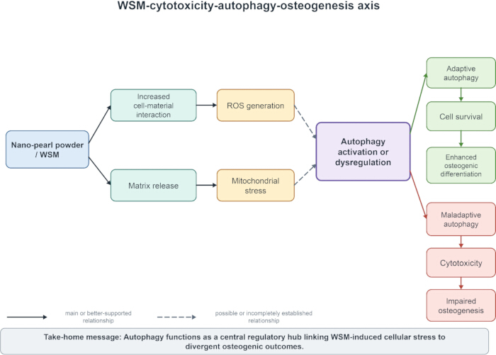
Figure 1: Evidence-mapped WSM-cytotoxicity-autophagy-osteogenesis framework. Solid arrows indicate relationships supported by pearl-specific studies, including selected WSM or nano-pearl effects on osteogenic markers. Dashed arrows indicate comparator-derived or hypothetical links, including ROS accumulation, mitochondrial injury, lysosomal burden, blocked autophagy, and reduced osteogenesis. Abbreviations: WSM = water-soluble matrix; ROS = reactive oxygen species. Please click here to view a larger version of this figure.
Challenges and limitations
This article is a narrative review rather than a systematic review. Studies were selected to map pearl-specific osteogenic evidence, evidence on nacre/WSM composition, scaffold and defect-repair models, and comparator nanoparticle studies relevant to oxidative stress, mitochondrial injury, and autophagy. No formal meta-analysis, risk-of-bias scoring, or exhaustive systematic search protocol was performed. Therefore, the review is best interpreted as an evidence map and research agenda rather than as a quantitative estimate of effect size.
Evidence strength differs by claim. Direct osteogenic activity in selected pearl-derived materials is moderately supported. WSM-specific effects are plausible but preparation-dependent. Scaffold-mediated benefit is only moderately supported and is confounded by architectural and carrier effects. In vivo repair is promising but remains limited by the duration of follow-up and safety endpoints. Pearl-specific high-dose cytotoxicity and autophagy-mediated injury remain weakly supported or hypothetical until flux, ROS, mitochondrial, inflammatory, and long-term in vivo data are collected.
Future perspectives
Short-term priorities are to standardize material characterization, identify active WSM components, and establish minimum requirements for the autophagy-flux assay. A recommended same-batch protocol should measure particle size, morphology, zeta potential, crystal phase, aggregation in medium, WSM release profile, calcium release, ROS, mitochondrial membrane potential, lysosomal function, autophagic flux, viability, ALP, Runx2, OPN, OCN, and mineralization. Reports should state dose metric, serum condition, exposure duration, normalization to cell number, and whether flux inhibitors such as bafilomycin A1 or chloroquine were used.
Long-term goals are to define personalized or defect-specific dose windows, evaluate long-term safety in large-animal models, compare degradation rate with bone-regeneration rate, and complete multicenter preclinical validation. Go/no-go criteria should include acceptable acute and delayed viability, absence of persistent ROS or mitochondrial injury, preserved or increased mineralization normalized to cell number, reproducible WSM release, absence of chronic inflammation or ectopic tissue response, acceptable heavy-metal and endotoxin profiles, and degradation products compatible with local and systemic metabolism (Figure 2).
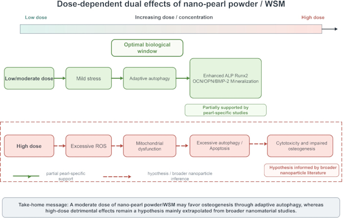
Figure 2: Dose-window model for pearl-derived biomaterials. The low-to-moderate exposure zone represents the putative osteogenesis-promoting window; the middle zone represents tolerance without clear benefit; the high or prolonged exposure zone represents a hypothesis requiring pearl-specific validation. Arrow labels indicate that particle size, aggregation, WSM release, and exposure duration can shift the zone boundaries. Please click here to view a larger version of this figure.
Conclusions
Selected pearl-derived and WSM-containing formulations show osteogenic potential in specific preclinical models, but this evidence cannot yet be generalized to all nano-pearl or WSM formulations without standardized dose, release, autophagic flux, and safety testing. The strongest direct evidence concerns osteogenic promotion in defined cell and scaffold systems, whereas evidence for high-dose pearl-specific toxicity, dysfunctional autophagy, immunogenicity, heavy-metal risk, and chronic biosafety remains weaker and often extrapolated from comparator materials. The field should therefore treat WSM and nano-pearl systems as promising but incompletely validated biomaterial candidates, with future studies prioritizing reproducible characterization, dose-window definition, and the parallel assessment of osteogenic and safety endpoints.
Disclosures
The authors have no conflicts of interest to declare.
Acknowledgements
The authors thank the Hainan Provincial Science and Technology Talent Innovation Project for support. This review was funded by the Hainan Provincial Science and Technology Talent Innovation Project (KJRC2023D25).
References
- Lamghari M, et al. Stimulation of bone marrow cells and bone formation by nacre: in vivo and in vitro studies. Bone. 1999;25(2 Suppl):91S-4S.
- Rousseau M, et al. The water-soluble matrix fraction from the nacre of Pinctada maxima produces earlier mineralization of MC3T3-E1 mouse pre-osteoblasts. Comp Biochem Physiol B Biochem Mol Biol. 2003;135(1):1-7.
- Heinemann F, Gummich M, Radmacher M, Fritz M. Modification of CaCO₃ precipitation rates by water-soluble nacre proteins. Mater Sci Eng C. 2011;31(2):99-105.
- Oliveira DV, et al. Identification of proteins with potential osteogenic activity present in the water-soluble matrix proteins from Crassostrea gigas nacre using a proteomic approach. ScientificWorldJournal. 2012;2012:765909.
- Sancho-Tomás M, et al. Calcium carbonate bio-precipitation in counter-diffusion systems using the soluble organic matrix from nacre and sea-urchin spine. Eur J Mineral. 2014;26(4):523-35.
- Li SG, et al. In vivo study on osteogenic efficiency of nHA/gel porous scaffold with nacre water-soluble matrix. Tissue Cell. 2024;88:102347.
- Ruan R, et al. Converting ocean nacre into bone mineral matrix composite for bone regeneration: in vitro and in vivo studies. Mater Des. 2024;245:113271.
- Liu B, et al. 3D printing nacre powder/sodium alginate scaffold loaded with PRF promotes bone tissue repair and regeneration. Biomater Sci. 2024;12(9):2418-33.
- Guo W, et al. Ionic substitution through bredigite doping for microstructure and performance adjustment in DLP 3D-printed TPMS porous HA bone scaffolds. Virtual Phys Prototyp. 2024;19(1).
- Guo W, et al. Biomimetic TPMS porous hydroxyapatite bone scaffolds doped with bioactive glass: digital light processing additive manufacturing, microstructure and performance. Compos Part A Appl Sci Manuf. 2025;193:108870.
- Guo W, et al. Niobium carbide-functionalized polylactic acid bone scaffold with near-infrared light-mediated shape memory and photothermal therapy for complex bone defects. Int J Biol Macromol. 2025;322(Pt 1):146578.
- Guo W, et al. Functionalized degradable soybean oil-based biomimetic porous scaffolds for complex bone defects: vat photopolymerization additive manufacturing, photothermal-mediated shape memory and tumor thermotherapy. Compos Sci Technol. 2026;265:111412.
- Guo W, et al. Multi-element CuFeSe₂ nanoparticle-functionalized 3D-printed polylactic acid scaffolds with photothermal shape memory and therapy for complex bone defects. Ceram Int. 2026. doi:10.1016/j.ceramint.2025.10.016.
- Chaturvedi R, Singha PK, Dey S. Water soluble bioactives of nacre mediate antioxidant activity and osteoblast differentiation. PLoS One. 2013;8(12).
- Xu J, et al. The osteoinductive effect of nano-nacre particles on MC-3T3 E1 preosteoblast through controlled release of water soluble matrix and calcium ions. Dent Mater J. 2019;38(6):981-6.
- Cheng Y, Zhang W, Fan H, Xu P. Water-soluble nano-pearl powder promotes MC3T3-E1 cell differentiation by enhancing autophagy via the MEK/ERK signaling pathway. Mol Med Rep. 2018;18(1):993-1000.
- Suwannasing C, et al. The osteoinductive effect of water-soluble matrix from nano-nacre particles of Haliotis diversicolor abalone on MC3T3-E1 osteoblasts. Appl Sci. 2025;15(6):2907.
- Bernier MC, El Kirat K, Besse M, Morandat S, Vayssade M. Preosteoblasts and fibroblasts respond differently to anatase titanium dioxide nanoparticles: a cytotoxicity and inflammation study. Colloids Surf B Biointerfaces. 2012;90:68-74.
- Inkielewicz-Stepniak I, et al. Titanium dioxide nanoparticles enhance production of superoxide anion and alter the antioxidant system in human osteoblast cells. Int J Nanomedicine. 2015;10:1095-107.
- Jin Y, et al. Oxidative stress-induced apoptosis of osteoblastic MC3T3-E1 cells by hydroxyapatite nanoparticles through lysosomal and mitochondrial pathways. RSC Adv. 2017;7(21):13010-8.
- Suh KS, Lee YS, Seo SH, Kim YS, Choi EM. Effect of zinc oxide nanoparticles on the function of MC3T3-E1 osteoblastic cells. Biol Trace Elem Res. 2013;155(2):287-94.
- Ye M, Shi B. Zirconia nanoparticles-induced toxic effects in osteoblast-like 3T3-E1 cells. Nanoscale Res Lett. 2018;13(1):353.
- Zielinska E, Tukaj C, Radomski MW, Inkielewicz-Stepniak I. Molecular mechanism of silver nanoparticles-induced human osteoblast cell death: protective effect of inducible nitric oxide synthase inhibitor. PLoS One. 2016;11(10).
- Andrée L, et al. Effect of hydroxyapatite nanoparticle crystallinity and colloidal stability on cytotoxicity. ACS Biomater Sci Eng. 2024;10(11):6964-73.
- Steckiewicz KP, Dmochowska M, Megiel E, Barcińska E, Inkielewicz-Stępniak I. Functionalization-dependent cytotoxicity of silver nanoparticles: a comparative study of chlorhexidine and metronidazole conjugates. Biomolecules. 2025;15(6):850.
- Xu J, Ze X, Zhao L, Sheng L, Ze Y. Titanium dioxide nanoparticles oral exposure induce osteoblast apoptosis, inhibit osteogenic ability and increase lipogenesis in mouse. Ecotoxicol Environ Saf. 2024;277:116367.
- Porter GC, et al. Endocytosed silver nanoparticles degrade in lysosomes to form secondary nanoparticle structures during expression of autophagy genes in osteogenic cells. Nanomedicine. 2021;33:102355.
- Chinese Pharmacopoeia Commission. Pharmacopoeia of the People’s Republic of China. Vol 1. Chinese Pharmacopoeia pearl powder quality standard. China Medical Science Press; Beijing; 2025.
- Wang X, Feng Z, Li J, Chen L, Tang W. High glucose induces autophagy of MC3T3-E1 cells via ROS-AKT-mTOR axis. Mol Cell Endocrinol. 2016;429:62-72.
- Liu W, et al. Dexamethasone-induced production of reactive oxygen species promotes apoptosis via endoplasmic reticulum stress and autophagy in MC3T3-E1 cells. Int J Mol Med. 2018;41(4):2028-36.
- Wang S, et al. Excessive production of mitochondrion-derived reactive oxygen species induced by titanium ions leads to autophagic cell death of osteoblasts via the SIRT3/SOD2 pathway. Mol Med Rep. 2020;22(1):257-64.
- Ruolan W, Liangjiao C, Longquan S. The mTOR/ULK1 signaling pathway mediates the autophagy-promoting and osteogenic effects of dicalcium silicate nanoparticles. J Nanobiotechnology. 2020;18(1):119.
- She C, Zhu LQ, Zhen YF, Wang XD, Dong QR. Activation of AMPK protects against hydrogen peroxide-induced osteoblast apoptosis through autophagy induction and NADPH maintenance: new implications for osteonecrosis treatment? Cell Signal. 2014;26(1):1-8.
- Zuo X, et al. Protective effect of photobiomodulation against hydrogen peroxide-induced oxidative damage by promoting autophagy through inhibition of PI3K/AKT/mTOR pathway in MC3T3-E1 cells. Oxid Med Cell Longev. 2022;2022:7223353.
- Shi Y, et al. Monotropein attenuates oxidative stress via Akt/mTOR-mediated autophagy in osteoblast cells. Biomed Pharmacother. 2020;121:109566.
- Chen F, et al. 5-(3′,4′-dihydroxyphenyl)-γ-valerolactone, a microbiota metabolite of flavan-3-ols, activates SIRT1-mediated autophagy to attenuate H₂O₂-induced inhibition of osteoblast differentiation in MC3T3-E1 cells. Free Radic Biol Med. 2023;208:309-18.
- Ling Z, et al. Copper-doped bioactive glass promotes matrix vesicles-mediated biomineralization via osteoblast mitophagy and mitochondrial dynamics during bone regeneration. Bioact Mater. 2025;46:195-212.
- Ha SW, Weitzmann MN, Beck GR Jr. Bioactive silica nanoparticles promote osteoblast differentiation through stimulation of autophagy and direct association with LC3 and p62. ACS Nano. 2014;8(6):5898-910.
- Kang C, et al. Involvement of autophagy in tantalum nanoparticle-induced osteoblast proliferation. Int J Nanomedicine. 2017;12:4323-33.
- Kang C, et al. Assessment of tantalum nanoparticle-induced MC3T3-E1 proliferation and underlying mechanisms. J Mater Sci Mater Med. 2021;32(11):133.
- Li L, et al. Nanotopography on titanium promotes osteogenesis via autophagy-mediated signaling between YAP and β-catenin. Acta Biomater. 2019;96:674-85.