We present a reproducible murine liver laceration model of profound hemorrhage integrating blood loss, trauma-induced coagulopathy, and survival outcomes. The model enables evaluation of hemostatic interventions, including recombinant factor VIIa and tranexamic acid, alongside coagulation, fibrinolytic, and mortality assessments, providing a cost-effective, scalable platform for mechanistic and translational hemorrhage research.
Method Article
A Murine Liver Laceration Model Simplifying Quantification of Trauma-Induced Hemorrhage Coagulopathy, Inflammation, and Survival
In This Article
Summary
Abstract
Uncontrolled hemorrhage remains a leading cause of preventable death in trauma patients, and trauma-induced coagulopathy (TIC) is a major determinant of early mortality. Experimental models that reliably recapitulate hemorrhage and associated coagulopathic responses are essential for mechanistic studies and evaluation of targeted interventions. Here, we describe a standardized murine model of profound hemorrhage using reproducible liver laceration (LL) causing hemorrhage, which can be quantified precisely, while also tracking simultaneously systemic coagulopathy, inflammatory markers, and mortality.
Mice undergo a standardized midline laparotomy followed by LL, and blood loss is quantified by blood-soaked sponges. Systemic markers of coagulation, fibrinolysis, and inflammation markers are collected by retro-orbital blood draw prior to and at pre-specified, serial intervals post-procedure. LL with blood loss reliably produces TIC (characterized by activated partial thromboplastin time (aPTT) prolongation, thrombin-antithrombin (TAT) complex increase, selective depletion of coagulation factors (F) V and VIII with activation of fibrinolysis). In parallel, inflammatory activation is evidenced by increased interleukin-6 (IL-6). Moreover, the model further allows survival analyses after midline incision repair.
As a translational application, rhFVIIa and TXA are used to prove that the model is sensitive to bleed rescue with pharmacologic modulation of coagulation and fibrinolytic parameters. Compared to large-animal models, this murine system offers an economical, versatile, and easily scalable opportunity to study innovative, targeted interventions for traumatic hemorrhage.
Introduction
Trauma is a leading cause of death and disability in young adults (age < 45 years)1,2, with uncontrolled hemorrhage accounting for a major cause of early mortality3. A critical factor of poor outcomes in severely injured patients is the development of trauma-induced coagulopathy (TIC)4,5. TIC is a complex, multifactorial dysregulation of hemostasis characterized by depletion of coagulation factors V and VIII6,7, dysregulated fibrinolysis7,8,9, and systemic inflammatory activation10. TIC contributes to ongoing bleeding and is associated with increased transfusion requirements11, organ failure5, and mortality despite advances in damage-control resuscitation and therapeutic interventions5.
Experimental models that reliably reproduce hemorrhagic shock with associated coagulopathic changes are therefore essential for mechanistic investigation and evaluation of targeted hemostatic interventions. Controlled hemorrhage models employ standardized blood withdrawal, which may not cause tissue injury, contributing to the systemic coagulopathic response observed in clinical trauma12. Therefore, hemorrhage models incorporating solid organ injury should provide greater clinical relevance, provided sustained bleeding and systemic responses can be quantitatively assessed13,14,15,16. More complex TIC models incorporate combinations of traumatic brain injury, long-bone or limb fractures, soft-tissue injury, laparotomy, and controlled hemorrhagic shock to reproduce multiple components of severe trauma17,18. Although these approaches provide a more comprehensive representation of polytrauma, their multiple injury procedures and physiological manipulations can increase technical complexity and experimental variability.
Despite decades of investigation, clinically relevant and reproducible experimental models of TIC remain limited and heterogeneous. Reviews have emphasized the need for robust models that more accurately reproduce the clinical sequence of tissue injury and hemorrhagic shock and support evaluation of therapeutic interventions17,19. Complex models requiring extensive instrumentation, including cardiac catheterization and continuous hemodynamic monitoring, can provide detailed physiological information but also require specialized equipment and additional procedural manipulation. To remove barriers to trauma research, simplifying the experimental approach while retaining the key features of TIC seems critical. Here, we describe a standardized murine model resulting in uncontrolled hemorrhagic shock14 using reproducible LL that induces profound bleeding and recapitulates key features of TIC. This model enables assessment of coagulation and inflammatory responses, including general coagulation measures (aPTT and thrombin generation), selective depletion of coagulation factors (FV and FVIII) as a hallmark of TIC, markers of fibrinolysis, and cytokine profiling. To demonstrate the sensitivity and relevance of this model to human trauma, we evaluate the effects of rhFVIIa (prohemostatic agent) and TXA (antifibrinolytic agent), used in trauma resuscitation, on hemorrhage severity, progression of coagulopathy, and survival outcomes.
The model further supports serial and terminal blood sampling, enabling longitudinal assessment of coagulation and inflammatory trajectories in relation to injury severity and therapeutic intervention. Together, the model provides a reproducible and scalable murine system for studying TIC in the setting of profound hemorrhage and for evaluating hemostatic and targeted pharmacologic interventions using clinically relevant endpoints, including blood loss, coagulopathy, fibrinolysis, inflammation, and survival.
Compared to previously published murine liver laceration models, including the protocol described Dyer et al.13, this method introduces several key methodological advances. These include quantitative gravimetric measurement of blood loss, standardization of injury severity through normalization of lacerated liver mass to body weight, and comprehensive profiling of TIC, fibrinolysis, and inflammation. Furthermore, the model enables serial sampling, pharmacologic modulation, and survival analyses detailing abdominal closure techniques, enabling physical recovery rather than termination. All procedures can be performed within a single experimental approach, providing a scalable and translational platform for studying hemorrhage and targeted hemostatic interventions.
Protocol
The Institutional Animal Care and Use Committee of the University of California San Diego (UCSD) approved all animal protocols. C57BL/6J mice were bred and maintained at the UCSD internal breeding facility. Both male and female mice, aged 8 to 10 weeks, were used in the experiments. A schematic overview of the experimental workflow and timeline is provided in Supplementary Figure 1.
NOTE: Sample sizes were determined based on preliminary experiments, expected variability of individual endpoints, and prior experience with the murine liver laceration model. Final sample sizes for individual experimental endpoints and experimental groups are provided in the corresponding figure legends. For liver weight measurements, data from multiple independent experiments were pooled to assess consistency across experimental cohorts. For coagulation assays performed in separate experimental runs, previously analyzed samples were included across runs to monitor inter-assay consistency and reproducibility.
1. Pre-experimental preparation
- Baseline sample collection (Day -1)
- Collect ~200 µL of blood through retro-orbital access from each mouse (8–10 weeks old) 24 h before the experiment to establish baseline parameters.
- Administer ~200 µL of sterile injectable-grade saline subcutaneously following blood collection.
- House the mice in clean cages under standard laboratory conditions, with a controlled temperature of 20–24 °C, relative humidity of 30%–70%, and a 12-h light/12-h dark cycle, with ad libitum access to food and water until the day of the experiment.
- Cage preparation (Day 0)
- Transfer the mouse to a clean cage lined with fresh paper towels on the day of the experiment.
- Remove food and water 15 min before the experiment and withhold them until the absorption sponges are removed after surgery.
2. Instrument and surgical field preparation
- Instrument sterilization
- Sterilize surgical instruments, cotton, applicators, and filter papers by autoclaving.
- Disinfect the surgical surface using 70% ethanol.
- Surgical table setup
- Arrange sterile instruments on a sterile drape.
- Prepare a stainless-steel container with 70% ethanol and sterile cotton for interim instrument cleaning.
- Activate a microbead sterilizer and allow it to reach 150 °C for between-animal sterilization.
3. Temperature maintenance preparation
- Isothermal pad preparation
- Activate the isothermal heating pad immediately before surgery according to the manufacturer’s instructions.
- Set the isothermal heating pad to maintain the mouse body temperature within a target range of 36–37 °C throughout anesthesia and surgery.
- Confirm uniform heat distribution across the pad surface.
- Place the pad on the surgical platform and cover with a sterile drape.
- Connect the surgical board to the isoflurane anesthesia system.
4. Anesthesia and pre-operative procedures
- Anesthesia induction
- Induce anesthesia by placing the mouse in an induction chamber with 2% isoflurane in 2 L/min oxygen.
- Confirm adequate anesthesia via toe pinch reflex.
- Record body weight before surgery.
- Analgesia and treatment administration
- Administer buprenorphine (3.25 mg/kg, subcutaneously) for analgesia.
- Prepare hemostatic agents as required.
- For treatment groups, administer recombinant human activated factor VII (rhFVIIa; 3 mg/kg) or tranexamic acid (10 mg/kg) via retro-orbital intravenous injection 5 min before injury.
- Administer equivalent volume (~100 µL) of injectable-grade sterile saline to control animals.
5. Surgical preparation
- Surgical site preparation
- Transfer the anesthetized mouse to the surgical board positioned on the isothermal pad.
- Maintain anesthesia via a nose cone using 1.5%–2% isoflurane in 2 L/min oxygen throughout the surgical procedure.
- Position the mouse supine and secure all four limbs with tape.
- Shave the abdominal region.
- Perform aseptic preparation using alternating povidone-iodine and 70% ethanol (three cycles).
6. Liver laceration procedure
- Pre-weighing materials
- Pre-weigh two 1.5 mL tubes, including one tube containing 0.5 mL of saline and one empty tube.
- Pre-weigh three primary and two secondary absorption triangles for each animal and record their combined mass.
- Pre-weigh one weigh boat for each animal for subsequent weighing of the excised liver tissue.
- Induction of liver injury
- Perform a midline laparotomy approximately 0.5 inch (12.7 mm) in length to expose the liver.
- Place two absorption triangles along the right and left abdominal walls, avoiding contact with the liver.
- Exteriorize the left liver lobe and identify the lobe margins to standardize the extent of tissue removal. Avoid unintended tissue injury.
- Using sharp scissors, lacerate approximately 75% of the lobe in a single controlled motion to ensure consistency across animals.
- Transfer the excised lacerated tissue into the pre-weighed empty tube for subsequent quantification of injury severity.
- Insert a third absorption triangle into the abdominal cavity to facilitate uniform blood collection during the hemorrhage phase.
- Rapid abdominal closure to maintain internal hemorrhage (critical step)
- Immediately close the abdominal wall within ~10–15 s using two surgical autoclips.
- Ensure precise alignment of the incision edges to achieve complete closure without gaps. Incomplete closure may result in external blood leakage and underestimation of blood loss.
- Apply consistent but gentle tension during closure, avoiding compression of abdominal contents, which may alter bleeding dynamics.
- Reinforce the closure with tissue adhesive (2–3 drops) to ensure a fully sealed abdominal compartment.
- Post-injury monitoring
- Weigh the excised liver tissue and normalize the measurement to body weight to quantify and confirm the extent of liver injury.
- Return the mouse to the cage placed on the isothermal pad.
- Allow hemorrhage to proceed for 60 min for the endpoint and survival cohort.
7. Blood collection and quantification of blood loss
- Blood sample collection and processing
- For the endpoint cohort, re-anesthetize the mouse with isoflurane at 60 min after liver laceration.
- For the survival cohort, re-anesthetize the mouse with isoflurane at 6 h and 7 days after liver laceration for blood collection.
- Collect blood via retro-orbital puncture into tubes containing sodium citrate solution (blood-to-anticoagulant ratio: 9:1) for plasma preparation.
- Centrifuge the samples at 2,000 × g for 10 min, followed by a second centrifugation at 12,000 × g for 5 min, to obtain platelet-poor plasma for downstream coagulation and fibrinolysis assays.
- Blood loss measurement
- Reopen the abdomen at 60 min after liver laceration for both the endpoint and survival cohorts and remove the absorption sponges containing blood.
- Transfer all blood-soaked sponges into the pre-weighed tube containing sterile saline.
- Use additional pre-weighed absorption sponges to collect residual blood within the abdominal cavity.
- Weigh the collection tube after blood collection to obtain the final tube weight.
- Calculate blood loss using the following gravimetric equation:
Blood loss (µL/g) = [(final tube weight − initial tube weight – absorption sponge weight) / mouse body weight] × 1000.
NOTE: All weights are expressed in grams. The factor of 1,000 converts grams of blood to microliters, assuming a blood density of approximately 1 g/mL.
- Survival and longitudinal outcome cohort
- Following blood loss measurement at 60 min, immediately close the incision using surgical staples and tissue adhesive to ensure a fully sealed abdominal compartment.
- Administer 400 µL of sterile saline subcutaneously before recovery.
- Return the mouse to standard housing and monitor it continuously during recovery from anesthesia and assess it at 2 h, 6 h, and 24 h after surgery, followed by daily monitoring through day 7.
8. Post-operative care
- Post-operative monitoring
- Monitor animals continuously during early recovery from anesthesia for signs of distress or morbidity.
- Record survival status and clinical observations at each scheduled assessment.
- Supportive care
- Administer 400 µL of sterile saline subcutaneously daily for 3 days.
- Provide analgesia (buprenorphine, 3.25 mg/kg, subcutaneously) every 72 h according to the approved animal care protocol.
- Maintain mice in standard housing with ad libitum access to food and water.
- Humane endpoints
- Monitor animals for predefined humane endpoints, including severe or persistent lethargy, inability to ambulate or obtain food or water, severe respiratory distress, unresponsiveness, or other signs of severe distress as specified in the approved institutional animal care protocol.
- Euthanize animals that meet the predefined humane endpoints in accordance with institutional guidelines.
- Continue survival monitoring through day 7 or until a humane endpoint is reached.
9. Endpoint procedures and tissue collection
- At predefined experimental endpoints, anesthetize mice and collect blood via retro-orbital puncture.
- Euthanize using CO₂ inhalation in accordance with institutional guidelines.
- Harvest organs and process tissues for histological analysis according to the specified downstream procedures.
10. Coagulation, fibrinolysis, and cytokine assays
NOTE: Reagents, assay kits, and equipment used for these procedures are listed in the Table of Materials.
- Measuring aPTT
- Record all clotting times using the coagulometer listed in the Table of Materials.
- Mix 25 µL of mouse plasma with 25 µL of aPTT reagent and incubate for 3 min at 37 °C. Initiate clotting by adding 25 µL of CaCl₂ (25 mM) prepared in HBS (20 mM HEPES, 147 mM NaCl, 3 mM KCl, pH 7.4), and record the clotting time.
- Measuring FV, FII, and FX activity
- Mix 5 µL of mouse plasma with 20 µL of the corresponding factor-deficient plasma and 25 µL of HBS containing 0.5% bovine serum albumin.
- Incubate for 1 min at 37 °C, and initiate clotting by adding Innovin reagent listed in the Table of Materials.
- Determine factor activity relative to the corresponding calibration/reference system.
- Measuring FVIII
- Determine FVIII using a chromogenic FVIII assay according to the manufacturer's instructions.
- Measuring fibrinogen
- Determine fibrinogen concentration using the Clauss method. Add 10 µL of mouse plasma to 40 µL of HEPES-buffered saline containing 0.01% bovine serum albumin and incubate for 3 min at 37 °C.
- Initiate clot formation by adding 25 µL of thrombin (25 U/mL), and determine fibrinogen concentration using the assay calibration.
- Measuring plasma biomarkers
- Measure plasma TAT complexes, PAP complexes, and D-dimer using the commercially available assay kits listed in the Table of Materials. Perform each assay according to the manufacturer's instructions.
- Use assay-specific standards, calibrators, and controls as specified by the manufacturers, and calculate analyte concentrations from the corresponding standard curves.
- Measuring plasma IL-6
- Measure plasma IL-6 concentrations at baseline and at 60 min and 6 h after liver laceration using the multiplex cytokine assay listed in the Table of Materials.
- Perform the assay according to the manufacturer's instructions and analyze the data using the manufacturer's Q-View software.
Results
The LL model results in reproducible and standardized injury severity in mice. Figure 1A demonstrates the consistency of lacerated liver weight when normalized to body weight (%BW), with a median injury of 1.4% BW across experimental groups. This consistency confirms reliable surgical induction and enables reproducibility between animals and across experimental conditions. The ability to standardize injury severity is critical for evaluating downstream physiological responses independent of variability in tissue damage.
Validation of the bleeding response and therapeutic modulation within the model is demonstrated in Figure 1B. Mice were pretreated with saline, the prehemostatic agent rhFVIIa, or the antifibrinolytic agent TXA, and blood loss was quantified and normalized to body weight (µL/g). Saline-treated mice exhibited substantial hemorrhage following LL, whereas rhFVIIa or TXA-treated mice demonstrated reduced blood loss. These findings demonstrate the ability of this model to detect pharmacologic modulation of hemorrhage and to evaluate hemostatic interventions under conditions of uncontrolled bleeding.
The model also reproduces key features of trauma-induced coagulopathy. As shown in Figure 1C, LL resulted in prolongation of aPTT, indicating impaired coagulation following injury. rhFVIIa or TXA treatment attenuated this prolongation, suggesting restoration of coagulation function. Similarly, thrombin generation, assessed by TAT complexes, was increased following LL and reduced with TXA treatment, whereas rhFVIIa does not reduce TAT levels (Figure 1D), suggesting modulation of coagulation activation.
Further characterization of coagulopathy is shown in Figure 2. LL resulted in selective depletion of coagulation factors V and VIII, consistent with TIC, and these changes were restored with rhFVIIa or TXA treatment (Figure 2A,B). In contrast, factors II and X were not significantly altered (Figure 2C,D), indicating that these changes occur in the context of preserved factors II and X, supporting a TIC-predominant rather than disseminated intravascular coagulation (DIC)-like phenotype.
The development of fibrinolysis following LL is demonstrated in Figure 3. Fibrinogen levels were reduced following injury, indicating consumption, and were not fully restored with rhFVIIa or TXA (Figure 3A). In contrast, fibrinolytic markers, including plasmin-antiplasmin (PAP) complexes and D-dimer, were elevated following LL. rhFVIIa reduced PAP complex levels but does not significantly alter D-dimer levels. TXA reduced both PAP complex and D-dimer levels following LL (Figure 3B,C).
Finally, systemic inflammatory response and survival outcomes are assessed in Figure 4. Plasma interleukin-6 (IL-6) levels are measured at 60 min and 6 h following LL in all treatment groups. IL-6 levels are elevated at early time points across all groups (Figure 4A). Survival over a 7-day period is assessed using Kaplan-Meier analysis. Seven-day survival is 75% in the saline group, 55% in the rhFVIIa, and 80% in the TXA-treated group. No significant difference is observed between saline and TXA groups, whereas survival is significantly reduced in the rhFVIIa group compared with saline and TXA groups (Figure 4B).
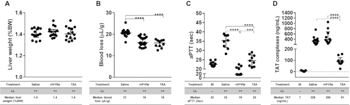
Figure 1: Validation of the liver laceration (LL) model: reproducibility of injury severity, quantitative blood loss, and induction of coagulopathy. Severe internal hemorrhage is induced by midline laparotomy followed by standardized LL. Mice are pretreated with saline (~100 µL) or recombinant human activated factor VII (rhFVIIa; 3 mg/kg), or tranexamic acid (TXA; 10 mg/kg) prior to injury to assess pharmacologic modulation of bleeding and coagulopathy. (A) Injury severity is quantified as lacerated liver weight normalized to body weight (%BW), demonstrating reproducible induction of standardized injury across animals. (B) Blood loss is quantified using a gravimetric method and normalized to body weight (µL/g), demonstrating measurable hemorrhage that is reduced with TXA treatment. (C) Activated partial thromboplastin time (aPTT) and (D) thrombin–antithrombin (TAT) complexes demonstrate reproducible induction of coagulopathy following LL, with modulation observed following rhFVIIa or TXA treatment. Data are presented as individual values with median and interquartile range; comparisons were performed using a nonparametric Mann–Whitney U test (n = 5–21). ****P ≤ 0.0001. Adapted with permission from Joseph et al.16. Please click here to view a larger version of this figure.
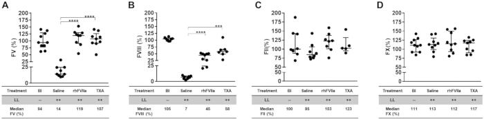
Figure 2: Validation of trauma-induced coagulopathy (TIC) following liver laceration (LL): selective depletion of coagulation factors. Severe internal hemorrhage is induced by midline laparotomy followed by standardized LL. Mice are pretreated with saline (~100 µL) or recombinant human activated factor VII (rhFVIIa; 3 mg/kg), or tranexamic acid (TXA; 10 mg/kg) prior to injury to assess pharmacologic modulation of coagulopathy. Coagulation parameters are assessed at 60 minutes following LL. (A) Factor V and (B) Factor VIII levels demonstrate reproducible depletion following LL, consistent with TIC. (C) Factor II and (D) Factor X remain relatively preserved, indicating selective coagulation factor depletion rather than global consumption. Data are presented as individual values with median and interquartile range (n = 7–10). Statistical comparisons were performed using a nonparametric Mann-Whitney U test. ***P ≤ 0.001; ****P ≤ 0.0001. Adapted with permission from Joseph et al.16. Please click here to view a larger version of this figure.
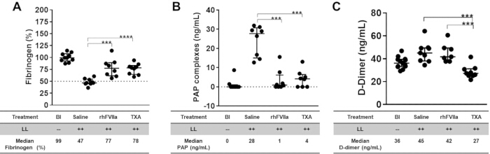
Figure 3: Validation of fibrinolytic activation following liver laceration (LL). Severe internal hemorrhage is induced by midline laparotomy followed by standardized LL. Mice are pretreated with saline (~100 µL), or recombinant human activated factor VII (rhFVIIa; 3 mg/kg), or tranexamic acid (TXA; 10 mg/kg) prior to injury to assess pharmacologic modulation of fibrinolysis. Fibrinolytic parameters are assessed at 60 minutes following LL. (A) Fibrinogen levels demonstrate consumption following injury, consistent with activation of fibrinolysis and coagulation factor depletion. (B) Plasmin–antiplasmin (PAP) complexes and (C) D-dimer levels are increased following LL, indicating active fibrin degradation and systemic fibrinolytic activation. Data are presented as individual values with median and interquartile range (n = 8–13). Statistical comparisons were performed using a nonparametric Mann-Whitney U test. **P ≤ 0.01; ***P ≤ 0.001; ****P ≤ 0.0001. Adapted with permission from Joseph et al.16. Please click here to view a larger version of this figure.
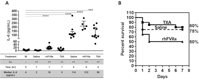
Figure 4: Validation of systemic inflammatory response and survival assessment following liver laceration (LL). Severe internal hemorrhage is induced by midline laparotomy followed by standardized LL. Mice are pretreated with saline (100 µL), recombinant human activated factor VII (rhFVIIa; 3 mg/kg), or tranexamic acid (TXA; 10 mg/kg) prior to injury. Following injury, the abdominal incision is closed using wound clips and tissue adhesive, and animals are returned to their home cages. Supportive care consists of daily subcutaneous administration of saline (400 µL) for the first three days post-injury. Plasma samples are collected at 60 min and 6 h following LL to assess systemic inflammatory activation. (A) Interleukin-6 (IL-6) levels demonstrate a sustained systemic inflammatory response following injury. Data are presented as individual values with median and interquartile range. Statistical comparisons were performed using a nonparametric Mann–Whitney U test (n = 9–13). (B) Seven-day survival is assessed using Kaplan-Meier analysis, demonstrating the capacity of the model to support longitudinal outcome-based endpoints (n = 20). ****P ≤ 0.0001. Adapted with permission from Joseph et al.16. Please click here to view a larger version of this figure.
Supplementary Figure 1: Schematic overview of the liver laceration model and experimental timeline. This figure summarizes the experimental workflow for the standardized murine liver laceration (LL) model of uncontrolled hemorrhage. Mice undergo midline laparotomy followed by reproducible LL of approximately 75% of the left liver lobe. Prior to injury, mice receive either saline (~100 µL, vehicle control) or recombinant human activated factor VII (rhFVIIa; 3 mg/kg), or tranexamic acid (TXA; 10 mg/kg), administered intravenously via retro-orbital injection 5 min before LL. Quantification of blood loss is performed using pre-weighed abdominal sponges placed intra-abdominally at the time of injury, retrieved 60 min post-LL, and weighed to determine total hemorrhage. The abdomen is then closed using wound clips with tissue adhesive. Blood samples are collected via retro-orbital access at 60 min in the endpoint cohort and 6 h and 7 d in the survival cohort. Plasma is analyzed for coagulation parameters, including activated partial thromboplastin time (aPTT), coagulation factor activity, thrombin-antithrombin (TAT) complexes, plasmin-α2-antiplasmin (PAP) complexes, and D-dimer at 60 min. Cytokine profiling is performed at 60 min and 6 h. Survival is monitored over a 7-day period. Adapted with permission from Joseph et al.16. Please click here to download this file.
Discussion
In this study, we present a reproducible murine model of profound hemorrhage using standardized LL that integrates quantitative blood loss assessment, characterization of TIC and inflammation, and survival analysis within a single experiment. The model produces consistent injury severity, as demonstrated by comparable lacerated liver weight normalized to body weight, while enabling reproducible evaluation of hemorrhage and systemic hemostatic responses across experimental groups. The use of a small-rodent model with standardized injury and quantitative outcome measures enables efficient assessment of multiple experimental endpoints within a single experimental framework, supporting reproducible and scalable investigation of TIC, without requiring technically complex procedures.
Importantly, this model recapitulates key features of TIC, including prolongation of general coagulation parameters (aPTT), increased thrombin generation as reflected by TAT complexes, selective depletion of coagulation factors V and VIII, and activation of fibrinolysis evidenced by elevated PAP complexes and D-dimer formation. These changes occur in the context of preserved factors II and X, supporting a TIC-predominant rather than disseminated intravascular coagulation phenotype. The ability to simultaneously quantify these parameters provides a translationally relevant framework for evaluating hemostatic dysregulation following traumatic injury.
A key strength of this model is the incorporation of survival as an outcome endpoint in the setting of traumatic hemorrhage. To test clinically relevant rescue strategies, we evaluated both rhFVIIa and TXA, two agents used or investigated for trauma resuscitation in humans. TXA demonstrated consistent effects on reducing blood loss, normalizing coagulopathy and fibrinolytic activation, translating into survival rescue. Conversely, rhFVIIa resulted in notable mortality associated with overcorrection of hemostatic parameters and incomplete fibrinolysis control16.
These findings highlight an important aspect of the model: survival is driven by a complex interplay of hemorrhage severity, coagulopathy, and systemic response, rather than isolated correction of individual hemostatic pathways. It appears that this hemorrhage model is sensitive to the integration of biochemical hemostatic parameters with survival, the latter being a critical outcome determinant in clinical practice. Although severe traumatic hemorrhage is the primary injury modeled in this system, secondary organ injury may also contribute to the outcome. In our previous study using this model, pulmonary microthrombi and fibrin deposition were observed in rhFVIIa-treated mice and were associated with increased mortality, suggesting a potential contribution of thromboinflammatory organ injury to outcome16.
Although a contemporaneous sham-operated group was not included in the present study, we previously demonstrated that laparotomy without LL did not result in TIC, demonstrating the need for additional rapid blood loss (shock) to produce the characteristic coagulation abnormalities observed with TIC14.
Several technical considerations are important for the reproducibility of this model. The extent and location of LL should be standardized, as variation in the amount of liver removed can substantially affect bleeding severity and the resulting coagulopathic response. In our protocol, ~75% of the left liver lobe is removed, with lacerated liver weight normalized to body weight, providing an objective measure of injury consistency. Rapid and secure abdominal closure is also critical to prevent external blood loss and maintain a contained intra-abdominal hemorrhage environment during the observation period. For accurate blood-loss measurement, pre-weighed sponges should be consistently recovered, using the same collection procedure and timing across experimental groups. These steps minimize operator-dependent variation and are particularly important because differences in initial blood loss can influence subsequent coagulopathy and survival. If excessive bleeding occurs before closure or blood is lost externally, the animal should be excluded.
A limitation of this model is that it is focused on organ injury, which does not mirror the heterogeneity of polytrauma, which may include long bone fractures, soft tissue crush trauma, and other complex shock states. However, this focused injury is also an intentional feature of the model, allowing the effects of profound hemorrhage and TIC to be studied without the additional procedural complexity and variability introduced by multiple injury modalities. In conclusion, this murine LL model provides a cost-effective (compared to large animal studies), robust, and scalable approach for studying trauma-related hemorrhage TIC and mortality. By combining a simple small-rodent experimental platform with standardized injury, quantitative blood-loss measurement, comprehensive hemostatic profiling, and survival assessment, the model provides a practical alternative to more extensively instrumented trauma models. It enables integrated assessment of blood volume loss, coagulation (aPTT, FII, FV, FVIII, FX, TAT), fibrinolysis (Fibrinogen, PAP, D-dimer), and survival endpoints in a clinically relevant context, well-suited for evaluating antifibrinolytic and targeted hemostatic interventions.
Disclosures
A.v.D. has received honoraria for participating in scientific advisory boards, consulting, and speaking engagements from BioMarin, Regeneron, Pfizer, Bioverativ/Sanofi, CSL Behring, Novo Nordisk, Spark Therapeutics, Genentech, and uniQure. A.v.D. is a cofounder of Hematherix LLC, a biotech company that is developing superFVa therapy for bleeding complications. B.C.J., M.L.C., and R.M. have nothing to disclose.
Acknowledgements
We thank Laurent Mosnier, PhD, for technical advice.
Materials
| Name | Company | Catalog Number | Comments |
|---|---|---|---|
| 25 G needle | BD Diagnostics | 305124 | |
| 30 G needle | BD Diagnostics | 305106 | |
| Anesthesia system | Patterson Scientific | 78915712 | |
| aPTT reagent - STA-C.K. Prest. 5 | Stago | 597 | |
| Autoclip | Fine Science Tools | 12022-19 | |
| Autoclip applicator | Fine Science Tools | 12020-09 | |
| Autoclip remover | Fine Science Tools | 12023-00 | |
| Bovine serum albumin | Sigma Aldrich | 10735078001 | |
| Calcium chloride | Fisher scientific | C79-500 | |
| Capillary tube – plain | Fisher Scientific | 22-260943 | |
| Cotton applicator | Medique | 19-090-701 | |
| Cotton balls | Fisher Scientific | 22-456-103 | |
| D-dimer ELISA kit | Invitrogen | EEL094 | |
| Delta Phase Isothermal Pad | Braintree Scientific | 50-195-4663 | |
| Ethanol | Fisher scientific | BP28184 | |
| Ethiqa | Fidelis | SKU 072117 | |
| Factor II-deficient plasma | Affinity Biologicals | FII-DP | |
| Factor V-deficient plasma | Affinity Biologicals | FV-DP | |
| Factor VIII-deficient plasma | Affinity Biologicals | FVIII-DP | |
| Factor X-deficient plasma | Affinity Biologicals | FX-DP | |
| Filter Paper, Extra Thick, 7 cm × 8.4 cm | Fisher Scientific | PI88605 | |
| Graefe forceps | Fine Science Tools | 11049-10 | |
| Induction chamber | Patterson Scientific | 78933385 | |
| Injectable grade saline | Hospira | 0409-4888-02 | |
| Innovin | Siemens Healthineers | 23-044-778 | |
| Isoflurane | Med Vet International | 50-304-2993 | |
| K824086 Chromogenix Coatest SP Factor VIII | Diapharma | K822585 | |
| Liqui Vet Rapid Tissue Adhesive | World Precision Instrument | 50-253-9340 | |
| Microbead sterilizer | Fisher scientific | 14-955-342 | |
| Microfuge tubes – 1.5 mL | Fisher Scientific | AM12400 | |
| Mouse cytokine multiplex kit | Quansys Biosciences | 111049MS-13P | |
| PAP complex assay kit | Biotechne | NBP3-06903 | |
| povidone-iodine | Med Vet International | 50-283-2277 | |
| Recombinant human activated factor VII | Novo Nordisk | 0169-7001 | |
| Sodium citrate | Fisher Scientific | S279-500 | |
| Surgical drape | Stoelting | 10-000-695 | |
| Surgical platform equaflow mouse 5 anesthesia manifold | Patterson Scientific | 78919268 | |
| Surgical scissors | Fine Science Tools | 91405-11 | |
| Tape | Fisher scientific | 15-901-5R | |
| TAT complex assay kit | Siemens Healthineers | 23-044-704 | |
| Thrombin | Invitrogen | PIRP43100 | |
| Tranexamic acid | Auromedics | 55150-0188-10 | |
| Trimmer | Stoelting | 10-000-792 | |
| WAG activated charcoal filtration system | Patterson Scientific | 78909457 | |
| Weighing boat | Fisher Scientific | 01-549-752 |
References
- Rhee P, et al. Increasing trauma deaths in the United States. Ann Surg. 2014;260(1):13-21.
- Kauvar DS, Lefering R, Wade CE. Impact of hemorrhage on trauma outcome: an overview of epidemiology, clinical presentations, and therapeutic considerations. J Trauma. 2006;60(6):S3-S11.
- Bernhardt IM, et al. Timing of trauma deaths due to uncontrolled bleeding have not changed in three decades: a multicenter study of patients in hemorrhagic shock. Am J Surg. 2025;250.
- Cohen MJ, Christie SA. Coagulopathy of trauma. Crit Care Clin. 2017;33(1):101-18.
- Kornblith LZ, Moore HB, Cohen MJ. Trauma-induced coagulopathy: the past, present, and future. J Thromb Haemost. 2019;17(6):852-62.
- Brohi K, et al. Acute traumatic coagulopathy: initiated by hypoperfusion: modulated through the protein C pathway? Ann Surg. 2007;245(5):812-8.
- Cohen MJ, et al. Early coagulopathy after traumatic brain injury: the role of hypoperfusion and the protein C pathway. J Trauma. 2007;63(6):1254-61; discussion 1261-2.
- Davenport RA, et al. Activated protein C drives the hyperfibrinolysis of acute traumatic coagulopathy. Anesthesiology. 2017;126(1):115-27.
- Moore HB, et al. Hyperfibrinolysis, physiologic fibrinolysis, and fibrinolysis shutdown: the spectrum of postinjury fibrinolysis and relevance to antifibrinolytic therapy. J Trauma Acute Care Surg. 2014;77(6):811-7; discussion 817.
- Johansson PI, et al. Traumatic endotheliopathy: a prospective observational study of 424 severely injured patients. Ann Surg. 2017;265(3):597-603.
- Vernon T, Morgan M, Morrison C. Bad blood: a coagulopathy associated with trauma and massive transfusion review. Acute Med Surg. 2019;6(3):215-22.
- Lie SL, Rognes IN. Human and animal models for studying hemorrhagic shock. Scand J Trauma Resusc Emerg Med. 2025;33(1):124.
- Dyer M, et al. Uncontrolled hemorrhagic shock modeled via liver laceration in mice with real time hemodynamic monitoring. J Vis Exp. 2017;(123).
- Joseph BC, et al. An engineered activated factor V for the prevention and treatment of acute traumatic coagulopathy and bleeding in mice. Blood Adv. 2022;6(3):959-69.
- Joseph BC, et al. Traumatic bleeding and mortality in mice are intensified by iron deficiency anemia and can be rescued with tranexamic acid. Res Pract Thromb Haemost. 2024;8(6):102543.
- Chumappumkal Joseph B, et al. The effects of recombinant human activated factor VII and tranexamic acid on traumatic bleeding and mortality in mice. Res Pract Thromb Haemost. 2026;10(3):103436.
- Ask A, et al. Spotlight on animal models of acute traumatic coagulopathy: an update. Transfus Apher Sci. 2022;61(2):103412.
- Mayer AR, et al. A systematic review of large animal models of combined traumatic brain injury and hemorrhagic shock. Neurosci Biobehav Rev. 2019;104:160-77.
- Frith D, Cohen MJ, Brohi K. Animal models of trauma-induced coagulopathy. Thromb Res. 2012;129(5):551-6.
Reprints and Permissions
This article has been published
Video Coming Soon