This review examines the molecular mechanisms underlying diabetes-associated heart failure after percutaneous coronary intervention (PCI) and highlights early multi-targeted therapeutic strategies, including SGLT2 inhibitors, GLP-1 receptor agonists, and metabolic modulators, within an integrated clinical implementation framework.
Review Article
Diabetes-Associated Heart Failure After Percutaneous Coronary Intervention: Molecular Mechanisms and Prospects for Early Multi-Targeted Intervention
In This Article
Summary
Abstract
Diabetes mellitus (DM) is a major independent risk factor for heart failure (HF) following percutaneous coronary intervention (PCI). Metabolic disturbances associated with DM, including insulin resistance, hyperglycemia, and altered substrate utilization, contribute to myocardial injury and adverse ventricular remodeling through multiple interconnected molecular pathways. This review systematically examines the pathophysiological mechanisms underlying post-PCI HF in patients with DM and coronary artery disease, with particular emphasis on cardiomyocyte metabolic dysfunction, inflammatory signaling, cell death pathways, and microvascular injury. The diabetic myocardium is characterized as being predisposed to maladaptive responses to PCI-related stressors, including ischemia-reperfusion injury, contrast-induced nephropathy, and stent-associated inflammation, thereby increasing the risk of HF. Building on these mechanistic insights, emerging biomarkers for early risk stratification and predictive modeling, together with multi-targeted therapeutic strategies, are reviewed. These include cardioprotective glucose-lowering agents such as sodium-glucose cotransporter 2 (SGLT2) inhibitors and glucagon-like peptide-1 receptor agonists (GLP-1RAs), myocardial metabolic modulators, and integrated multidisciplinary management approaches. The integration of clinical, molecular, and imaging biomarkers may facilitate the early identification of high-risk patients and support the implementation of personalized therapeutic interventions. Collectively, the available evidence provides a mechanistic and clinically relevant framework for preventing the progression of HF and improving cardiovascular outcomes in patients with DM undergoing PCI.
Introduction
Diabetes mellitus (DM) is a common metabolic disorder characterized by persistent hyperglycemia, insulin resistance (IR), and dyslipidemia, each of which adversely affects cardiovascular health1,2. Among its cardiovascular complications, coronary artery disease (CAD) is particularly common in patients with DM and contributes substantially to global morbidity and mortality. DM and CAD interact through metabolic disturbances that intensify atherosclerosis, endothelial dysfunction, inflammation, and myocardial remodeling3. These pathophysiological alterations collectively increase the risk of adverse cardiovascular events—including myocardial infarction (MI) and heart failure (HF)—particularly after percutaneous coronary intervention (PCI), a common revascularization strategy in the management of CAD4.
Epidemiological studies have demonstrated a higher incidence of major adverse cardiovascular events (MACE) in patients with diabetes undergoing PCI. At the molecular level, diabetes-induced metabolic disturbances create a milieu conducive to myocardial injury and dysfunction5. Hyperglycemia and IR result in altered substrate utilization, mitochondrial dysfunction, and increased oxidative stress in cardiomyocytes. Inflammation and immune activation play pivotal roles in the development of cardiovascular complications associated with diabetes6. In clinical settings, patients with CAD and diabetes who undergo PCI tend to experience poorer in-hospital and long-term outcomes, including higher rates of HF and mortality. Notably, the combination of preexisting DM and acute PCI-related insults (e.g., reperfusion injury and microembolization) produces a “double-hit” phenomenon that accelerates postprocedural HF. The pathophysiology of post-PCI HF in patients with DM can be conceptualized as a “two-hit” model: the “first hit” represents chronic diabetic metabolic/inflammatory priming, and the “second hit” comprises acute PCI-related stressors (balloon-induced ischemia, reperfusion injury, mechanical vessel trauma, distal microembolization, and stent-induced inflammation). In the diabetic myocardium—where mitochondria are dysfunctional, antioxidant defenses are depleted, and the NLRP3 inflammasome is primed—the same degree of PCI stress triggers a disproportionately amplified injury response. This review systematically examines the molecular mechanisms linking DM to post-PCI HF and discusses current evidence supporting multi-targeted early intervention strategies (Figure 1).
Review and Perspective
Metabolic dysregulation and cardiomyocyte dysfunction in diabetes
Hyperglycemia and IR
Hyperglycemia profoundly disrupts the energy metabolism of cardiomyocytes by suppressing glucose oxidation, increasing free fatty acid oxidation, reducing ATP production efficiency, and promoting mitochondrial dysfunction7,8,9. Enhanced fatty acid oxidation, mediated by PPARα upregulation, intensifies lipid accumulation and oxidative stress10,11,12. IR impairs myocardial glucose metabolism by reducing GLUT4 translocation and glucose uptake13. PCI-induced transient ischemia further downregulates GLUT4 and insulin signaling, creating an energy deficit that predisposes patients to post-PCI low cardiac output syndrome and HF. IR shifts myocardial substrate use towards fatty acids. Although fatty acids normally contribute substantially to cardiac energy production, excessive dependence on their oxidation increases lipid accumulation and the generation of reactive oxygen species (ROS), thereby promoting lipotoxicity and mitochondrial dysfunction14,15,16.
These substrate metabolic abnormalities were first mechanistically validated in H9c2 cardiomyocyte lines and db/db diabetic rodent models, which provide causal experimental evidence linking hyperglycemia-lipotoxicity to cardiac dysfunction. Retrospective clinical FDG-PET cohort observations in type 2 diabetic PCI patients support correlative associations between sustained insulin resistance and impaired myocardial glucose utilization; however, large-scale human interventional trials targeting PPARα or myocardial fatty acid metabolism to reverse post-PCI cardiac injury are still lacking to confirm direct clinical causality.
AGE-RAGE Axis
Advanced glycation end products accumulate in the diabetic myocardium and bind RAGE, triggering oxidative stress, inflammation, and fibrosis17,18. PCI-induced vessel trauma amplifies AGE-RAGE signaling, promoting neointimal hyperplasia, microvascular dysfunction, and post-PCI heart failure with preserved ejection fraction (HFpEF). AGE-RAGE signaling activates NF-κB, increasing IL-6 and TNF-α, recruiting M1 macrophages, and intensifying ischemia-reperfusion (I/R) injury19,20,21. Soluble RAGE attenuates these effects. The AGE-RAGE axis promotes cardiac fibroblast differentiation and extracellular matrix deposition via the PI3K/Akt and NF-κB pathways22. Dapagliflozin and liraglutide inhibit AGE-RAGE-mediated injury23.
Myocardial biopsy samples from patients with diabetic heart failure confirm elevated tissue AGE deposition as a clinical correlate, while knockout animal models establish a direct causal role for RAGE signaling in cardiac remodeling. No phase 2/3 human clinical trials have yet demonstrated that selective inhibition of the AGE-RAGE pathway reduces HF events after PCI, meaning causal human clinical evidence remains preliminary.
Inflammatory responses and PCI-specific mechanisms
Chronic inflammation and the NLRP3 inflammasome
Chronic low-grade inflammation in the diabetic myocardium is driven by pro-inflammatory cytokines, including TNF-α and IL-6, which activate the NF-κB and MAPK signaling pathways24. Activation of the NLRP3 inflammasome promotes the release of IL-1β and IL-18, leading to pyroptotic cell death25. During PCI, patients with diabetes exhibit an exaggerated acute-phase inflammatory response characterized by elevated IL-6 and C-reactive protein (CRP) levels, contributing to myocardial depression and microvascular obstruction. A predominance of pro-inflammatory M1 macrophages further sustains chronic inflammation26. In addition, activation of the cyclic GMP-AMP synthase–stimulator of interferon genes (cGAS–STING) pathway by mitochondrial DNA leakage amplifies cytokine production and inflammatory signaling27.
During PCI, ischemia-reperfusion (I/R) injury activates both NF-κB and the NLRP3 inflammasome. In the diabetic myocardium, pre-existing NLRP3 priming and mitochondrial dysfunction amplify myocardial injury through several mechanisms: (1) excessive reactive oxygen species (ROS) production; (2) rapid NLRP3 inflammasome activation; (3) pre-existing endothelial dysfunction that predisposes to the no-reflow phenomenon; and (4) distal coronary microembolization28. Consequently, patients with diabetes experience larger infarct sizes and higher rates of no-reflow following PCI.
Importantly, this inflammatory response is not unique to diabetes. Genetic polymorphisms involving IL-6, IL-1β, and NLRP3 influence individual susceptibility to inflammatory injury. Consequently, not all patients with diabetes develop post-PCI HF, with an estimated incidence of 20%–30% at 5 years, whereas some individuals without diabetes but with a pro-inflammatory phenotype may develop similar complications29.
This shared systemic inflammatory amplification cascade can also be observed in patients with COVID-19 complicated by heart failure, serving as an illustrative parallel clinical example of identical NF-κB/NLRP3-driven cardiac remodeling pathways rather than an unrelated pathogenic process30. These findings support the concept that inflammation represents a common final pathway linking diverse metabolic, infectious, and ischemic insults.
NF-κB–NLRP3 crosstalk and fibrosis
NF-κB and the NLRP3 inflammasome form a positive feedback loop in which NF-κB upregulates NLRP3 expression, whereas NLRP3-mediated IL-1β release further enhances NF-κB signaling31. This bidirectional crosstalk promotes cardiac fibrosis and myocardial dysfunction following PCI32. Accordingly, systemic inflammatory biomarkers, including the systemic immune-inflammation index (SII) and the fibrinogen-to-albumin ratio (FAR), have emerged as important predictors of adverse post-PCI outcomes (Figure 2)33.
Transforming growth factor-β1 (TGF-β1) promotes myocardial fibrosis through both Smad-dependent and p38 MAPK signaling pathways34. PCI-induced vascular injury activates circulating fibroblasts, and in the diabetic myocardium, upregulation of TGF-β1 accelerates diffuse myocardial fibrosis and the development of heart failure with preserved ejection fraction (HFpEF)35,36. Galectin-3 further promotes inflammation and acts synergistically with TGF-β1 to exacerbate fibrotic remodeling37.
The reciprocal NF-κB–NLRP3 positive feedback loop driving pyroptosis and cardiac fibrosis is definitively validated in diabetic I/R mouse models and primary human cardiac macrophage cell culture. In clinical diabetic PCI cohorts, circulating IL-1β, TNF-α, SII, and FAR serve as indirect surrogate readouts reflecting pathway activation. However, selective NLRP3 inhibitors have only demonstrated cardiac-protective effects in preclinical studies; prospective randomized human trials demonstrating that NLRP3 blockade reduces post-PCI HF hospitalizations are still ongoing, meaning this therapeutic mechanism remains proposed rather than clinically confirmed in humans.
PCI-specific mechanisms of myocardial injury
PCI introduces several forms of myocardial injury that are exacerbated in patients with diabetes. These include: (1) ischemia-reperfusion (I/R) injury, which is intensified by mitochondrial dysfunction and excessive reactive oxygen species (ROS) production; (2) microvascular obstruction and the no-reflow phenomenon, occurring in approximately 25%–40% of patients with diabetes presenting with ST-segment elevation myocardial infarction (STEMI), primarily due to distal embolization, endothelial injury, and vasospasm; (3) stent-associated inflammation, which promotes exaggerated neointimal hyperplasia; (4) coronary microembolization, resulting in cumulative myocardial injury because of impaired reparative capacity; and (5) contrast-induced nephropathy, which contributes to systemic inflammation and volume overload, thereby further increasing the risk of HF.
Cardiomyocyte death pathways
Mitochondrial apoptosis
Mitochondrial apoptosis is characterized by the loss of mitochondrial membrane potential (Δψm), opening of the mitochondrial permeability transition pore (mPTP), and subsequent cytochrome c release38. PCI-induced I/R injury triggers mPTP opening, whereas chronic ROS exposure primes diabetic mitochondria for enhanced susceptibility to apoptosis, resulting in greater cytochrome c release and poorer recovery of left ventricular ejection fraction (LVEF)39. In addition, dysregulated mitochondrial fission and fusion dynamics, together with impaired mitophagy, further exacerbate apoptotic cell death40.
Endoplasmic reticulum stress and apoptosis
Endoplasmic reticulum (ER) stress induced by hyperglycemia, advanced glycation end products (AGEs), and lipotoxicity activates the unfolded protein response (UPR) through the PERK, IRE1α, and ATF6 signaling pathways41. PCI-related I/R injury further exacerbates ER stress, and in diabetic cardiomyocytes with chronically activated UPR signaling, this initially adaptive response shifts toward apoptosis via upregulation of C/EBP homologous protein (CHOP)42,43,44.
Autophagy dysfunction
Diabetes impairs autophagic activity through downregulation of the Klotho/SIRT1 signaling pathway45. During PCI-induced I/R injury, efficient autophagic clearance is essential for removing damaged cellular components; however, impaired autophagic flux in the diabetic myocardium results in the accumulation of dysfunctional organelles, thereby exacerbating myocardial stunning and HF46. Metformin has been shown to enhance autophagic activity47, whereas sodium-glucose cotransporter 2 (SGLT2) inhibitors suppress excessive autophagy-associated cell death through inhibition of the sodium–hydrogen exchanger 1 (NHE1; Table 1)48.
Clinical risk factors, biomarkers, and AI-based prediction
Clinical characteristics
HbA1c exhibits a U-shaped association with MACE, whereby both levels below 6.5% and poor glycemic control are associated with an increased risk49. hs-CRP and estimated glomerular filtration rate predict the risk of HF at 1 year50. Elevated angio-Index of Microcirculatory Resistance predicts a high risk of MACE at 2 years; the no-reflow phenomenon is more prevalent in patients with diabetes51.
Biomarkers
Established routine clinical biomarkers
NT-proBNP and cardiac troponin are guideline-endorsed, standardized circulating markers of myocardial injury and ventricular wall stress52. For post-PCI diabetic patients, NT-proBNP >300 pg/mL at the 30-day outpatient follow-up serves as a validated cutoff for elevated long-term HF and mortality risk. Serial detection at pre-PCI baseline, 24 h post-procedure, and 90-day follow-up demonstrates excellent intra-individual assay reproducibility. When added to traditional clinical risk-scoring systems, NT-proBNP improves the AUC for predicting 5-year MACE by approximately 0.10–0.12, providing consistent incremental prognostic value. High-sensitivity troponin measured within 24 h after PCI identifies acute microvascular myocardial injury; persistent troponin elevation beyond 72 h independently predicts early HF decompensation. hs-CRP is routinely tested to reflect baseline systemic inflammation, with a widely accepted clinical threshold of >2 mg/L for elevated cardiovascular risk.
Investigational novel inflammatory, fibrotic and metabolic biomarkers
Ceramides and proteomic panels identify high-risk individuals53; ANGPTL7 links IR to HF54; GDF-15 and galectin-3 reflect fibrosis55,56; and epicardial adipose thickness and miRNAs (miR-223-3p) are emerging markers57. The systemic immune-inflammation index (SII) and fibrinogen-to-albumin ratio (FAR) are derived from routine blood counts to quantify the chronic inflammatory burden. These inflammatory composite indices (SII, FAR), circulating cytokines (IL-1β, TNF-α), fibrotic markers (galectin-3, GDF-15), and microRNAs are only supported by single-center retrospective PCI cohort data. No universal, multi-ethnic validated cutoff thresholds have been established for clinical risk stratification. Detection protocols lack standardized commercial assays, limiting cross-laboratory reproducibility. Furthermore, these novel biomarkers provide only modest incremental predictive gains when combined with the gold-standard troponin + NT-proBNP panel, and none have been incorporated into ACC/ESC clinical practice guidelines for routine screening.
Acute injury biomarkers (troponin, IL-6) require sampling within 24 h post-PCI to capture procedural I/R damage, whereas chronic inflammatory and fibrotic markers (SII, FAR, NT-proBNP, galectin-3) are best quantified at 30 and 90 days to track sustained adverse myocardial remodeling.
AI-based risk prediction
Current standardized risk stratification tools recommended by cardiovascular societies include GRACE, SYNTAX, and chronic heart failure linear regression risk scores. These models integrate age, glycemic status, procedural data, troponin, and NT-proBNP values via fixed linear equations, have undergone large-scale multi-registry external validation, and feature universally accepted risk cutoffs for routine bedside clinical use.
Machine learning algorithms (random forest, gradient boosting, SVM, and XGBoost) outperform conventional models by 15%–20% in predictive accuracy, with area under the curve values of 0.85–0.92 and 0.70–0.78 for traditional risk scores58. SHAP enhances interpretability. Models incorporate glycemic variability, inflammatory trajectories, and procedural variables and can detect nonlinear interactions that conventional clinical assessment may overlook. Parizad et al. demonstrated that AI-based risk prediction improves cardiovascular risk stratification by 15%–20% compared with multivariate regression alone58.
Despite superior internal predictive performance in retrospective training cohorts, all currently reported machine learning prediction frameworks remain investigational tools rather than clinically deployable systems. Critical translational gaps persist: most AI models lack external validation across multi-center, multi-ethnic PCI registries; no standardized online calculators or guideline recommendations exist for AI risk scoring; prospective clinical trials verifying their real-world prognostic benefit over conventional scores are absent. A clear distinction exists between regression-based risk tools ready for daily patient care and exploratory AI algorithms that require further validation before widespread adoption.
Multi-target early intervention strategies
Sodium-glucose cotransporter 2 (SGLT2) inhibitors and glucagon-like peptide-1 receptor agonists (GLP-1RAs): CVOT data and clinical implementation
Sodium-glucose cotransporter 2 (SGLT2) inhibitors may improve myocardial metabolism by enhancing ketone body utilization, reducing inflammation by inhibiting NF-κB signaling, and suppressing NHE1 activity to limit autophagy-associated cell death59,60. The EMPA-REG trial demonstrated a 38% reduction in cardiovascular death, DAPA-HF reported a 26% reduction in worsening HF or cardiovascular death, and the DELIVER trial showed significant clinical benefits in patients with HFpEF61,62. Furthermore, early initiation of SGLT2 inhibitors (within ≤72 h after PCI) was associated with a 32% reduction in the risk of HF hospitalization in a real-world registry (2024–2026). The SELECT trial demonstrated that semaglutide reduced major adverse cardiovascular events (MACE) by 20%, although its effect on HF outcomes was more modest (hazard ratio [HR]: 0.86)63. Overall, SGLT2 inhibitors provide greater protection against HF-related outcomes, whereas glucagon-like peptide-1 receptor agonists (GLP-1RAs) offer superior benefits for weight reduction and glycemic control64.
A practical periprocedural strategy includes: (1) continuing therapy in clinically stable patients; (2) initiating treatment 24–72 h after PCI in treatment-naïve patients; (3) deferring therapy in hemodynamically unstable patients; and (4) exercising caution in patients with an estimated glomerular filtration rate (eGFR) <30 mL/min/1.73 m2. SGLT2 inhibitors do not increase the risk of contrast-induced acute kidney injury (CI-AKI); instead, meta-analyses have demonstrated neutral or protective effects65. GLP-1RAs are generally safe; however, gastrointestinal adverse effects may interfere with adequate oral intake.
Distinguishing early post-PCI myocardial stunning from HFpEF is clinically important. Myocardial stunning typically peaks within 24–48 h, is self-limiting, and is characterized by regional wall motion abnormalities. In contrast, HFpEF is persistent and is characterized by preserved left ventricular ejection fraction (LVEF) and diastolic dysfunction. A pulmonary capillary wedge pressure (PCWP) >15 mmHg beyond 72 h strongly suggests true HF rather than transient myocardial stunning66 (Table 2).
Metabolic modulators and comprehensive management
Trimetazidine and ranolazine promote a metabolic shift from fatty acid oxidation to glucose oxidation, thereby improving myocardial energy efficiency67. Gut microbiota-targeted therapeutic strategies are also emerging as promising approaches for improving cardiometabolic health68. Comprehensive management should integrate glycemic, lipid, and blood pressure control with lifestyle modification and participation in cardiac rehabilitation programs69,70. Multidisciplinary care addressing cardiovascular-kidney-metabolic (CKM) syndrome is essential for optimizing outcomes in this high-risk population (Figure 3).
Molecular imaging for precision management of post-PCI HF in patients with diabetes
Molecular imaging techniques, particularly positron emission tomography (PET) and cardiac magnetic resonance (CMR) imaging, are valuable tools for evaluating myocardial metabolic abnormalities, inflammation, and fibrosis in patients with HF and ischemic heart disease. PET using fluorine-18 fluorodeoxyglucose (18F-FDG) enables visualization and quantification of myocardial glucose uptake, serving as a surrogate marker of metabolic activity and myocardial viability71,72. When combined with myocardial perfusion imaging, PET can identify dysfunctional yet viable myocardium (hibernating myocardium) that may benefit from revascularization. In patients with diabetes following PCI, serial 18F-FDG PET imaging may facilitate monitoring of myocardial metabolic recovery and identify patients with persistent metabolic inflexibility who remain at high risk of HF.
Hybrid PET/MRI systems can simultaneously assess myocardial perfusion, metabolism, and fibrosis, providing a comprehensive evaluation of cardiovascular risk. Complementary CMR techniques, particularly parametric mapping and magnetic resonance spectroscopy, provide high-resolution structural and functional information while enabling noninvasive assessment of myocardial fibrosis and edema, both of which are closely associated with diabetic cardiomyopathy.
Emerging molecular imaging tracers targeting specific metabolic pathways, including carbon-11 palmitate for evaluating fatty acid metabolism, are expanding the scope of myocardial metabolic assessment. Additional novel tracers are under development to facilitate more precise phenotyping of HF subtypes and personalized therapeutic monitoring73,74.
For imaging inflammation and fibrosis, PET tracers such as gallium-68 DOTATATE (68Ga-DOTATATE) specifically detect arterial inflammation by targeting somatostatin receptor subtype 2 expressed on activated macrophages within atherosclerotic plaques75. Likewise, Ga-labeled fibroblast activation protein inhibitor-04 (68Ga-FAPI-04) PET detects active myocardial fibroblast proliferation several weeks before late gadolinium enhancement (LGE) becomes detectable, thereby providing an early therapeutic window for antifibrotic intervention76. CMR with LGE and T1 mapping further enables quantification of diffuse myocardial fibrosis and extracellular volume (ECV) expansion, facilitating the detection of early myocardial remodeling before overt clinical manifestations develop77.
Despite their considerable diagnostic potential, several barriers currently limit routine clinical implementation. Cost remains a major limitation, with a single 18F-FDG PET/MRI examination costing approximately US$3,000–5,000 in the United States and 68Ga-DOTATATE PET requiring an additional US$4,000–6,000. Availability is largely restricted to tertiary academic centers because 68Ga tracers require on-site cyclotron production. Furthermore, reimbursement for these advanced imaging modalities in the post-PCI setting has not yet been established, and image interpretation requires specialized expertise that is not widely available.
To address these limitations, a tiered imaging strategy is proposed. Tier 1 (widely available) consists of echocardiography with strain imaging and NT-proBNP measurement for initial risk stratification in all patients with diabetes who develop HF symptoms following PCI. Tier 2 (selective availability) incorporates CMR with T1 mapping and LGE in high-risk patients (age >65 years, multiple comorbidities, or unexplained symptoms) to quantify fibrosis and evaluate microvascular dysfunction. Tier 3 (specialized centers) includes 18F-FDG PET/CT in patients with persistent symptoms despite negative Tier 2 findings to assess myocardial metabolism and inflammation. Tier 4 (investigational) comprises 68Ga-DOTATATE or 68Ga-FAPI-04 PET in patients with refractory HF who are candidates for novel anti-inflammatory therapies, enabling quantification of macrophage-mediated inflammation and fibroblast activation.
For patients undergoing Tier 2–4 evaluation, high-risk candidates include those with post-PCI HF symptoms (New York Heart Association [NYHA] class ≥II) and either LVEF 40%–50%, unexplained dyspnea despite preserved LVEF, or NT-proBNP >300 pg/mL without an identifiable cause. Initial assessment should begin with CMR incorporating T1 mapping and LGE. Patients demonstrating myocardial fibrosis (ECV >30% or LGE >5% of left ventricular mass) should subsequently undergo a Tier 3 evaluation.
Quantitative PET assessment uses standardized uptake values (SUVs). On 18F-FDG PET, an SUV >3.0 indicates active inflammation, an SUV of 2.0–3.0 indicates borderline inflammatory activity, and an SUV <2.0 suggests quiescent disease. On 68Ga-DOTATATE PET, an SUV >2.5 correlates with macrophage activation and predicts responsiveness to anti-inflammatory therapy.
Therapeutic decisions should be guided by imaging findings. High inflammatory activity (SUV >3.0) supports initiation or optimization of anti-inflammatory therapies, including SGLT2 inhibitors, colchicine, or IL-1β inhibitors. Marked myocardial fibrosis (ECV >35%) supports optimization of antifibrotic therapy with SGLT2 inhibitors and GLP-1RAs. Patients with minimal inflammatory or fibrotic activity should receive standard guideline-directed HF management, whereas 68Ga-FAPI-04 uptake >4.0 supports early initiation of SGLT2 inhibitors with close monitoring for HF progression.
Therapeutic monitoring should include repeat imaging after 6–12 months. A reduction in SUV >30% or a decrease in ECV >3% indicates an adequate therapeutic response, whereas a smaller reduction (<30% SUV reduction) suggests the need for treatment escalation through the addition of GLP-1RAs, colchicine, or IL-1β inhibitors. Although resource-intensive, this framework provides a practical roadmap for integrating molecular imaging into clinical practice and will require multidisciplinary collaboration among cardiologists, radiologists, nuclear medicine specialists, and health economists to establish cost-effective clinical applications78.
Clinical translation and implementation challenges
Current international guidelines from the American College of Cardiology (ACC), American Heart Association (AHA), and European Society of Cardiology (ESC) (2024–2026) recommend SGLT2 inhibitors for patients with type 2 diabetes mellitus (T2DM) and established cardiovascular disease, irrespective of baseline glycemic control (Class I, Level A evidence). Similarly, GLP-1RAs receive a Class I, Level A recommendation for patients with T2DM and established atherosclerotic cardiovascular disease. Although the optimal timing of therapy initiation following PCI remains under investigation, current evidence supports early initiation (within 72 h of PCI) in hemodynamically stable patients.
A structured risk-stratification strategy should be adopted for patients with diabetes undergoing PCI. Patients aged >65 years should be considered for early advanced imaging. Those with HbA1c >8% or <6.5% require optimization of glycemic control. Patients with an eGFR <60 mL/min/1.73 m2 require close renal monitoring and medication adjustment. Patients with NT-proBNP >300 pg/mL should undergo echocardiography and receive guideline-directed medical therapy. Those with diabetes and an LVEF <50% should receive SGLT2 inhibitor therapy. Patients with a prior HF hospitalization require comprehensive HF management, whereas those with an angiography-derived index of microcirculatory resistance (angio-IMR) >40 should be considered to have significant microvascular dysfunction and may particularly benefit from SGLT2 inhibitor therapy.
The timing of medical therapy relative to PCI is also critical. SGLT2 inhibitors should be initiated before PCI or within 72 h afterward based on observational and registry evidence. GLP-1RAs should be initiated before PCI or within 7 days after PCI according to current observational data. Antiplatelet therapy should be initiated immediately before PCI (Class I, Level A evidence), whereas statin therapy should be initiated or intensified immediately before PCI or within 24 h afterward (Class I, Level A evidence). Cardiac rehabilitation should begin within 2 weeks following PCI (Class I, Level A evidence).
Despite these advances, several barriers continue to impede clinical implementation. Post-PCI HF remains underdiagnosed because symptoms are often attributed to the procedure itself, delaying recognition. Diagnostic uncertainty persists because distinguishing myocardial stunning, HFpEF, and heart failure with reduced ejection fraction (HFrEF) often requires specialized testing. Additional challenges include the high cost and limited availability of advanced imaging and novel therapies; inconsistent guideline recommendations across cardiology and endocrinology societies; reduced adherence due to complex polypharmacy; healthcare disparities that limit access for underserved populations; and fragmented multidisciplinary care.
To overcome these barriers, several implementation strategies are proposed, including structured screening protocols incorporating routine NT-proBNP assessment at 30 and 90 days after PCI, integrated care pathways involving cardiology, endocrinology, and primary care, implementation of clinical decision-support tools within electronic health record systems, comprehensive patient education to improve symptom recognition and self-management, quality improvement initiatives that monitor post-PCI HF outcomes as performance indicators, and expanded use of telemedicine to facilitate remote monitoring of HF symptoms and biomarkers.
Conclusions
Heart failure (HF) following percutaneous coronary intervention (PCI) in patients with diabetes mellitus (DM) and coronary artery disease (CAD) is a complex, multifactorial condition driven by interconnected mechanisms, including metabolic dysregulation, chronic inflammation, cardiomyocyte death, and microvascular dysfunction. Advances in mechanistic research have shifted the understanding of post-PCI HF from a complication attributable solely to ischemic injury to one resulting from the combined effects of persistent metabolic stress and maladaptive molecular responses. This evolving perspective has facilitated the identification of early biomarkers and the development of integrated risk prediction models, bringing the field closer to precision cardiovascular medicine.
Future management strategies should focus on the early implementation of multi-target therapeutic interventions within comprehensive, patient-centered care frameworks. Emerging cardioprotective therapies, including sodium-glucose cotransporter 2 (SGLT2) inhibitors, glucagon-like peptide-1 receptor agonists (GLP-1RAs), and myocardial metabolic modulators, have the potential to complement guideline-directed therapy and support personalized treatment strategies. Effective management will require close interdisciplinary collaboration among cardiologists, endocrinologists, and primary care physicians to optimize cardiovascular and metabolic outcomes.
By advancing our understanding of the molecular mechanisms underlying post-PCI HF, integrating clinical, molecular, and imaging biomarkers for early risk stratification, and implementing multimodal therapeutic approaches, clinicians may improve early detection, reduce the risk of HF progression, and ultimately enhance long-term cardiovascular outcomes in this high-risk population.
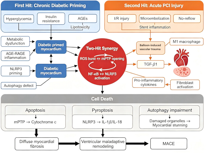
Figure 1: Schematic illustration of the integrated “two-hit” model for post-PCI heart failure in patients with diabetes and coronary artery disease. This schematic visualizes two sequential pathogenic insults driving myocardial dysfunction. The first hit refers to chronic diabetic metabolic and inflammatory priming of cardiomyocytes, encompassing hyperglycemia, insulin resistance, AGE-RAGE signaling, mitochondrial dysfunction, and baseline NLRP3 inflammasome priming. The second hit covers all acute PCI-specific stressors, including ischemia-reperfusion injury, distal microembolization, stent-mediated inflammation, and the no-reflow phenomenon. Preconditioned diabetic myocardium amplifies inflammatory and oxidative stress through the NF-κB–NLRP3 positive feedback loop, which activates three distinct cardiomyocyte death programs: apoptosis, pyroptosis, and defective autophagy. Collectively, these interconnected signaling cascades trigger diffuse myocardial fibrosis, adverse ventricular remodeling, and subsequent development of HFpEF, HFrEF, and major adverse cardiovascular events (MACE). Abbreviations: AGEs = advanced glycation end products; CAD = coronary artery disease; HFpEF = heart failure with preserved ejection fraction; HFrEF = heart failure with reduced ejection fraction; I/R = ischemia-reperfusion; MACE = major adverse cardiovascular events; NLRP3 = NOD-like receptor pyrin domain-containing 3; PCI = percutaneous coronary intervention; RAGE = receptor for AGEs; ROS = reactive oxygen species; T2DM = type 2 diabetes mellitus. Please click here to view a larger version of this figure.
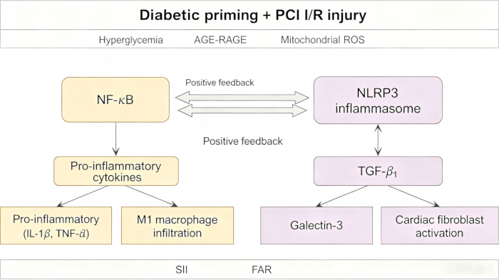
Figure 2: Schematic illustration of bidirectional NF-κB–NLRP3 inflammasome crosstalk driving myocardial inflammatory and fibrotic remodeling in diabetic patients post-PCI. Chronic diabetic metabolic priming (including hyperglycemia and AGE-RAGE signaling) combined with acute PCI-induced ischemia-reperfusion (I/R) injury jointly activates NF-κB and NLRP3 inflammasome, forming a self-amplifying positive feedback loop. Activated NF-κB upregulates the transcription of the pro-inflammatory mediators TNF-α and IL-1β, thereby facilitating M1 macrophage recruitment within cardiac tissue. Meanwhile, NLRP3 inflammasome activation further elevates TGF-β1 and galectin-3 expression, synergistically triggering fibroblast proliferation and diffuse myocardial fibrosis. Systemic immune-inflammation index (SII) and fibrinogen-to-albumin ratio (FAR) serve as peripheral circulating surrogate markers reflecting the overall activity of this inflammatory cascade. Although COVID-19 induces analogous systemic inflammatory signaling via identical axes, it is only referenced as a comparative clinical example rather than a core pathogenic pathway for post-PCI diabetic heart failure. Abbreviations: AGEs = advanced glycation end products; FAR = fibrinogen-to-albumin ratio; I/R = ischemia-reperfusion; IL = interleukin; NF-κB = nuclear factor κB; NLRP3 = NOD-like receptor pyrin domain-containing 3; PCI = percutaneous coronary intervention; ROS = reactive oxygen species; SII = systemic immune-inflammation index; TGF-β1 = transforming growth factor-β1; TNF-α = tumor necrosis factor-α. Please click here to view a larger version of this figure.
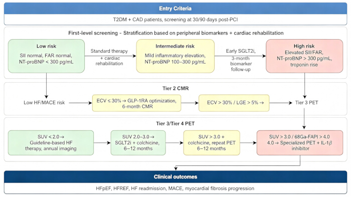
Figure 3: Stepwise clinical decision algorithm for risk stratification and individualized intervention in patients with type 2 diabetes mellitus (T2DM) complicated with coronary artery disease (CAD) after percutaneous coronary intervention (PCI). All enrolled subjects undergo standardized biomarker screening at 30 and 90 days post-procedure. Based on predefined cut-off values of systemic inflammatory indices and cardiac biomarkers, patients are stratified into low-, intermediate-, and high-risk subgroups with corresponding routine management and early targeted medication strategies. High-risk individuals receive tiered cardiac imaging assessments, including cardiac magnetic resonance (CMR) and positron emission tomography (PET). Quantitative extracellular volume (ECV) and standardized uptake value (SUV) thresholds guide further anti-inflammatory and anti-fibrotic therapies. This hierarchical screening and treatment framework ultimately predicts long-term cardiovascular adverse outcomes, including heart failure with preserved ejection fraction (HFpEF), heart failure with reduced ejection fraction (HFrEF), heart failure readmission, and major adverse cardiovascular events (MACE). Abbreviations: CAD = coronary artery disease; CMR = cardiac magnetic resonance; ECV = extracellular volume; FAR = fibrinogen-to-albumin; FAPI = fibroblast activation protein inhibitor; GLP-1RA = glucagon-like peptide-1 receptor agonist; HFpEF = heart failure with preserved ejection fraction; HFrEF = heart failure with reduced ejection fraction; LGE = late gadolinium enhancement; MACE = major adverse cardiovascular events; NT-proBNP = N-terminal pro-B-type natriuretic peptide; PCI = percutaneous coronary intervention; PET = positron emission tomography; SGLT2i = sodium-glucose cotransporter 2 inhibitor; SII = systemic immune-inflammation index; SUV = standardized uptake value; T2DM = type 2 diabetes mellitus. Please click here to view a larger version of this figure.
| Mechanism | Key Mediators/Pathways | PCI-Specific Trigger | Myocardial Consequences | Potential Interventions |
| Metabolic dysregulation | PPARα, AMPK, ACC, GLUT4 | I/R-induced energy deficit | Energy deficit, oxidative stress, mitochondrial dysfunction | Metabolic modulators, SGLT2 inhibitors |
| AGE-RAGE axis | RAGE, NF-κB, ROS | Stent-induced inflammation | Inflammation, fibrosis, endothelial dysfunction | sRAGE, antioxidants, dapagliflozin |
| Inflammatory signaling | NF-κB, NLRP3, IL-1β, IL-18 | I/R, microembolization | Pyroptosis, cytokine storm, adverse remodeling | NLRP3 inhibitors, colchicine, anti-IL-1β |
| Apoptosis | mPTP, cytochrome c, caspases | I/R-induced mitochondrial permeabilization | Cardiomyocyte loss, contractile dysfunction | mPTP blockers, caspase inhibitors |
| Autophagy dysfunction | Klotho/SIRT1, LC3, p62 | Accumulation of damaged mitochondria during reperfusion | Damaged mitochondria accumulation | Autophagy enhancers (metformin, exercise) |
| Microvascular dysfunction | eNOS, endothelin-1, glycocalyx | Microembolization, no-reflow | No-reflow, reduced perfusion, HF progression | SGLT2 inhibitors, GLP-1RAs |
| Fibrosis | TGF-β, Smad3, galectin-3 | Stent-induced fibroblast activation | Ventricular stiffening, diastolic dysfunction | TGF-β inhibitors, galectin-3 blockers |
Table 1: Summary of Major Pathophysiological Mechanisms, Key Mediators, PCI-Specific Triggers, and Potential Therapeutic Targets.
| Drug Class | Representative Agents | Primary Cardioprotective Mechanisms | Effect on HF | Preferred Population | Key Trial Outcomes |
| SGLT2 inhibitors | Empagliflozin, Dapagliflozin | Metabolic reprogramming (ketone utilization), NHE1 inhibition, anti-inflammatory, anti-fibrotic | ↓ HF hospitalization, ↓ CV death | HFrEF, HFpEF, post-MI | EMPA-REG: 38% CV death reduction |
| GLP-1 receptor agonists | Liraglutide, Semaglutide | Anti-inflammatory, endothelial function, plaque stabilization, weight loss | ↓ MACE (non-significant HF benefit) | ASCVD, obesity | SELECT: 20% MACE reduction |
| DPP-4 inhibitors | Sitagliptin, Linagliptin | Mild incretin elevation, modest anti-inflammatory | Neutral (no major HF benefit) | Glycemic control without high CV risk | EXAMINE, SAVOR-TIMI: neutral |
Table 2: Comparison of SGLT2 Inhibitors, GLP-1 Receptor Agonists, and DPP-4 Inhibitors for Heart Failure Prevention Post-PCI.
Disclosures
The authors have no conflicts of interest to disclose.
Acknowledgements
This work was supported by Noncommunicable Chronic Diseases-National Science and Technology Major Project: Comparative Effectiveness Study of Traditional Chinese Medicine Intervention on Comorbidity of Ischemic Cardio-Cerebrovascular Diseases and Diabetes Mellitus. No. 2023ZD0505604. This work was supported by Weifang Science and Technology Bureau (Project No. 2024YX022).
References
- Siam NH, et al. Diabetes mellitus and cardiovascular disease: Exploring epidemiology, pathophysiology, and treatment strategies. Rev Cardiovasc Med. 2024;25(12):436.
- Chakraborty S, et al. Cardiometabolic risk factors associated with type 2 diabetes mellitus: A mechanistic insight. Clin Med Insights Endocrinol Diabetes. 2023;16:11795514231220780.
- Juricic S, et al. Molecular and pathophysiological mechanisms leading to ischemic heart disease in patients with diabetes mellitus. Int J Mol Sci. 2025;26(9).
- Sud M, et al. Low-density lipoprotein cholesterol and adverse cardiovascular events after percutaneous coronary intervention. J Am Coll Cardiol. 2020;76(12):1440-1450.
- Hussain S, et al. Hyperglycemia induces myocardial dysfunction via epigenetic regulation of JunD. Circ Res. 2020;127(10):1261-1273.
- Anagnostopoulou L, et al. Intersecting molecular pathways in cardiovascular disease and diabetes mellitus: Emerging roles of inflammation and therapeutics. Diabetes Metab Res Rev. 2026;42(4):e70167.
- Dludla PV, et al. The combination effect of aspalathin and phenylpyruvic acid-2-O-β-D-glucoside from Rooibos against hyperglycemia-induced cardiac damage: An in vitro study. Nutrients. 2020;12(4).
- Cividini F, et al. Ncor2/PPARα-dependent upregulation of MCUb in the type 2 diabetic heart impacts cardiac metabolic flexibility and function. Diabetes. 2021;70(3):665-679.
- Vargas-Delgado AP, et al. Renal and cardiovascular metabolic impact caused by ketogenesis of the SGLT2 inhibitors. Int J Mol Sci. 2023;24(4).
- Song X, et al. Mitochondria fission accentuates oxidative stress in hyperglycemia-induced H9c2 cardiomyoblasts in vitro by regulating fatty acid oxidation. Cell Biol Int. 2024;48(9):1378-1391.
- Rudokas MW, et al. Mitochondrial network remodeling of the diabetic heart: Implications to ischemia-related cardiac dysfunction. Cardiovasc Diabetol. 2024;23(1):261.
- Murthy D, et al. CD24 negativity reprograms mitochondrial metabolism to PPARα- and NF-κB-driven fatty acid β-oxidation in triple-negative breast cancer. Cancer Lett. 2024;587:216724.
- Honka H, et al. Therapeutic manipulation of myocardial metabolism: JACC state-of-the-art review. J Am Coll Cardiol. 2021;77(16):2022-2039.
- Liang L, et al. Discovery and identification of natural alkaloids with potential to impact insulin resistance syndrome in Cyclocarya paliurus (Batal) leaves by UPLC-QTOF-MS combined with HepG2 cells. Food Res Int. 2024;190:114545.
- Wang K, et al. Role of epigenetic regulation in myocardial ischemia/reperfusion injury. Pharmacol Res. 2021;170:105743.
- Yang B, et al. Salidroside pretreatment alleviates ferroptosis induced by myocardial ischemia/reperfusion through mitochondrial superoxide-dependent AMPKα2 activation. Phytomedicine. 2024;128:155365.
- Wang Y, et al. MD2 activation by direct AGE interaction drives inflammatory diabetic cardiomyopathy. Nat Commun. 2020;11(1):2148.
- Wang X, et al. Engineered cardiac tissues: A novel in vitro model to investigate the pathophysiology of mouse diabetic cardiomyopathy. Acta Pharmacol Sin. 2021;42(6):932-941.
- Li M, et al. The investigation of potential mechanism of Fuzhengkangfu Decoction against diabetic myocardial injury based on a combined strategy of network pharmacology, transcriptomics, and experimental verification. Biomed Pharmacother. 2024;177:117048.
- Liu J, et al. An anti-RAGE chimeric antibody alleviates CCl4-induced liver fibrosis via the RAGE/NF-κB pathway in mice. Biomed Pharmacother. 2024;181:117737.
- Zhang L, et al. Plantamajoside attenuates cardiac fibrosis via inhibiting the AGEs-activated RAGE/autophagy/EndMT pathway. Phytother Res. 2023;37(3):834-847.
- Mayyas F, Omeish A. Comparison of the cardioprotective effects of liraglutide, dapagliflozin, and their combination in a rat model of diabetes induced by streptozotocin. Life Sci. 2025;375:123721.
- Liu M, et al. Mechanisms of inflammatory microenvironment formation in cardiometabolic diseases: Molecular and cellular perspectives. Front Cardiovasc Med. 2024;11:1529903.
- Romacho T, et al. Visfatin/eNampt induces endothelial dysfunction in vivo: A role for Toll-like receptor 4 and the NLRP3 inflammasome. Sci Rep. 2020;10(1):5386.
- Wang X, et al. ETS1-THBS1 axis regulates macrophage polarization and exacerbates myocardial injury in diabetic cardiomyopathy. J Cardiovasc Pharmacol. 2025;86(3):270-280.
- He M, et al. Beta-hydroxybutyrate ameliorates cardiac fibrosis in diabetic cardiomyopathy rats via regulating macrophage polarization. Diabetes Res Clin Pract. 2025;229:112461.
- Cai G, et al. Targeting the cGAS-STING pathway to modulate immune inflammation in diabetes and cardiovascular complications: Mechanisms and therapeutic insights. Curr Issues Mol Biol. 2025;47(9).
- Peng M, et al. Cathelicidin-WA ameliorates diabetic cardiomyopathy by inhibiting the NLRP3 inflammasome. Cell Cycle. 2021;20(21):2278-2290.
- Prakash S, Choudhury P, Bisht S. Diabetic cardiomyopathy and COVID-19: Intersecting pathways and amplified cardiovascular risk. Front Pharmacol. 2025;16:1683159.
- Kaur N, et al. Mechanisms and therapeutic prospects of diabetic cardiomyopathy through the inflammatory response. Front Physiol. 2021;12:694864.
- Kim J, et al. IκBζ controls NLRP3 inflammasome activation via upregulation of the Nlrp3 gene. Cytokine. 2020;127:154983.
- Mai W, et al. Berberine inhibits Nod-like receptor family pyrin domain containing 3 inflammasome activation and pyroptosis in nonalcoholic steatohepatitis via the ROS/TXNIP axis. Front Pharmacol. 2020;11:185.
- Bian X, et al. The combined effect of systemic immune-inflammation index and type 2 diabetes mellitus on the prognosis of patients undergoing percutaneous coronary intervention: A large-scale cohort study. J Inflamm Res. 2023;16:6415-6429.
- Eisinger K, Girke P, Buechler C, Krautbauer S. Adipose tissue depot-specific expression and regulation of fibrosis-related genes and proteins in experimental obesity. Mamm Genome. 2024;35(1):13-30.
- Zhang Y, et al. TGF-β1-containing exosomes from cardiac microvascular endothelial cells mediate cardiac fibroblast activation under high-glucose conditions. Biochem Cell Biol. 2021;99(6):693-699.
- Ding H, et al. MicroRNA-195-5p downregulation inhibits endothelial mesenchymal transition and myocardial fibrosis in diabetic cardiomyopathy by targeting Smad7 and inhibiting transforming growth factor beta 1-Smads-Snail pathway. Front Physiol. 2021;12:709123.
- Liu T, Yang F. Galectin-3 in acute myocardial infarction: From molecular mechanisms to clinical translation. Front Mol Biosci. 2025;12:1730173.
- Chen P, et al. Mitochondrial dysfunction: A promising therapeutic target for liver diseases. Genes Dis. 2024;11(3):101115.
- Bekhite M, et al. The role of ceramide accumulation in human induced pluripotent stem cell-derived cardiomyocytes on mitochondrial oxidative stress and mitophagy. Free Radic Biol Med. 2021;167:66-80.
- Liu JX, et al. Disturbance of mitochondrial dynamics and mitophagy in sepsis-induced acute kidney injury. Life Sci. 2019;235:116828.
- Xue M, Irshad Z, Rabbani N, Thornalley PJ. Increased cellular protein modification by methylglyoxal activates endoplasmic reticulum-based sensors of the unfolded protein response. Redox Biol. 2024;69:103025.
- Sebastian SA, Padda I, Johal G. Cardiovascular-kidney-metabolic (CKM) syndrome: A state-of-the-art review. Curr Probl Cardiol. 2024;49(2):102344.
- Klimontov VV, Saik OV, Korbut AI. Glucose variability: How does it work? Int J Mol Sci. 2021;22(15).
- Patel J, Parab SB, Doshi GM. Endoplasmic reticulum stress and the unfolded protein response in ischemic nephropathy: Pathogenic mechanisms and emerging therapeutic strategies. Pathol Res Pract. 2026;279:156367.
- Tian JH, et al. Zonisamide, an antiepileptic drug, alleviates diabetic cardiomyopathy by inhibiting endoplasmic reticulum stress. Acta Pharmacol Sin. 2021;42(3):393-403.
- Kapadia P, et al. Effect of anti-hyperglycemic drugs on endoplasmic reticulum (ER) stress in human coronary artery endothelial cells. Eur J Pharmacol. 2021;907:174249.
- Chien CY, et al. Diabetes upregulates oxidative stress and downregulates cardiac protection to exacerbate myocardial ischemia/reperfusion injury in rats. Antioxidants (Basel). 2020;9(8).
- Qiu Z, et al. Activation of the Klotho/SIRT1 signaling pathway attenuates myocardial ischemia-reperfusion injury in diabetic rats. Shock. 2024;62(3):447-456.
- Osorio-Llanes E, et al. Effects of metformin on ischemia/reperfusion injury: New evidence and mechanisms. Pharmaceuticals (Basel). 2023;16(8).
- Jiang K, et al. Cardioprotective mechanism of SGLT2 inhibitor against myocardial infarction is through reduction of autosis. Protein Cell. 2022;13(5):336-359.
- Yang T, et al. Increased risk of adverse cardiovascular events by strict glycemic control after percutaneous coronary intervention (HbA1c <6.5% at 2 years) in type 2 diabetes mellitus combined with acute coronary syndrome: A 5-year follow-up study. Curr Med Res Opin. 2021;37(9):1517-1528.
- Liu Y, et al. Association of estimated glomerular filtration rate (eGFR) and high-sensitivity C-reactive protein (hs-CRP) with the risk of new-onset atrial fibrillation in patients with diabetes. J Inflamm Res. 2025;18:91-103.
- Mateoc T, et al. SGLT2 inhibitors: From structure-effect relationship to pharmacological response. Int J Mol Sci. 2025;26(14).
- White A, et al. Body mass index, left ventricular wall stress, and NT-proBNP in elderly adults. JACC Adv. 2026;5(2):102564.
- Zhang Y, et al. Evaluation of large-scale plasma proteomics for prediction of heart failure in individuals with a full range of glucose metabolism profiles. Eur J Prev Cardiol. 2026;33(2):175-186.
- Song Y, et al. Plasma ANGPTL7 links insulin resistance to heart failure: Unveiling a novel fibro-metabolic axis in type 2 diabetes. Diabetes Metab Res Rev. 2026;42(3):e70153.
- Rochette L, et al. GDF15 and cardiac cells: Current concepts and new insights. Int J Mol Sci. 2021;22(16).
- Parizad R, et al. Artificial intelligence for cardiovascular risk prediction: An umbrella review of applications and translational challenges. Vasc Health Risk Manag. 2026;22:590502.
- Lee HJ, et al. Systemic proinflammatory-profibrotic response in aortic stenosis patients with diabetes and its relationship with myocardial remodeling and clinical outcome. Cardiovasc Diabetol. 2023;22(1):30.
- Zhu J, et al. Association of epicardial adipose tissue with left ventricular strain and MR myocardial perfusion in patients with known coronary artery disease. J Magn Reson Imaging. 2023;58(5):1490-1498.
- Mbarek L, et al. Predicting 3-month poor functional outcomes of acute ischemic stroke in young patients using machine learning. Eur J Med Res. 2024;29(1):494.
- Solomon SD, et al. Dapagliflozin in heart failure with preserved and mildly reduced ejection fraction: Rationale and design of the DELIVER trial. Eur J Heart Fail. 2021;23(7):1217-1225.
- Ma Z, et al. Cardiovascular adverse events associated with epidermal growth factor receptor tyrosine kinase inhibitors in EGFR-mutated non-small cell lung cancer: Systematic review and network meta-analysis. BMJ. 2025;390:e082834.
- Raparelli V, et al. Sex differences in cardiovascular effectiveness of newer glucose-lowering drugs added to metformin in type 2 diabetes mellitus. J Am Heart Assoc. 2020;9(1):e012940.
- Marketou M, et al. Effects of sodium-glucose cotransporter-2 inhibitors on cardiac structural and electrical remodeling: From myocardial cytology to cardiodiabetology. Curr Vasc Pharmacol. 2022;20(2):178-188.
- Yu P, et al. Dapagliflozin ameliorates myocardial ischemia/reperfusion injury by modulating EGFR signaling and targeting NCOA4-mediated ferritinophagy. Life Sci. 2026;391:124254.
- Vergès B, et al. Protection against stroke with glucagon-like peptide-1 receptor agonists: A comprehensive review of potential mechanisms. Cardiovasc Diabetol. 2022;21(1):242.
- Alshanwani A, Kashour T, Badr A. Anti-diabetic drugs GLP-1 agonists and DPP-4 inhibitors may represent potential therapeutic approaches for COVID-19. Endocr Metab Immune Disord Drug Targets. 2022;22(6):571-578.
- Mangoura SA, Ahmed MA, Zaka AZ. New insights into the pleiotropic actions of dipeptidyl peptidase-4 inhibitors beyond glycaemic control. touchREV Endocrinol. 2024;20(2):19-29.
- Spoladore R, et al. Metabolic approaches for the treatment of dilated cardiomyopathy. J Cardiovasc Dev Dis. 2023;10(7).
- Fitzgerald J, Parker JA, Danias PG. F-18 fluorodeoxyglucose SPECT for assessment of myocardial viability. J Nucl Cardiol. 2000;7:382-387.
- Hartikainen S, et al. Cardiometabolic disorders affect myocardial [18F]fluorodeoxyglucose uptake: Impact on diagnostic PET/CT imaging. EJNMMI Res. 2025;15:80.
- Saraste A, Knuuti J. PET imaging in heart failure: The role of new tracers. Heart Fail Rev. 2017;22(4):501-511.
- Tian Y, et al. Application of radionuclide myocardial imaging in the diagnosis and treatment of heart failure with preserved ejection fraction. Rev Cardiovasc Med. 2025;26(10):41231.
- Tarkin JM, et al. Detection of atherosclerotic inflammation by 68Ga-DOTATATE PET compared to [18F]FDG PET imaging. J Am Coll Cardiol. 2017;69(14):1774-1791.
- Kong L, et al. Scanner-generated native T1 mapping: A novel approach for assessing myocardial fibrosis in coronary heart disease. Front Cardiovasc Med. 2025;12:1553919.
- Zhang X, et al. Positron emission tomography molecular imaging for phenotyping and management of lymphoma. Phenomics. 2022;2(2):102-118.
- Zuo Y, et al. Multiparametric cardiac 18F-FDG PET in humans: Kinetic model selection and identifiability analysis. IEEE Trans Radiat Plasma Med Sci. 2020;4(6):759-767.