A modified technique combining an orbital septal fascial flap with extended orbital fat release was associated with correction of lower eyelid bags and midface depression, with a low complication rate and favorable aesthetic outcomes in a single-center retrospective cohort.
Research Article
Orbital Septal Fascial Flap with Extensive Release of a Long Orbital Fat Flap for Correcting Lower Eyelid Bags Associated with Midface Depression
In This Article
Summary
Abstract
Conventional lower blepharoplasty remains constrained by limited fat availability and insufficient transposition distance in patients with severe tear trough deformity and midfacial depression. This study aimed to assess the clinical outcomes of a technique combining an orbital septal fascial flap with extensive release of an elongated orbital fat flap (>2 cm). A total of 121 patients treated from December 2019 to March 2025 were enrolled for retrospective analysis. Of these, 68 cases underwent a transconjunctival approach (median age 28 years), while 53 cases with obvious skin laxity received a transcutaneous approach (median age 45 years). With precise dissection of the prezygomatic space, a pedicled orbital septal fascial flap was harvested to cover the 2.0–2.5 cm vascularized fat flap advanced into the midfacial recipient site. The rates of complete tear trough effacement (Barton Grade 0) were 89.7% and 92.5% in the two groups, respectively, with no statistically significant intergroup difference (P = 0.607). Given the substantial baseline differences between groups, this should not be interpreted as evidence of equivalence. Median operative time was 61 min vs. 125 min, and median recovery time was 11 days vs. 15 days (both P < 0.001). The overall complication rates were 1.47% and 3.77% (P = 0.581). No surgical site infection or permanent lower eyelid ectropion occurred. Over a median follow-up of 8.0 months, no cases of diplopia, sensory nerve injury, enophthalmos, or fat necrosis/displacement were recorded. However, these events were not assessed with a pre-specified standardized protocol. This composite flap technique is designed to address the anatomical constraints of conventional fat transposition. The fascial flap may provide additional microcirculatory support and mechanical fixation, and was associated with favorable aesthetic outcomes and a low complication rate. These findings from a single-center retrospective cohort require confirmation in prospective, controlled studies.
Introduction
Traditional lower blepharoplasty has occupied a central role in facial rejuvenation surgery for decades. However, early surgical paradigms were often confined to the simple excision or basic release of eyelid fat pads, severely neglecting the intricately intertwined pathological relationship between lower eyelid aging and midfacial volume loss. In clinical practice, the synergistic correction of lower eyelid aging and midfacial depression has persistently remained a highly challenging technical difficulty in plastic surgery. Based on an in-depth review of periorbital aging pathology and advanced anatomical theories, this study systematically extends the suborbicularis oculi fat transposition (SOFT) concept within the surgical domain.
We describe and analyze the clinical outcomes of the "orbital septal fascial flap combined with the extensive release of an elongated orbital fat flap (>2 cm)." By precisely dissecting and utilizing the anatomical plane of the prezygomatic space, this technique applies the concept of extensive release to overcome the physical length limitations of traditional fat transposition. It is intended to achieve the dual objectives of tension-free repositioning of orbital fat and structural restoration of ptotic deep midfacial volume within a single surgical procedure.
Through a retrospective analysis of a cohort of patients with lower eyelid aging treated at the Department of Plastic and Aesthetic Surgery, First Affiliated Hospital of Guangxi Medical University, between December 2019 and March 2025, this report details the surgical indications, preoperative evaluation criteria, and follow-up outcomes of this surgical modality in patients presenting with infraorbital bags accompanied by severe tear trough and midfacial depression deformities. The study describes short-term morphological outcomes and complication rates of this combined procedure, which reconstructs the middle fascial support of the periorbital region and the lid-cheek junction, addressing orbital fat herniation, tear trough deformity, and midfacial depression within a single operation (Figure 1).
The aging of the lower eyelid region is a complex, multidimensional biological process involving multiple tissue layers. Its characteristic clinical manifestations primarily include progressive skin laxity caused by the rupture of elastic fibers, lower eyelid retraction, orbital fat herniation (commonly known as eye bags) secondary to the attenuation of the orbital septum and muscle tension, as well as midfacial depression and tear trough concavity accompanying midfacial tissue atrophy, and the exposure of the lid-cheek junction, which is the most definitive hallmark of aging1. Among these signs of aging, the pathological understanding of the tear trough deformity has undergone a long and tortuous evolution2. As early as 1961, anatomists Duke-Elder and Wybar, in their work System of Ophthalmology, first described this anatomical depression located inferior to the medial canthus and extending obliquely outward and downward as the "nasojugal fold". In 1969, Robert S. Flowers, a pioneer of modern plastic surgery, formally introduced the modern clinical term "Tear Trough Deformity"3, and over subsequent decades of research, completely revolutionized the medical community's traditional understanding of its etiology. The connection between these soft tissues and the underlying skeleton, coupled with age-related orbital bone resorption and the aforementioned soft-tissue degeneration, gradually results in a senescent appearance of midfacial depression, tear troughs, and medial fat pad herniation1.
Regarding the surgical correction of lower eyelid aging, as early as 2,000 years ago, the scholar Sushruta first described lower eyelid surgery in the Sushruta Samhita (Susruta Tantra), while Bourguet was the first to describe a transconjunctival approach for the excision of lower eyelid fat3. However, traditional surgical approaches could only alleviate the issue of eye bags but were unable to treat the tear trough deformity and even risked exacerbating the concavity. Therefore, surgical techniques required improvement to fully satisfy the aesthetic demands of rejuvenation. In 1981, Loeb first released lower eyelid orbital fat, combining fat repositioning with cheek fat to create a smoother lid-cheek contour4. In the 20th century, scholars such as Hamra and Barton investigated new anatomical positions for repositioning fat and the orbital septum, reaffirming the significant contribution of this technique to youthful cheek remodeling5,6. This technique not only reduces puffiness in eye bags but also uses orbital fat to correct the concavity of the lower eyelid. Jin Yunbo applied the SOFT technique to release a fat flap exceeding 2 cm, which not only filled the defect at the lid-cheek junction but also augmented midfacial depression to a certain extent7.
Addressing the clinical dilemmas of "insufficient fat volume" or "inadequate transposition distance" faced by traditional orbital fat release surgeries in patients with severe midfacial depression, our research team, based on the expanded SOFT theory in 2019, established and standardized a combined technique at the Department of Plastic and Aesthetic Surgery of the First Affiliated Hospital of Guangxi Medical University: the orbital septal fascial flap combined with the extensive release of an elongated orbital fat flap (>2 cm). We evaluated its safety and efficacy in achieving the anticipated aesthetic outcomes.
Protocol
All procedures involving human participants were reviewed and approved by the Medical Ethics Committee of the First Affiliated Hospital of Guangxi Medical University (Approval No.: 2026-E0435; date of approval: 13 May 2026). Ethics approval was obtained for the retrospective use of these data. Written informed consent for the surgical procedures, the use of clinical data, and the publication of identifiable photographs was obtained from all participants. This study was conducted in accordance with the Declaration of Helsinki.
General clinical data
Clinical data were collected from 121 patients who underwent treatment with an orbital septum fascial flap combined with orbital fat release at the outpatient clinic of the Department of Plastic and Reconstructive Surgery, First Affiliated Hospital of Guangxi Medical University. All procedures were performed by the same surgeon between December 2019 and March 2025. All patients presented with lower eyelid bags accompanied by pronounced tear trough deformity, and their preoperative evaluations met the surgical indications for orbital fat release.
A total of 121 cases (242 eyes) were included in this study. Among these, 53 cases underwent a transcutaneous approach, comprising 50 females (94.3%) and 3 males (5.7%), with a median age of 45 years (IQR, 41–49). One case in this subgroup had undergone lower eyelid surgery 4 years prior to enrollment; because the exclusion criterion specifies no lower eyelid or periorbital plastic surgery within the past 2 years, this patient was not excluded. The remaining 52 transcutaneous cases and all 68 transconjunctival cases were primary surgeries. The transconjunctival group comprised 64 females (94.1%) and 4 males (5.9%), with a median age of 28 years (IQR 25–33; mean 29.03 ± 5.49). Comprehensive clinical records, including standardized photographs, were complete for all patients. Patients with severe underlying diseases, surgical contraindications, infectious diseases, or psychiatric disorders were excluded preoperatively.
Inclusion and exclusion criteria
Inclusion criteria: Patients diagnosed with Type II or Type III lower eyelid bags, according to the Summary of the First Academic Conference on Ophthalmic Plastic and Orbital Diseases of the Ophthalmological Society of the Chinese Medical Association; Patients aged > 18 years, with complete clinical data and the ability to tolerate surgery; Patients with clear consciousness and normal communication capabilities; Patients and their families who provided informed consent.
Exclusion criteria: Presence of malignant tumors, severe organ dysfunction, or severe systemic diseases; Drug addiction or dependence on alcohol/tobacco; History of lower eyelid or periorbital plastic surgery within the past 2 years; History of ocular or periocular trauma within the past six months.
Surgical technique (Figure 2 and Figure 3)
Preoperative preparation and anesthesia
All procedures were performed under local anesthesia on an outpatient basis. Anesthesia consisted of 2% lidocaine hydrochloride injection combined with epinephrine at a 1:500,000 concentration, administered via infraorbital nerve block, zygomaticofacial nerve block, and local infiltration anesthesia. The surgical field was prepared with povidone-iodine solution (0.5%) applied three times, and the head and face were draped to expose the full facial region while maintaining a sterile field. Preoperative marking with methylene blue delineated the tear trough, the lower eyelid fat herniation, and the midfacial depression to be corrected.
For the transconjunctival approach, the patient was instructed to keep the eyes closed, and a small square gauze was placed to protect the upper eyelid and cornea. For the transcutaneous approach, the patient was instructed to keep the eyes open to facilitate assessment of eyelid contour and protect the globe. Sharp dissection was performed using a single-use monopolar needle-tip electrocautery electrode, which also provided intraoperative hemostasis.
In the transconjunctival group, the distal fat flap was fixated to the skin at the pre-marked points using 6-0 nylon sutures, secured externally with a petrolatum gauze bolster. In the transcutaneous group, the distal flap was fixated to the periosteum using 5-0 polydioxanone (PDS) absorbable sutures. Postoperatively, ofloxacin eye drops (0.3%) were instilled four times daily. The external bolster was maintained until suture removal, and the transcutaneous incision was cleansed daily. Patients were advised to avoid strenuous activity and bending for 1 week, and skin sutures were removed at 7 days. Standardized follow-up evaluations were scheduled at postoperative day 30, and at 3, 6, and 12 months.
Surgical technique for the Type II study group
The patient was instructed to keep their eyes closed. The conjunctiva was exposed, and a small square gauze was used to protect the upper eyelid. A transconjunctival incision of approximately 15 mm was made at the lower margin of the tarsal plate using a needle-tip electrocautery. Dissection was performed closely along the deep surface of the orbicularis oculi muscle using mosquito forceps. While keeping the orbital septum intact, the dissection proceeded to just above the arcus marginalis, and an eyelid retractor was applied to expose the orbital rim. Using needle-tip electrocautery, the tear trough ligament and the orbicularis retaining ligament were sharply released from medial to lateral up to the preoperatively marked position. This loosened the orbicularis oculi muscle, exposed the levator labii superioris alaeque nasi, and provided access to the prezygomatic space. A nerve dissector was then used to dissect the area according to the preoperatively marked fat fixation sites. To ensure adequate release, the endpoint of this dissection was approximately 7 mm above the nasal ala. After achieving meticulous hemostasis, the orbital septum was opened at the arcus marginalis and dissected to form a complete fan-shaped orbital septum fascial flap (as shown in Figure 3). The preoperatively marked protruding fat, specifically the medial and central compartments of orbital fat, was fully released to create a fan-shaped orbital fat flap measuring approximately 2 cm to 2.5 cm. The entire procedure was performed delicately to avoid injury to the inferior oblique muscle, and care was taken to ensure the harvested fat flap retained its vascular pedicle, which was carefully preserved at the base to maintain perfusion from the infraorbital artery branches.
The orbital septum fascial flap was laid flat on the inner side of the fat flap to reinforce the middle-layer structure, and then sutured into place. The distal portion of the fat flap was engaged using a 6-0 nylon suture. The fat flap, now covered by the orbital septum fascial flap, was pulled down into the premaxillary space. The sutures were passed through the skin at the pre-marked fixation points. After confirming a satisfactory aesthetic appearance of the midface, the sutures were secured externally with a petrolatum gauze bolster.
Surgical technique for the Type III study group
The patient was instructed to keep their eyes open. The skin was incised at the designed incision site, 2 mm below the lower margin of the tarsal plate. Using ophthalmic scissors, a skin flap was undermined between the skin and the orbicularis oculi muscle down to 4–5 mm from the lower eyelid margin. A needle-tip electrocautery was used to incise the orbicularis oculi muscle to enter the preseptal space. Dissection was performed closely along the deep surface of the orbicularis oculi muscle (i.e., the preseptal space) using mosquito forceps. While keeping the orbital septum intact, the dissection proceeded to just above the arcus marginalis, and an eyelid retractor was applied to expose the orbital rim. Using needle-tip electrocautery, the tear trough ligament and the orbicularis retaining ligament were sharply released from medial to lateral to loosen the orbicularis oculi muscle, forming a muscle flap. Entry was gained into the premaxillary space and prezygomatic space. A nerve dissector was used for dissection within these spaces, with the endpoint of dissection approximately 7 mm above the lower margin of the nasal ala. After achieving meticulous hemostasis, the orbital septum was opened at the arcus marginalis and dissected to form a complete fan-shaped orbital septum fascial flap (as shown in Figure 3). The medial, central, and lateral compartments of orbital fat were fully released to create a fan-shaped orbital fat flap measuring approximately 2 cm to 2.5 cm. The orbital septum fascial flap was laid flat on the inner side of the fat flap and sutured into place. The fat flap was positioned to cover the midface depression. After confirming a satisfactory aesthetic result, the distal portion was fixated to the periosteum using 5-0 PDS absorbable sutures. The patient was instructed to open their mouth and look straight ahead to evaluate the amount of excess skin to be excised. The excess skin (approximately 2–4 mm) was marked with methylene blue and removed using ophthalmic scissors. The orbicularis oculi muscle flap was retracted upward to lift the midface, and the main ligament was sutured and fixated to the orbital rim. The orbicularis oculi muscle and skin were then sutured in precise anatomical alignment.
Outcome measures
Comparison of baseline data and objective intraoperative and perioperative parameters between the two groups
The operative duration, length of the prepared fat flap, and recovery time of the ocular appearance were recorded for patients in both groups. Criteria for normal recovery of ocular appearance: The periorbital skin exhibited no erythema, edema, or inflammation; the eye contour maintained a normal, natural curvature; there was no obvious scarring in the periorbital or adjacent areas; eyelid dynamics during blinking were natural; there was no alteration in the palpebral fissure size during facial expressions, and patients reported no sensation of pulling or heaviness around the eyes.
Assessment of tear trough deformity (Barton grading) and inter-rater reliability
Tear trough deformity was graded using the Barton classification preoperatively and at postoperative day 30. Grading was performed independently by two certified oculoplastic surgeons (with 12 and 8 years of experience, respectively), neither of whom had participated in the index surgeries or the initial assessments. Both assessors were fully blinded to surgical approach, postoperative time point, patient demographics, and the original scores. Standardized clinical photographs (frontal, 45-degree oblique, and lateral views) were de-identified and presented in random order using REDCap (version 13.0). Inter-rater reliability was formally evaluated in a stratified random subsample of 25 patients (20.7% of the cohort). Agreement was quantified using the weighted kappa coefficient with bootstrap-estimated 95% confidence intervals, interpreted according to Landis and Koch benchmarks. Preoperative weighted kappa was 0.78 (95% CI 0.62–0.91) and postoperative weighted kappa was 0.82 (95% CI 0.69–0.94), indicating substantial agreement.
Intra-group and inter-group evaluation of surgical efficacy (complete resolution rate of the tear trough)
The Barton grading system for tear troughs was utilized to evaluate the tear trough condition of patients in both groups preoperatively and postoperatively (at day 30). Grade 0: No obvious demarcation line from the medial to the lateral orbital rim; the contour at the lid-cheek junction is smooth, without a transitional outline. Grade I: Mild depression or shadowing is visible at the medial orbital rim, while the contour of the lid-cheek junction at the lateral orbital rim remains smooth. Grade II: A discernible demarcation line is present at the lid-cheek junction, with moderate fat bulging observed above the line. Grade III: An obvious demarcation line is present at the lid-cheek junction, and the area between the orbit and the cheek exhibits a step-like bulging deformity. A higher Barton grade indicates a more pronounced tear trough deformity.
Postoperative safety and complication assessment
Complication data were recorded up to 6 months postoperatively and included incidences of incisional hematoma, incisional infection, and lower eyelid ectropion, which were the three complications systematically captured by a pre-specified protocol. In addition, the following events were reviewed through clinical follow-up records: diplopia, sensory nerve injury, enophthalmos, and fat necrosis or displacement. None of these four events was observed during the follow-up period.
Beyond the 30-day and 6-month endpoints (Figure 4 and Figure 5), selected cases with extended follow-up illustrated the durability of the combined technique. A representative 45-year-old male patient who underwent the transcutaneous approach maintained a smooth lower eyelid contour, resolved eye bags, and an effaced tear trough at 18 months postoperatively (Figure 6). A representative 35-year-old female patient who underwent the transconjunctival approach showed comparable, stable restoration of the lid-cheek junction at 36 months postoperatively (Figure 7). These observations are consistent with the overall cohort, in which standardized comparative photographs were available up to 36 months (Figure 8), supporting the short- to mid-term stability of the procedure. Nevertheless, because standardized comparative photographs at ≥2 years were not available for the majority of patients, long-term durability remains to be confirmed in prospective studies with structured long-term follow-up.
Statistical methods and data processing
Study groups: Data were extracted and compared as two independent samples: the transconjunctival approach group (n = 68) and the transcutaneous approach group (n = 53), yielding a total cohort of 121 cases. Given that many patients presented with bilateral involvement, totaling 242 eyes across 121 patients, all analyses were conducted at the patient level, yielding 121 independent observations. For efficacy evaluation, the worse eye, defined as the higher-grade eye according to the Barton classification, was used to represent the patient-level outcome, adhering to the worst-eye principle. For safety evaluation, complications were coded on a binary basis at the patient level, meaning a patient was recorded as having a specific complication if it occurred in either eye.
Handling of missing data: Variables such as "aesthetic satisfaction" and some postoperative follow-up evaluations were recorded in only a small subset of the sample (validity rate < 10%), indicating severe non-random missingness. In accordance with statistical standards for this analysis, these variables were excluded from quantitative inter-group hypothesis testing to avoid introducing strong selection bias.
Normality testing and descriptive statistics: The Shapiro-Wilk test indicated that all continuous variables (age, operative duration, fat flap length, and recovery time) exhibited non-normal distributions (P < 0.05). Consequently, continuous quantitative data are uniformly expressed as the median and interquartile range M (P25, P75). Categorical and ordinal data are expressed as frequencies and percentages n (%).
Statistical tests: Inter-group comparisons of independent continuous and ordinal variables were performed using the Mann-Whitney U test. Intra-group comparisons of ordinal data before and after surgery (rating downgrades) were evaluated using the Wilcoxon signed-rank test. For inter-group comparisons of nominal categorical variables, Fisher's exact test was uniformly applied due to the presence of expected frequencies < 5.
Significance level and software: All statistical tests were two-sided, with the significance level set at α = 0.05. P-value < 0.05 was considered to indicate a statistically significant difference. Continuous and ordinal comparisons were performed using IBM SPSS Statistics (version 26.0). Inter-rater reliability was analyzed using R software (version 4.3.0) with the irr package. Effect sizes for the primary outcome are reported as absolute risk differences with 95% confidence intervals.
Results
Comparison of baseline characteristics and intraoperative objective parameters between the two groups
The data indicate substantial baseline differences between the two groups, reflecting the fact that the choice of surgical approach was driven by the patient's presenting anatomy rather than by randomization. The transcutaneous approach group, which specifically targeted patients with severe conditions accompanied by "skin laxity," demonstrated a significantly higher median age than the transconjunctival approach group (45 years vs. 28 years, P < 0.001). Furthermore, all patients in the transcutaneous group presented with severe (Type III) tear trough deformities (100% vs. 8.8%), indicating a marked difference in baseline characteristics between the two groups.
Regarding anatomical and temporal parameters, the lengths of the harvested fat flaps dissected via the two approaches were comparable (P = 0.278). However, because the transcutaneous approach requires additional excision of lax skin followed by meticulous suturing and suspension, its median operative time (125 min) and recovery time (15 days) were significantly longer than the 61 min and 11 days observed in the transconjunctival approach group (both P < 0.001), as shown in Table 1.
Intra-group and inter-group evaluation of surgical efficacy (Rate of complete tear trough effacement)
Longitudinal efficacy: Postoperative tear trough conditions in all patients demonstrated substantial improvement. Intra-group comparisons of preoperative and postoperative grading in both groups showed highly significant statistical differences (Wilcoxon test, P < 0.001).
Cross-sectional endpoint comparison: Although the transcutaneous approach group involved a more challenging patient cohort characterized by older age, 100% severe (Type III) deformities, and concurrent tissue aging and laxity, the proportion of patients achieving a "Grade 0 (no depression, complete effacement)" outcome postoperatively in this group was 92.5%. Compared with the transconjunctival approach group, which predominantly consisted of mild-to-moderate cases (89.7%), no statistically significant difference in the final morphological outcome was detected between the groups (P = 0.607; absolute risk difference -2.7%, 95% CI -12.9% to 7.4%). Because the groups differed substantially at baseline and no adjustment was performed, this finding should not be interpreted as evidence that the two techniques are equivalent, as shown in Table 2.
Postoperative safety and complication assessment
During the follow-up period (median 8.0 months; IQR 6.0–11.0; range 6–36 months), no postoperative local infections were observed in the entire cohort. The overall complication rate was low (1.47% in the transconjunctival group and 3.77% in the transcutaneous group). Fisher's exact test revealed no statistically significant difference between the two groups (P = 0.581). In the transcutaneous approach group, two elderly patients experienced transient lower eyelid ectropion (records indicate complete resolution to normal anatomical position after 4 and 6 months, respectively, without permanent deformity); these were managed conservatively, as shown in Table 3.
Beyond the three systematically recorded complications, the following events were not observed during the follow-up period (Table 3): diplopia (0/121), sensory nerve injury (0/121), enophthalmos (0/121), and fat necrosis or displacement (0/121).
DATA AVAILABILITY:
The de-identified data of this study are provided in Supplementary File 1.
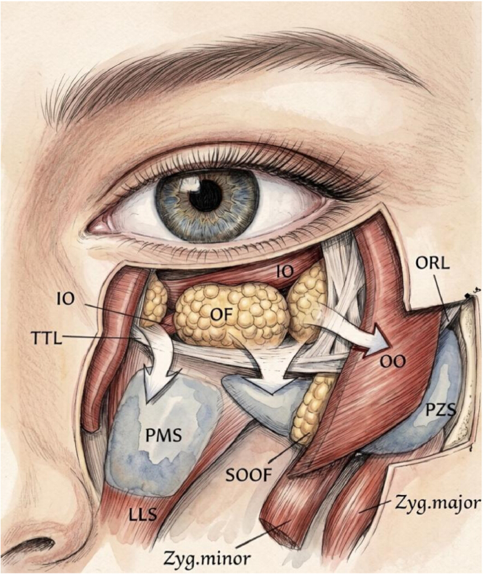
Figure 1: Detailed anatomy of the periorbital fat pads, ligaments, and potential spaces. This anatomical illustration presents the key structures of the midfacial periorbital region, explicitly delineating the fat pads, muscles, retaining ligaments, and two critical potential spaces. The deep anatomical relationships are revealed following the incision and dissection of the orbicularis oculi (OO) muscle. Abbreviations: IO: Inferior oblique, TTL: Tear trough ligament, ORL: Orbicularis retaining ligament, OO: Orbicularis oculi muscle, PZS: Prezygomatic space, SOOF: Suborbicularis oculi fat, OF: Orbital fat, PMS: Premaxillary space, Zyg. minor: Zygomaticus minor muscle, Zyg. major: Zygomaticus major muscle, LLS: Levator labii superioris muscle. Please click here to view a larger version of this figure.
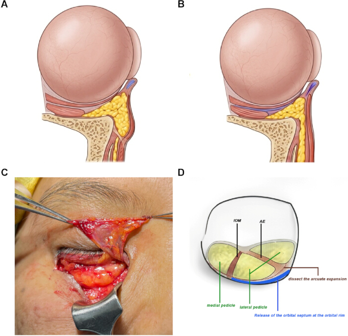
Figure 2: Schematic diagram of the key surgical steps. (A) Herniation of orbital fat. (B) Release of the tear trough ligament and tension-free repositioning of the fat pedicle into the tear trough and midfacial depression. (C) Intraoperative illustration of the orbital septal flap dissection. (D) Schematic showing the incision of the orbital septum at the arcus marginalis. Please click here to view a larger version of this figure.
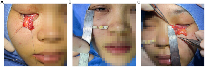
Figure 3: Intraoperative preparation and measurement of the orbital fat flap. (A) Adequate intraoperative exposure and mobilization of the pedicled orbital fat flap. (B) Surface measurement of the recipient site (tear trough depression) to determine the required length and extent of filling. (C) Precise intraoperative measurement of the dimensions (length and width) of the prepared orbital fat flap under direct vision, ensuring adequate, tension-free coverage and filling of the recipient site. Please click here to view a larger version of this figure.
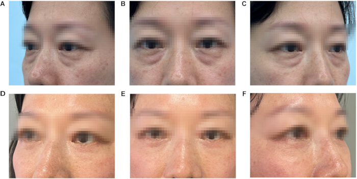
Figure 4: Typical case: A 49-year-old female patient who underwent the transcutaneous approach using an orbital septal fascial flap combined with extensive release of an elongated orbital fat flap. Preoperative and postoperative comparison. (A–C) Preoperative photographs (A: left oblique lateral view, B: anterior view, C: right oblique lateral view), showing obvious lower eyelid fat herniation (bags under the eyes) and tear trough depression deformity. (D–F) Corresponding photographs taken at 6 months postoperatively, showing a smooth lower eyelid contour, elimination of eye bags, and significant improvement of the tear trough depression. Please click here to view a larger version of this figure.
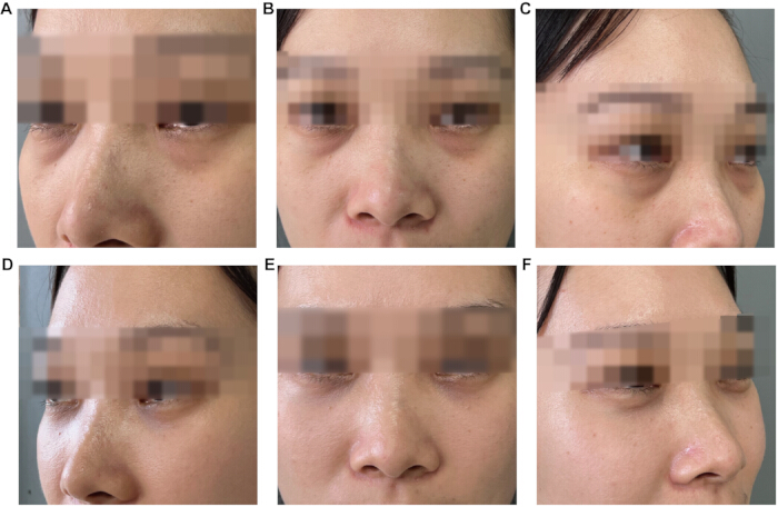
Figure 5: Typical case: A 39-year-old female patient who underwent the transconjunctival approach using an orbital septal fascial flap combined with extensive release of an elongated orbital fat flap. Preoperative and postoperative comparison. (A–C) Preoperative photographs (A: left oblique lateral view, B: anterior view, C: right oblique lateral view), showing obvious lower eyelid fat herniation (bags under the eyes) and tear trough depression deformity. (D–F) Corresponding photographs taken at 6 months postoperatively, showing a smooth lower eyelid contour, elimination of eye bags, and significant improvement of the tear trough depression. Please click here to view a larger version of this figure.
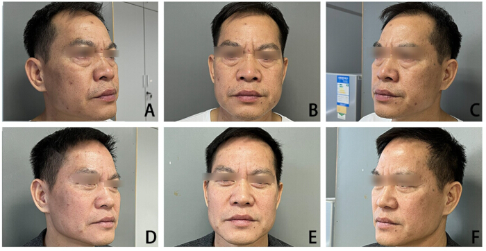
Figure 6: Long-term follow-up case: A 45-year-old male patient who underwent the transcutaneous approach using an orbital septal fascial flap combined with extensive release of an elongated orbital fat flap. Preoperative and 18-month postoperative comparison. (A–C) Preoperative photographs A: right oblique lateral view, B: anterior view, C: left oblique lateral view, showing prominent lower eyelid fat herniation, tear trough depression, and midfacial volume loss with associated skin laxity. (D–F) Corresponding photographs taken at 18 months postoperatively, showing sustained smoothing of the lower eyelid contour, elimination of eye bags, effacement of the tear trough depression, and stable midfacial volume restoration. Please click here to view a larger version of this figure.
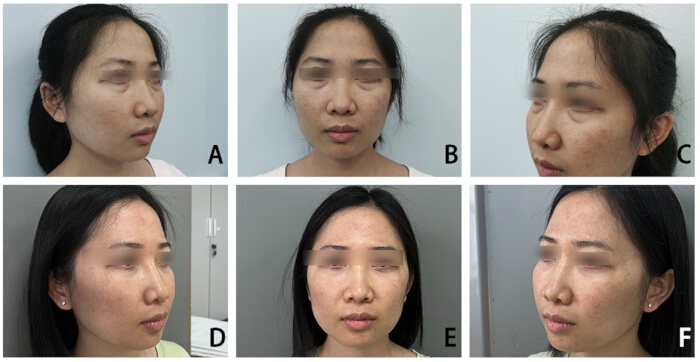
Figure 7: Long-term follow-up case: A 35-year-old female patient who underwent the transconjunctival approach using an orbital septal fascial flap combined with extensive release of an elongated orbital fat flap. Preoperative and 36-month postoperative comparison. (A–C) Preoperative photographs, A: right oblique lateral view, B: anterior view, C: left oblique lateral view, showing obvious lower eyelid fat herniation and tear trough depression deformity. (D–F) Corresponding photographs taken at 36 months postoperatively, showing durable smooth lower eyelid contour, elimination of eye bags, and significant improvement of the tear trough depression with stable long-term aesthetic outcomes. Please click here to view a larger version of this figure.
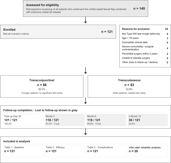
Figure 8: Study flow diagram. A total of 145 patients who underwent screening for the combined orbital septal fascial flap with extensive orbital fat release between December 2019 and March 2025 were assessed for eligibility; 24 were excluded for the reasons indicated, and 121 patients were enrolled. Patients were assigned to the transconjunctival (n = 68) or transcutaneous (n = 53) approach on the basis of presenting anatomy. Follow-up completion was recorded at postoperative day 30 (121/121, 100.0%), 3 months (118/121, 97.5%), 6 months (115/121, 95.0%), and ≥12 months (30/121, 24.8%); the maximum available follow-up was 36 months. The de-identified analytic dataset comprised all 121 patients for baseline, efficacy, and complication analyses, and 25 patients for inter-rater reliability analysis. Please click here to view a larger version of this figure.
| Parameters | Transconjunctival Approach Group(n = 68) | Transcutaneous Approach Group (n = 53) | Test Statistic | P-value |
| Gender, n (%) | PFisher = 1.000 | 1 | ||
| Female | 64 (94.1%) | 50 (94.3%) | ||
| Male | 4 (5.9%) | 3 (5.7%) | ||
| Age (y), M (P25, P75) | 28.0 (24.8, 33.0) | 45.0 (41.0, 49.0) | Z = -9.027 | < 0.001 |
| Preoperative Tear Trough Type, n (%) | Z = -9.914 | < 0.001 | ||
| Type II (Moderate) | 62 (91.2%) | 0 (0.0%) | ||
| Type III (Severe) | 6 (8.8%) | 53 (100.0%) | ||
| Fat Flap Length (cm), M (P25, P75) | 2.20 (2.10, 2.30) | 2.10 (2.00, 2.30) | Z = -1.087 | 0.278 |
| Operative Time (min), M (P25, P75) | 61.0 (58.0, 65.0) | 125.0 (120.0, 128.0) | Z = -9.424 | < 0.001 |
| Recovery Time (days), M (P25, P75) | 11.0 (9.0, 13.0) | 15.0 (14.0, 17.0) | Z = -6.467 | < 0.001 |
Table 1: Comparison of baseline characteristics and perioperative objective parameters between the two groups
| Parameters | Transconjunctival Approach Group (n = 68) | Transcutaneous Approach Group (n = 53) | Test Statistic (Z) | P-value |
| Final Postoperative Tear Trough Grading, n (%) | Z = -0.519 | 0.607 | ||
| Grade 0 (Complete effacement) | 61 (89.7%) | 49 (92.5%) | ||
| Grade I (Minimal residual) | 7 (10.3%) | 4 (7.5%) | ||
| Intra-group Pre- vs. Postoperative Comparison (P-value) | < 0.001 | < 0.001 | Wilcoxon Test | - |
Table 2: Evaluation of postoperative clinical morphological outcomes between the two groups.
| Type of Complication | Transconjunctival Approach Group (n = 68) | Transcutaneous Approach Group (n = 53) | Inter-group Fisher's Exact Test P-value |
| Overall complication rate | 1/68 (1.47%) | 2/53 (3.77%) | 0.581 |
| Local hematoma | 1/68 (1.47%) | 0/53 (0.0%) | 1 |
| Transient lower eyelid ectropion | 0/68 (0.0%) | 2/53 (3.77%) | 0.19 |
| Postoperative infection | 0/68 (0.0%) | 0/53 (0.0%) | - |
| Diplopia | 0/68 (0.0%) | 0/53 (0.0%) | - |
| Sensory nerve injury | 0/68 (0.0%) | 0/53 (0.0%) | - |
| Enophthalmos | 0/68 (0.0%) | 0/53 (0.0%) | - |
| Fat necrosis/displacement | 0/68 (0.0%) | 0/53 (0.0%) | - |
Table 3: Incidence of postoperative adverse reactions and complications between the two groups, n (%).
Discussion
In orbital fat release surgery, the selection of the prezygomatic space as the recipient site for fat transposition is primarily based on its unique anatomical advantages8. As a natural loose areolar space in the midface, its anatomical planes are distinct and easily separated via blunt dissection. Compared to restricted anatomical spaces such as the subperiosteal space, the suborbicularis oculi fat (SOOF) layer, the suborbicularis oculi layer, and the supraorbicularis oculi layer, the prezygomatic space provides a greater area for extension. This allows the "super-released" orbital fat flap to fully extend within it, providing a broad physical accommodating platform for correcting midface volume loss9. This plane is also referred to as the ideal "safe zone" for orbital fat flap transposition.
Restricted by the orbital septum and the distribution of vascular bundles, traditional techniques usually allow only the harvesting of a fat flap 1.5 cm to 2 cm in length during dissection. Although a flap of this length can improve tear trough deformity, it is difficult for it to reach the anterolateral maxillary region to address midface volume loss. This results in residual postoperative issues such as "cheek hollowing" or "insufficient midface volume"9. Previous studies have indicated that when the length of the fat flap extends beyond 2 cm, it can improve not only the tear trough but also midface depressions caused by maxillary aging. However, when the fat flap length is extended beyond 2 cm, the risk to its distal blood supply increases significantly. Traditional periosteal fixation methods often fail to effectively maintain distal nutritional supply, which may increase the risk of fat absorption or necrosis, a recognized challenge in clinical application. When managing the orbital septum fascia, our surgical technique opens the fascia at the orbital rim and reflects it superiorly. This anatomical strategy not only exposes the inferior oblique muscle that is originally hidden beneath the orbital septum, but also allows the surgeon to directly visually assess the specific volume of herniated orbital fat. This may reduce the risk of accidental injury associated with blind release3. No postoperative diplopia was recorded in the 121 cases; however, ocular motility and diplopia were not assessed with a standardized protocol, so this should be interpreted with caution. Compared to traditional septal fat repositioning techniques, this method, guided by the "super-release" concept, is intended to improve the precision of the release extent through clear visual feedback.
Furthermore, the orbital septum fascia is dissected and trimmed to form a pedicled, fan-shaped fascial flap, which is then folded over to cover the surface of the elongated fat flap. Because the fascial flap is vascularized, we hypothesize that it may provide additional microcirculatory support to the distal end of the fat flap7 and thereby reduce the risk of distal ischemic necrosis; consistent with the principle that engineered microvascular networks can sustain perfusion under controlled fluidic conditions10, this proposed "biological dressing" effect was not directly verified in the present study and remains a hypothesis. The fascial flap also provides physical wrapping and, through its flexibility and tensile properties, may help restrain the movement of the fat flap, potentially reducing fat flap retraction or displacement caused by gravity or the pull of facial expression muscles11. It may also counteract the negative pulling forces brought about by postoperative scar contracture12.
In the cases of this study, comparing preoperative and postoperative photographs with a follow-up of 6 to 12 months, no severe complications were recorded. The tear trough deformity was substantially improved in the great majority of cases. In terms of postoperative structural stability and mechanical support, the fascial flap is proposed to function as an internal supportive layer12. Traditional orbital fat release surgery primarily focuses on release and filling, and can be regarded as a "single-tissue transposition" technique. Our surgical technique converts this into a "composite fat-fascia tissue flap" transfer, aiming to address the three practical challenges of blood supply, fixation, and graft survival13, while achieving simultaneous tear trough correction and midface volume restoration7. Such objectives—albeit in anatomically distinct reconstructive settings—have also motivated broader tissue- engineering strategies employing biological matrices to support tissue integration14.
Viewed against established fat-transposition techniques, the present composite flap is an incremental extension rather than a new paradigm. The SOFT procedure first used the prezygomatic space as a low-morbidity plane for transconjunctival pedicled fat transposition with a >2 cm flap7, a concept echoed by the "super-released" orbital fat technique9, extended transconjunctival release of the tear-trough and orbicularis-retaining ligaments12, and the orbital septal fat flap13. This method adopts the same prezygomatic recipient plane and an elongated (>2 cm) pedicled flap, but adds a vascularized orbital septal fascial flap for mechanical wrapping and, hypothetically, adjunctive microcirculation—distinguishing it from fat-only repositioning7,9,12,13 and from internal fixation such as guiding-needle-assisted redistribution, which anchors orbital fat to deep periosteum through a buried guiding needle without transferring a fascial component15. The fascial flap's putative vascularization and anti-displacement benefits remain unverified histologically or radiologically. Because this single-center, retrospective, non-randomized series assigned the surgical approach by each patient's presenting anatomy rather than by randomization, it is susceptible to selection bias: transcutaneous patients were older and presented with more advanced (Type III) deformities and skin laxity, whereas transconjunctival candidates were younger with milder disease, and no concurrently enrolled comparator7,15 was available. Thus, any apparent advantage of the composite flap over existing methods cannot be inferred from this study, and the comparative interpretation above should be regarded as hypothesis-generating, requiring prospective, multi-center, randomized validation with standardized long-term follow-up.
Since 2019, our team has performed 121 procedures using this composite technique. As a retrospective, single-center study, several limitations must be acknowledged. The two approach groups differed substantially at baseline and were not randomized; the surgical approach was selected on the basis of the patient's presenting anatomy, so between-group comparisons reflect different clinical populations and may be affected by selection bias, and no adjustment for baseline differences was performed. The definition of "recovery time" relied partly on the subjective judgment of return to a "normal" periorbital appearance. The sample size was 121 cases, but there was no prospective randomized comparison with other mainstream procedures, such as simple fat repositioning or fat grafting, so the level of evidence is limited. The follow-up period is insufficient to assess long-term durability; although the longest available postoperative photographs in this cohort extend to 36 months, standardized comparative photographs at ≥ 2 years were not available for the majority of patients, and longer structured follow-up is planned. Finally, the hypothesis that fascial-flap coverage supports postoperative blood supply is based on anatomical reasoning and clinical observation and lacks direct imaging or histological confirmation; such objective verification is a priority for future research.
In conclusion, the orbital septal fascial flap combined with extensive release of an elongated orbital fat flap is a technique intended to address three practical limitations of conventional orbital fat transposition: limited transposition distance, distal blood supply, and fixation. In this single-center retrospective cohort, the technique was associated with a high rate of complete tear trough effacement (92.5% and 89.7% in the two groups) and a low complication rate over 6–12 months of follow-up. Because this was an unadjusted, retrospective comparison of two clinically distinct groups, these findings should be regarded as preliminary and hypothesis-generating. They require validation through prospective, multi-center, controlled studies, longer-term follow-up, standardized patient-reported outcomes, and direct histological or imaging-based verification of the proposed vascularization mechanism.
Disclosures
The authors declare that there are no potential conflicts of interest with respect to the research, authorship, and publication of this article.
Acknowledgements
We sincerely thank the Medical Ethics Committee of The First Affiliated Hospital of Guangxi Medical University for the ethical review and official approval of this study (Approval No.: 2026-E0435; approved 13 May 2026). We are grateful to all enrolled patients for their voluntary participation and valuable cooperation in this clinical research. We also acknowledge the support from the Department of Medical Cosmetology.
Materials
| Name | Company | Catalog Number | Comments |
|---|---|---|---|
| 5-0 PDS absorbable suture | Johnson Medical Shanghai | W9073H | Periosteal fixation of fat flap |
| 6-0 nylon suture | Ethicon LLC | J570G | Temporary external fixation of fat flap distal end |
| Canon EOS 90D Digital Camera | Canon Inc., Tokyo, Japan | EOS 90D | Standardized clinical photography for preoperative and postoperative documentation |
| Eyelid retractor | Shinva Surgical Instruments | ZO14602T | Exposure of orbital rim and surgical field |
| Lidocaine hydrochloride injection 2% | Sinopharm Rongsheng | H20023546 | Local anesthesia |
| Methylene blue injection | Jichuan Pharmaceutical Group | H32024781 | Preoperative skin marking and redundant skin demarcation |
| Mosquito forceps | Shinva Surgical Instruments | ZO14602R | Blunt dissection along the preseptal space |
| Needle-tip electrocautery | Kangjia Medical Devices | 20152010742 | Sharp tissue dissection and intraoperative hemostasis |
| Nerve dissector | Shinva Surgical Instruments | A2618K | Release of tear trough ligament and dissection of prezygomatic space |
| Ofloxacin eye drops | Unither Nanjing | Q4835N | Postoperative anti-infective prophylaxis |
| Ophthalmic scissors | Henan Dongken Medical Technology Co., Ltd. | 20250007 | Skin flap undermining and redundant skin excision |
| Petrolatum (Vaseline) gauze | Zhende Medical | M1509V | External fixation bolster |
| Povidone-iodine solution | Yuang Technology | S7142P | Surgical site disinfection |
| R software (version 4.3.0; irr package) | R Foundation for Statistical Computing, Vienna, Austria | Open-source v4.3.0 | Inter-rater reliability analysis |
| SPSS Statistics (version 26.0) | IBM Corp., Armonk, NY, USA | - | Statistical analysis |
References
- Christodoulou S, Tzamalis A, Tsinopoulos I, Ziakas N. Surgical and non-surgical approach for tear trough correction: Fat repositioning versus hyaluronic acid fillers. J Pers Med. 2024;14(11):1096.
- Wong CH, Hsieh MKH, Mendelson B. The tear trough ligament: anatomical basis for the tear trough deformity. Plast Reconstr Surg. 2012;129(6):1392-1402.
- Bhattacharjee K, Ghosh S, Ugradar S, Azhdam AM. Lower eyelid blepharoplasty: An overview. Indian J Ophthalmol. 2020;68(10):2075-2083.
- Loeb R. Fat pad sliding and fat grafting for leveling lid depressions. Clin Plast Surg. 1981;8(4):757-776.
- Hamra ST. Arcus marginalis release and orbital fat preservation in midface rejuvenation. Plast Reconstr Surg. 1995;96(2):354-362.
- Barton FE Jr, Ha R, Awada M. Fat extrusion and septal reset in patients with the tear trough triad: a critical appraisal. Plast Reconstr Surg. 2004;113(7):2115-2123.
- Jin Y, Gao W, Teo HMT, Lin X. Transconjunctival lower blepharoplasty using midcheek spaces for orbital fat transposition (S.O.F.T.). Ann Plast Surg. 2021;86(6):620-626.
- Cotofana S, Lachman N. Anatomy of the facial fat compartments and their relevance in aesthetic surgery. J Dtsch Dermatol Ges. 2019;17(4):399-413.
- Huang S, Xiao H, Hu H, Cen Y. Transconjunctival lower eyelid blepharoplasty with "super released" orbital fat in correction of tear trough and palpebromalar groove depression. Zhongguo Xiu Fu Chong Jian Wai Ke Za Zhi. 2023;37(6):713-716.
- Yue T, Yang H, Wang Y, Jiang N, Yin H, Lu X, Liu N, Xu Y. A Vascularized Multilayer Chip Reveals Shear Stress-Induced Angiogenesis in Diverse Fluid Conditions. Cyborg Bionic Syst. 2025;6:Article 0207. https://doi.org/10.34133/cbsystems.0207
- Alghoul M. Blepharoplasty: Anatomy, planning, techniques, and safety. Aesthet Surg J. 2019;39(1):10-28.
- Wong CH, Mendelson B. Extended transconjunctival lower eyelid blepharoplasty with release of the tear trough ligament and fat redistribution. Plast Reconstr Surg. 2017;140(2):273-282.
- Huahui Z, Yuxiang Z, Zhihao L, et al. A new method of orbital septal fat flap in transconjunctival approach. Aesthetic Plast Surg. 2025;49(4):1065-1073.
- Wang T, Jiang M, Wu S, Zhang K, Del Rey RR, Wei R, Rodríguez RA. Efficacy of decellularized extracellular matrix (dECM) for articular cartilage repair in osteoarthritis (OA): a systematic review and meta-analysis. J Orthop Surg Res. 2025 May 16;20(1):467. doi: 10.1186/s13018-025-05881-2. PMID: 40380305; PMCID: PMC12083026.
- Wu K, Hou J, Chen J, et al. Guiding needle-assisted redistribution of orbital fat in transconjunctival lower eyelid blepharoplasty. J Cosmet Dermatol. 2025;24(9):e70446.