This study demonstrated that LUS and serum miR-486-3p were elevated in neonates with acute respiratory distress syndrome (ARDS). Their combined assessment improved the prediction of disease severity and prognosis, and miR-486-3p was found to regulate ARDS model cell function by targeting Delta-Like Canonical Notch Ligand 4.
Research Article
Prognostic Value of Lung Ultrasound Score and Serum miR-486-3p in Neonatal Acute Respiratory Distress Syndrome
* These authors contributed equally
In This Article
Summary
Abstract
This study aimed to assess the prognostic value of the lung ultrasound score (LUS) and serum miR-486-3p in neonatal acute respiratory distress syndrome (ARDS) and to explore the underlying mechanism. A total of 112 neonates with ARDS and 100 healthy infants born during the same period were enrolled in the study and healthy control groups, respectively. Each neonate underwent lung ultrasound within 6 h of admission to the intensive care unit and was evaluated for LUS. Real-time quantitative PCR was used to quantify gene expression, while receiver operating characteristic (ROC) curve and Pearson's correlation analyses were performed to evaluate prognostic performance and correlations. Furthermore, an in vitro ARDS model was established using HPMECs to investigate the underlying mechanism of miR-486-3p. A dual-luciferase reporter assay was performed to verify the target relationship between miR-486-3p and Delta-Like Canonical Notch Ligand 4 (DLL4). The results revealed that LUS and serum miR-486-3p were significantly upregulated in neonates with ARDS. ROC curve analysis showed that both miR-486-3p and LUS could predict the neonatal prognosis of ARDS (AUC = 0.883 and 0.873, sensitivity: 96.4% and 89.3%, specificity: 77.4% and 73.8%, respectively), and miR-486-3p combined with LUS had a better prediction effect (AUC = 0.929, sensitivity: 92.9%, specificity: 84.5%). Mechanistically, miR-486-3p targeted DLL4 to regulate the biological functions of ARDS model cells. Taken together, the combined assessment of LUS and serum miR-486-3p may serve as a promising approach for evaluating disease severity and predicting prognosis in neonates with ARDS. Further multi-center validation is still needed to confirm its general applicability.
Introduction
Neonatal acute respiratory distress syndrome (ARDS) is a life-threatening critical respiratory disorder primarily characterized by impaired alveolar surfactant function, which poses great risks to neonatal survival1,2. Despite the development of various treatment modalities, the overall prognosis for affected neonates remains unsatisfactory3. Early identification of disease severity and accurate prognostic assessment are therefore essential to optimize clinical interventions and improve patient outcomes. Once ARDS develops, neonatal pulmonary function is severely impaired, accompanied by disrupted ventilation and gas exchange4. Lung ultrasound can be considered the primary imaging method for monitoring ventilation in ARDS5. It has become a preferred bedside imaging tool for monitoring pulmonary lesions in ARDS, and the lung ultrasound score (LUS) has been widely applied to diagnose neonatal acute respiratory diseases and evaluate disease severity and prognosis in critical care settings6. However, single imaging or laboratory testing has inherent defects and cannot fully meet the clinical assessment demand7. Therefore, exploring novel biomarkers to establish a combined evaluation system is of great clinical significance.
MicroRNA (miRNA) represents a novel class of gene regulatory factors that play a pivotal role in the pathogenesis of ARDS8. For instance, downregulated miR-141-3p is closely associated with disease severity in pulmonary fibrosis and lung injury9. miR-124-3p is a protective factor for ARDS by targeting p6510. Previous high-throughput sequencing studies have confirmed that miR-486-3p is significantly upregulated in patients with ARD11. Recent research has highlighted the hsa_circ_0006892/miR-486-3p axis as a potential molecular mechanism implicated in the development of ARDS12. These findings suggest that miR-486-3p is closely linked to ARDS. Nonetheless, the prognostic and predictive value of miR-486-3p regarding the severity and outcome of neonatal ARDS remains to be fully elucidated.
Currently, chest radiography, blood gas analysis, and single biomarker detection are the mainstream methods for evaluating neonatal ARDS. Chest imaging involves radiation exposure and cannot provide real-time, repeated monitoring. Blood gas analysis reflects only acute respiratory function and lacks predictive value for long-term prognosis. Single microRNA detection provides objective quantitative results but cannot reflect real-time morphological changes of lung tissue. Compared with these conventional approaches, the combination of LUS and serum miR-486-3p integrates the advantages of bedside imaging and molecular detection.
This study focused on neonates who met ARDS criteria rather than on classic surfactant-deficiency RDS. Thus, this study seeks to assess whether baseline LUS and serum miR-486-3p predict short-term mortality in neonatal ARDS. This research aims to provide valuable data to enhance clinical assessment of disease conditions and short-term prognostic evaluations in neonatal ARDS.
Protocol
This study received ethical approval from the Medical Ethics Committee of The First People's Hospital of Yongkang, in strict adherence to the Declaration of Helsinki.
Subjects
This single-center, observational cohort study with an in vitro experimental component was conducted between January 2022 and December 2023. A total of 112 consecutive neonates diagnosed with ARDS were enrolled at The First People's Hospital of Yongkang. For comparative purposes, healthy infants born during the same timeframe, with no history of asphyxia or intrauterine distress, were recruited as control subjects.
Inclusion criteria:
All participants met the diagnostic criteria for neonatal acute respiratory distress syndrome as outlined in the Montreux guidelines (2017 edition). The diagnosis was substantiated by imaging and clinical assessments: X-ray examinations revealed diffuse opacities in both lungs consistent with pulmonary edema, and echocardiography showed no evidence of left atrial hypertension, thereby excluding cardiogenic pulmonary edema. Additional criteria included acute onset of symptoms and the need for mechanical ventilation for more than 3 days, with no severe extrapulmonary infections. Informed consent was duly obtained from the families of the neonates.
Exclusion criteria:
Neonates with primary alveolar surfactant deficiency, congenital heart disease, metabolic disorders, or other significant underlying conditions were excluded. Furthermore, infants with lung or chest wall malformations or other major congenital anomalies were excluded. Neonatal RDS was excluded.
Clinical data
Gestational age, sex, birth weight at admission, and maternal characteristics were documented for all participants. Each neonate underwent lung ultrasound within 6 h of admission to the intensive care unit and was evaluated for LUS. The LUS was scored on a scale with a maximum of 36 points, whereby a higher score indicated greater severity of pulmonary symptoms. Blood samples were collected immediately upon admission as part of the initial clinical assessment and prior to any major therapeutic intervention (e.g., surfactant re-dosing, escalation of ventilation mode, or initiation of vasoactive agents). Blood samples from healthy neonates were obtained at the time of their enrollment. For serum isolation, whole blood was placed in serum separator tubes and kept stationary at room temperature for 30 min to allow complete clot formation. Samples were then centrifuged at 1500 x g for 15 min at 4 °C. The upper clear serum supernatant was carefully aspirated, aliquoted into sterile cryogenic vials, and immediately stored in an ultra-low-temperature freezer at -80 °C for subsequent experiments. Only clear, non-hemolyzed serum samples were used for subsequent experiments. Used blood collection tubes and residual blood were discarded as medical biological waste in accordance with institutional regulations.
Grouping
All neonates were categorized into two distinct groups: the mild group (grades I–II) and the severe group (grades III–IV), based on the initial chest X-ray findings and the severity of the condition assessed 24 h post-admission. Chest X-ray interpretations revealed grade I, characterized by decreased lung transparency bilaterally, with small particles and reticular shadows within the lung parenchyma. Grade II indicated that the lesions extended into the middle and outer lung zones, with evidence of the air bronchogram sign. In grade III, there was a significant reduction in transparency across both lungs, resulting in obscured margins of the heart and diaphragm. Grade IV was marked by pronounced air bronchogram signs, culminating in a near-total opacification resembling a white lung appearance.
Neonates diagnosed with ARDS received a comprehensive management regimen following their admission to the neonatal intensive care unit. This regimen encompassed mechanical ventilation, sedation, pulmonary surfactant administration, anti-inflammatory therapies, nutritional support, expectoration assistance, and fluid replacement measures. The neonatal diagnosis of ARDS served as the starting point for the study. The endpoints were defined as either successful patient recovery and discharge or patient demise resulting from ineffective treatment. Based on clinical outcomes, participants were classified into two groups: the survival group and the death group.
Cell culture and treatment
Human pulmonary microvascular endothelial cells (HPMECs) were cultured in DMEM supplemented with 10% fetal bovine serum, 100 U/mL penicillin, and 100 µg/mL streptomycin. Cells were maintained at 37 °C in a humidified incubator containing 5% CO₂. To establish an ARDS cell model, the cells were treated with lipopolysaccharide (LPS) at a concentration of 1 mg/L13,14. Successful model establishment was confirmed by observing slight cellular shrinkage under an inverted microscope following LPS treatment. Following completion of the experiments, spent culture medium, dead cells, and disposable culture materials were discarded as biologically hazardous waste.
Cell transfection and grouping
HPMECs were seeded into culture plates and incubated until 70% confluence before transfection. Transfection complexes were prepared according to the manufacturer's instructions. Cells were divided into groups: blank control, miR-486-3p mimic, mimic negative control, miR-486-3p inhibitor, inhibitor negative control, miR-486-3p mimic + DLL4 overexpression plasmid (OE-DLL4), and miR-486-3p mimic + empty overexpression plasmid (OE-NC). Transfection complexes were added to the corresponding wells and incubated for 4–6 h. The medium was replaced with fresh complete medium, and culturing was continued for 24–48 h for subsequent experiments. Successful transfection was indicated by stable cell confluence without excessive cell death.
Real-time quantitative PCR
Total RNA was extracted from serum samples and cultured cells using an RNA extraction reagent. RNA concentration and purity were measured using a spectrophotometer, and samples with an A260/A280 ratio of 1.8–2.1 were considered suitable for further analysis. Complementary DNA (cDNA) was synthesized using a reverse transcription kit. Quantitative PCR was subsequently performed using a real-time PCR system with the following cycling conditions: an initial denaturation at 95 °C for 3 min, followed by 40 cycles of 95 °C for 10 s and 60 °C for 30 s. For microRNA detection, the relative expression of miR-486-3p was calculated using the 2-ΔΔCt method with U6 snRNA serving as the internal reference. For mRNA quantification, DLL4 expression was normalized to GAPDH mRNA by the identical 2-ΔΔCt algorithm. The primer sequences were listed as follows: miR-486-3p forward: 5'-GTATGACGGGGCAGCTCAGTA-3' and miR-486-3p reverse: 5'-CAGTGCGTGTCGTGGAGT-3', U6 forward: 5'-GGAACGATACAGAGAAGATTAGC-3' and U6 reverse: 5'-TGGAACGCTTCACGAATTTGCG-3'. DLL4 forward: 5'-AGGTGCCACTTCGGTTACAC-3' and DLL4 reverse: 5'-GGGAGAGCAAATGGCTGATA-3', GAPDH forward: 5'-GCAAATTCCATGGCACCGT-3' and GAPDH reverse: 5'-TCGCCCCACTTGATTTTGG-3'.
CCK-8 cell proliferation assay
Transfected cells were seeded into 96-well plates at a density of 1 × 10³ cells per well and incubated for 24, 48, and 72 h. The culture medium was then aspirated, and the cells were washed twice with 1× PBS. Subsequently, 100 µL of fresh 1× PBS and 10 µL of CCK-8 solution were added to each well, and the plates were incubated at 37 °C for 2 h. Absorbance was measured at 450 nm using a microplate reader, and cell proliferation was evaluated based on the absorbance values. Successful assay performance was indicated by time-dependent changes in absorbance among the experimental groups.
Flow cytometry apoptosis assay
Cells were digested with 0.25% trypsin, and the resulting cell suspension was collected and centrifuged at 1500 × g for 5 min at 4 °C. The supernatant was discarded, and the cell pellets were washed three times with pre-cooled 1× PBS. The cells were then resuspended in 500 µL of 1x Binding Buffer, followed by the addition of 5 µL Annexin V-FITC and 5 µL propidium iodide (PI). After gentle mixing, the samples were incubated for 40 min at room temperature in the dark. Cell apoptosis was measured by flow cytometry, and the data were analyzed with the appropriate flow cytometry analysis software. Successful assay performance was indicated by a low apoptosis rate in the normal control group and a marked increase in apoptosis in the ARDS model group.
Luciferase reporter assay
Potential target genes of miR-486-3p were identified using the online TargetScan database (http://www.targetscan.org/vert_72/). Following identification of the predicted binding sites, wild-type (DLL4-WT) and mutant (DLL4-MUT) luciferase reporter plasmids containing the corresponding binding sequences were constructed. The DLL4-WT and DLL4-MUT vectors were co-transfected into HPMECs together with miR-486-3p mimics or inhibitors, respectively. Relative luciferase activity was subsequently measured using a dual-luciferase reporter assay kit. Bioluminescence signals were detected with a luminometer according to the manufacturer's instructions. Successful target validation was indicated by a significant reduction in luciferase activity in the DLL4-WT group following miR-486-3p overexpression.
Detailed information on all reagents, consumables, and laboratory equipment used in this study was provided in the accompanying Table of Materials.
Statistical methods
Statistical analyses and data visualizations were performed utilizing SPSS version 26.0 and GraphPad Prism version 9.0. Measurement data were presented as mean ± standard deviation, with comparisons between the means of two independent samples assessed using an independent t-test. For comparisons across three or more groups, one-way analysis of variance (ANOVA) and repeated-measures ANOVA were adopted, followed by Tukey’s post-hoc multiple comparison test to identify intergroup differences. Categorical data were expressed as counts (percentages), and intergroup comparisons were analyzed employing the chi-square test. The Wilcoxon Mann-Whitney test was applied for non-parametric data. Additionally, the ROC curve was constructed to evaluate the predictive value of serum miR-486-3p expression levels and LUS in determining mortality in pediatric patients with ARDS. Pearson's correlation analysis was used to examine correlations. All experimental groups had three independent biological replicates and technical replicates. The statistical unit was defined as an independent experiment, with each biological replicate representing a separate cell culture batch derived from different passages, and technical replicates were used to reduce intra-experimental variation. Data from technical replicates were averaged first, and then statistical analysis was performed based on the mean values of biological replicates. Statistical significance was set at p < 0.05.
Results
Comparable baseline data among groups
The general characteristics of the infants included in the study are presented in Table 1. There were no significant differences in baseline demographic characteristics between the two groups: gestational age (36.07 ± 2.71 vs. 35.72 ± 2.64 weeks, p = 0.347), male sex proportion (59.0% vs. 66.1%, p = 0.290), birth weight (3.39 ± 0.57 vs. 3.33 ± 0.55 kg, p = 0.437), maternal age (29.18 ± 4.00 vs. 29.66 ± 4.05 years, p = 0.387), and delivery method (vaginal: 54.0% vs. 58.0%; cesarean: 46.0% vs. 42.0%, p = 0.554), indicating that these groups were comparable. Based on chest X-ray evaluation, in the ARDS group, 72 cases (64.3%) were classified as mild (grade I-II), while 40 cases (35.7%) were classified as severe (grade III-IV).
ARDS severity correlates with patient prognosis
The comparative analysis of survival outcomes between the group of children with ARDS who survived and those who succumbed is presented in Table 2. Among the 112 neonates diagnosed with ARDS, 84 cases (75%) survived while 28 cases (25%) resulted in mortality. Notably, there were no statistically significant differences in infant age, sex, birth weight, maternal age, mode of delivery, amniotic fluid characteristics, etiology, or other variables between the two groups (p > 0.05). However, a significant difference in ARDS severity was observed (p < 0.01), highlighting a critical prognostic factor.
miR-486-3p and LUS Are Elevated in ARDS. RT-qPCR analysis revealed that serum miR-486-3p levels were significantly elevated in neonates with ARDS compared with healthy controls. Moreover, within the ARDS cohort, the severe group had strikingly higher levels than the mild group (P < 0.001) (Figure 1A). Furthermore, LUS was higher in the severe ARDS group than in the mild ARDS group (p < 0.001) (Figure 1B). When dividing ARDS patients by clinical outcome, the death group showed markedly higher LUS and miR-486-3p levels than the survival group (Figure 1C,D). A strong positive correlation was found between LUS and serum miR-486-3p in non-surviving patients (r = 0.832, p <0.001) (Figure 1E).
Combined detection improves prognostic prediction
Based on the ROC analysis protocol, we evaluated the prognostic performance of single and combined indicators. The AUC values of LUS and miR-486-3p for predicting short-term mortality were 0.873 (95% CI: 0.809-0.936) and 0.883 (95% CI: 0.822-0.944), respectively. The sensitivity values were 89.3% and 96.4%, while the specificity values were 73.8% and 77.4%, respectively. The optimal cut-off values for LUS and miR-486-3p were 15.500 and 1.465, respectively. Furthermore, the AUC for the combined predictive model was 0.929 (95% CI: 0.883-0.975), demonstrating a sensitivity of 92.9% and a specificity of 84.5% with the optimal cut-off value of 1.659 (Figure 1F).
miR-486-3p upregulation impairs cell function
Following the cell culture and LPS intervention protocol, we constructed an in vitro ARDS cell model using HPMECs. qPCR results showed that miR-486-3p expression increased gradually over time after LPS stimulation (Figure 2A). miR-486-3p was overexpressed or knocked down to investigate its effects on ARDS model cells (Figure 2B). In line with the CCK-8 and flow cytometry assay protocols, cell proliferation and apoptosis were detected. miR-486-3p overexpression significantly suppressed cell proliferation and promoted cell apoptosis. Conversely, miR-486-3p knockdown alleviated LPS-induced cell damage (Figure 2C,D).
miR-486-3p downregulates DLL4 expression
According to the plasmid construction and dual-luciferase reporter assay protocol, we verified the interaction between miR-486-3p and DLL4. Bioinformatics prediction confirmed a conserved binding site between miR-486-3p and the 3’UTR of DLL4 (Figure 3A). The luciferase activity of the DLL4 wild-type group was significantly reduced after miR-486-3p overexpression, while no obvious change was observed in the mutant group (Figure 3B), proving that DLL4 is a direct target gene of miR-486-3p. In cell models, DLL4 expression decreased in a time-dependent manner after LPS treatment (Figure 3C). Manipulating miR-486-3p expression also negatively regulated DLL4 levels (Figure 3D).
In clinical samples, DLL4 expression was lower in ARDS neonates than in healthy controls, and further decreased in the death group (Figure 3E–F). A negative correlation between miR-486-3p and DLL4 was observed in non-surviving patients (r = -0.795, P < 0.001) (Figure 3G). All qPCR tests for DLL4 were performed with three technical replicates, and the results were stable.
DLL4 reverses miR-486-3p-mediated cell injury
Following the co-transfection protocol using the miR-486-3p mimic and the DLL4 overexpression plasmid, we carried out rescue experiments. qPCR confirmed that DLL4 expression was effectively restored after OE-DLL4 transfection (Figure 4A). Functional tests showed that overexpression of DLL4 partially reversed the inhibitory effect of miR-486-3p on cell proliferation and its pro-apoptotic effect (Figure 4B,C). These results indicated that miR-486-3p partially modulates cell activity by targeting DLL4.
Data Availability:
All data generated or analyzed during this study are provided as Supplementary File 1.
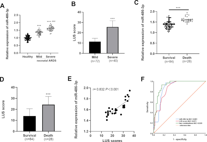
Figure 1: Comparison of LUS and serum miR-486-3p level between groups. (A) Serum miR-486-3p levels in healthy neonates and ARDS neonates. (B) LUS values in healthy neonates and ARDS neonates. (C) Serum miR-486-3p levels in the survival group and the death group. (D) LUS values in the survival group and the death group. (E) Correlation analysis between serum miR-486-3p and LUS in non-surviving ARDS neonates. (F) ROC curves of single LUS, single miR-486-3p, and combined detection for predicting neonatal ARDS prognosis. All quantitative data are presented as mean ± standard deviation; error bars represent standard deviation. *** = p <0.001, Compared with the healthy group; ### = p <0.001, Compared with mild ARDS group. Please click here to view a larger version of this figure.
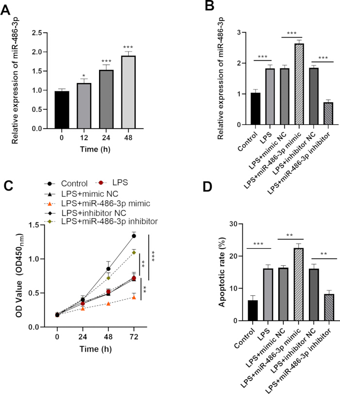
Figure 2: Effect of miR-486-3p on ARDS model cells. (A) Time-dependent changes of miR-486-3p expression in HPMECs after LPS treatment. (B) Relative expression of miR-486-3p after overexpression and knockdown intervention. (C) Cell proliferation activity detected by CCK-8 assay after altering miR-486-3p expression. (D) Cell apoptosis rate detected by flow cytometry after altering miR-486-3p expression. All data are presented as mean ± standard deviation; error bars represent standard deviation. Statistical markers: * = p <0.05, ** = p <0.01, *** = p <0.001. Please click here to view a larger version of this figure.
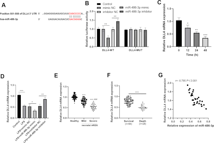
Figure 3: Verification of the targeting relationship between miR-486-3p and DLL4. (A) Schematic diagram of the predicted binding site between miR-486-3p and the 3’UTR of DLL4. (B) Dual luciferase reporter gene assay confirmed the targeting relationship between miR-486-3p and DLL4. (C) Dynamic changes of DLL4 expression in HPMECs after LPS treatment. (D) DLL4 expression after miR-486-3p overexpression or knockdown. (E) Serum DLL4 levels in healthy neonates and ARDS neonates. (F) DLL4 levels in the survival group and the death group of ARDS neonates. (G) Correlation analysis between serum miR-486-3p and DLL4 in non-surviving ARDS neonates. All quantitative data are shown as mean ± standard deviation; error bars represent standard deviation. Statistical markers: * = p <0.05, ** = p <0.01, *** = p <0.001. versus the healthy group, ### = p <0.001, versus the mild ARDS group. Please click here to view a larger version of this figure.
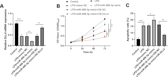
Figure 4: Effect of miR-486-3p targeting DLL4 on ARDS model cells. (A) Detection of DLL4 expression after co-transfection of miR-486-3p mimic and DLL4 overexpression plasmid. (B) Changes in cell proliferation activity after DLL4 rescue. (C) Changes in cell apoptosis rate after DLL4 rescue. All data are presented as mean ± standard deviation; error bars represent standard deviation. Statistical markers:* = p <0.05, ** = p <0.01, *** = p <0.001. Please click here to view a larger version of this figure.
| Variable | Healthy (n=100) | Neonatal ARDS (n=112) | p-value |
| Gestational age (weeks) | 36.07 ± 2.71 | 35.72 ± 2.64 | 0.347 |
| Sex (male, %) | 59 (59.0 %) | 74 (66.1 %) | 0.29 |
| Birth weight (kg) | 3.39 ± 0.57 | 3.33 ± 0.55 | 0.437 |
| Maternal age (years) | 29.18 ± 4.00 | 29.66 ± 4.05 | 0.387 |
| Delivery method | 0.439 | ||
| Vaginal | 54 (54.0 %) | 65 (58.0 %) | |
| Cesarean | 46 (46.0 %) | 47 (42.0 %) | |
| ARDS grades | --- | ||
| I-II | --- | 72 (64.3 %) | |
| III-IV | --- | 40 (35.7 %) |
Table 1: Baseline characteristics of the included infants. This table summarizes the baseline demographic information of healthy neonates and neonates with ARDS. Data are presented as mean ± standard deviation for continuous variables, and number (percentage) for categorical variables. P-values indicate between-group statistical differences. Abbreviations: ARDS = acute respiratory distress syndrome.
| Variable | Survival (n=84) | Death (n=28) | p-value |
| Gestational age (weeks) | 35.95 ± 2.58 | 35.04 ± 2.76 | 0.112 |
| Sex (male, %) | 53 (63.1 %) | 21 (75.0%) | 0.253 |
| Birth weight (kg) | 3.37 ± 0.58 | 3.23 ± 0.42 | 0.248 |
| Maternal age (years) | 29.49 ± 3.82 | 30.18 ± 4.74 | 0.438 |
| Delivery method | 0.444 | ||
| Vaginal | 47 (56.0 %) | 18 (64.3 %) | |
| Cesarean | 37 (44.0 %) | 10 (35.7 %) | |
| Abnormal amniotic fluid | 6 (9.5 %) | 4 (14.3 %) | 0.485 |
| ARDS grades | 0.001** | ||
| I-II | 61 (72.6 %) | 11 (39.3 %) | |
| III-IV | 23 (27.4 %) | 17 (60.7%) | |
| Etiology | 0.827 | ||
| Choking | 33 (39.3 %) | 12 (42.9 %) | |
| Pneumonia | 17 (20.2 %) | 6 (21.4 %) | |
| Sepsis | 12 (14.3 %) | 2 (7.1 %) | |
| Meconium aspiration syndrome | 22 (26.2 %) | 8 (28.6 %) |
Table 2: Comparison of clinical data between the survival group and the death group in ARDS neonates. This table compares clinical indicators between surviving and non-surviving ARDS neonates. Continuous data are shown as mean ± standard deviation, and categorical data are expressed as number (percentage). p-values indicate statistical significance between the two groups. ** = p<0.01. Abbreviations: ARDS = acute respiratory distress syndrome.
Supplementary File 1: Raw clinical and experimental datasets supporting the findings of this study, including individual-level measurements for lung ultrasound score (LUS), serum miR-486-3p expression, DLL4 expression, and cell-based assay results.Please click here to download this file.
Discussion
Neonatal ARDS is a leading cause of respiratory failure and early mortality among critically ill neonates15. Early and accurate evaluation of disease severity and prognosis is the prerequisite for individualized intervention and mortality reduction. A growing body of evidence confirms that microRNAs participate in the pathological progression of ARDS and serve as promising circulating biomarkers16. Research indicates that miR-155 is markedly elevated in septic patients suffering from acute lung injury/ARDS17. Additionally, studies have revealed an increase in serum miR-629-5p levels among children diagnosed with ARDS18. Serum miR-34a is closely related to the severity and prognosis of neonatal RDS19. New studies demonstrate miR-375 as a diagnostic and prognostic biomarker for neonatal ARDS20. Similar to these reported molecules, serum miR-486-3p can be stably detected in peripheral blood via quantitative PCR and is suitable for rapid screening in neonatal intensive care units (NICUs). Compared with miR-375 and miR-34a, which mainly reflect early disease onset, miR-486-3p expression correlates more closely with the severity of pulmonary lesions and dynamically changes alongside lung injury progression. When combined with LUS, it achieves higher prognostic accuracy for short-term mortality. Nevertheless, miR-486-3p exhibits relatively low disease specificity, and relevant clinical research evidence remains limited compared with that for well-established miRNA biomarkers. Collectively, miR-486-3p has the potential to serve as a marker for auxiliary diagnosis or prognostic prediction of neonatal ARDS.
LUS is a mature bedside imaging indicator that evaluates pulmonary aeration based on ultrasonic characteristics of lung tissue21. Notably, LUS serves as an effective quantitative metric for gauging the extent of alveolar ventilation22. Research has corroborated its utility in evaluating both the severity and prognosis of neonatal ARDS23. Consistent with previous studies, our clinical study revealed that LUS measurements in neonates with ARDS were significantly elevated compared with those in healthy neonates. Furthermore, among the affected neonates, the severe subgroup demonstrated markedly higher LUS values than the mild subgroup, underscoring a direct relationship between elevated LUS readings and the severity of atelectasis, pulmonary edema, and lung consolidation24. Our findings further indicated that patients who eventually died also showed remarkably higher LUS than survivors. A strong positive correlation between LUS and serum miR-486-3p was observed in non-surviving neonates, further supporting the synergistic value of the two indicators. Each method has inherent characteristics: LUS is radiation-free, portable, and suitable for real-time bedside monitoring, yet its interpretation relies heavily on operator experience and is subject to subjective bias. Serum miR-486-3p detection provides objective, repeatable quantitative results but cannot reflect real-time morphological changes in lung tissue. In clinical practice, this combined approach is recommended for routine use in neonatal intensive care units. It is suitable for early risk stratification of neonates with suspected or confirmed ARDS, regular dynamic assessment of disease progression, and auxiliary judgment of short-term clinical prognosis. However, this method is not intended to replace definitive imaging or etiological examinations; it shows limited specificity when applied to neonates complicated with other inflammatory lung diseases. In addition, as our research was conducted at a single center, its applicability to populations from different regions remains to be verified.
Clinically, we observed that elevated LUS values, combined with high serum miR-486-3p levels, were associated with poor prognosis in neonatal ARDS, suggesting that miR-486-3p may be closely linked to severe lung vascular injury and impaired vascular repair capacity in the immature lung. Our in vitro cell experiments complemented this clinical observation by demonstrating that miR-486-3p directly modulates endothelial cell proliferation and apoptosis, which are core processes governing vascular integrity and repair in the neonatal lung. Moreover, research has demonstrated that miRNAs play crucial roles in various cellular processes25,26. It is well-established that miRNAs primarily exert their effects through post-transcriptional regulation of target genes, predominantly involving miRNA splicing and the repression of protein translation27. The DLL4 gene plays an important role in vascular development and angiogenesis28. Research has indicated that the microRNA-30 family targets DLL4 to modulate endothelial cell behavior during angiogenesis29. These findings suggest that endothelial DLL4 modulation may be associated with lung vascular injury in neonatal ARDS. In our investigation, we identified and validated the targeting relationship between miR-486-3p and DLL4. Subsequent analyses revealed that elevated miR-486-3p expression significantly inhibited the proliferation of LPS-induced HPMECs while promoting apoptosis. Importantly, DLL4 expression could partially reverse the effects mediated by miR-486-3p. This suggests that miR-486-3p targets DLL4 to affect HPMECs function and regulate the progression of neonatal ARDS. However, whether it directly affects pulmonary vascular dysfunction and contributes to ARDS remains to be further verified. Notably, DLL4 only partially reversed the cellular changes induced by miR-486-3p. As a typical miRNA, miR-486-3p modulates multiple downstream target genes. Future studies will focus on identifying more target genes to clarify the full mechanism of miR-486-3p in neonatal ARDS.
Finally, it is worth noting that strict standardization of key protocol steps ensures reliable, repeatable data. We unified sample collection procedures, adopted standardized cell culture and transfection protocols, and included independent replicates to minimize experimental error. Common operational issues and solutions are summarized as follows: For LUS assessment, standardize the 12-region scanning protocol and unified scoring rules, and adjust neonatal posture to obtain clear images. For serum miRNA detection, avoid repeated freeze-thaw cycles, verify RNA purity (A260/A280 = 1.8–2.1), and include technical replicates. For LPS-induced cell models, use freshly prepared 1 mg/L LPS and stable culture conditions. Optimize cell confluence and reagent ratio during transfection, and replace medium to reduce cytotoxicity. Ensure complete cell lysis and standardized operation for dual-luciferase assays.
Collectively, compared with conventional methods including chest imaging, blood gas analysis, and single biomarker testing, the combined LUS/miR-486-3p strategy is radiation-free, low-cost, and easy to perform, with improved predictive performance. It acts as a reliable complementary prognostic tool for neonatal ARDS. Clinically, this method can be applied for early risk stratification and dynamic condition monitoring in neonatal intensive care units. It can also serve as a unified evaluation index for multi-center studies. Mechanistically, the validated miR-486-3p/DLL4 axis provides new directions for exploring pathological mechanisms and targeted therapy. However, this combined approach also has inherent technical limitations. LUS results vary among operators, and miR-486-3p lacks disease specificity for inflammatory lung diseases. This method supports only single-time-point detection and cannot replace definitive imaging or etiological examinations.
Despite the promising findings, several limitations merit attention. First, the single-institution design restricts the generalizability of our conclusions. Second, we lacked an external validation cohort to verify the robustness of the LUS/miR-486-3p panel’s predictive performance. Third, serum miR-486-3p was measured only at a single time point, failing to capture its dynamic association with disease progression. Fourth, the absence of supplementary protein analyses and in vitro/in vivo validation limits the robustness of our mechanistic inferences. Finally, the specific role of miR-486-3p in the pathophysiology of neonatal ARDS remains incompletely elucidated and warrants further investigation. In conclusion, the increased LUS and the elevated serum levels of miR-486-3p in neonates diagnosed with ARDS serve as significant reference points in clinical practice. The synergistic evaluation of these two biomarkers may facilitate the accurate prediction of both the severity and prognosis of neonatal ARDS, thereby demonstrating certain clinical application value.
Disclosures
Grammarly was used for English expression optimization, grammar checking and format standardization. All research ideas, experiments, data, and conclusions are original work of the authors.
Materials
| Name | Company | Catalog Number | Comments |
|---|---|---|---|
| 0.2 mL PCR Tubes | Axygen | PCR-02-C | |
| 0.25% Trypsin-EDTA Solution | Gibco (Thermo Fisher) | 25200056 | |
| 1.5 mL Microcentrifuge Tubes | Axygen | MCT-150-C | |
| 5% CO2 Humidified Cell Culture Incubator | Thermo Fisher | 3111 | |
| 6-Well Cell Culture Plates | Corning | 3516 | |
| 96-Well Cell Culture Plates | Corning | 3599 | |
| Absolute Ethanol | Sigma-Aldrich | E7023 | |
| Annexin V-FITC Apoptosis Detection Kit | BD Biosciences | 556547 | |
| Benchtop Centrifuge | Eppendorf | 5810R | |
| Biological Safety Cabinet | Thermo Fisher | 1300 Series A2 | |
| CCK-8 Cell Counting Kit | Dojindo Molecular Technologies | CK04 | |
| Disposable Sterile Blood Collection Tubes | BD Vacutainer | 367815 | |
| DLL4 Overexpression Plasmid (OE-DLL4) | GenePharma | EX-M02151 | |
| DLL4-MUT Luciferase Reporter Plasmid | GenePharma | PGL3-DLL4-MUT | |
| DLL4-WT Luciferase Reporter Plasmid | GenePharma | PGL3-DLL4-WT | |
| DMEM High Glucose Medium | Gibco (Thermo Fisher) | 11965092 | |
| Dual-Luciferase Reporter Assay Kit | Biolab | BL101A | |
| Enzyme-Linked Immunosorbent Assay (ELISA) Reader | Thermo Fisher | Multiskan FC | |
| Fetal Bovine Serum (FBS) | Gibco (Thermo Fisher) | 10099141C | |
| Filter Pipette Tips (10 μL, 200 μL, 1000 μL) | Axygen | TF-10-R-S, TF-200-R-S, TF-1000-R-S | |
| Flow Cytometer | BD Biosciences | FACSCalibur | |
| Flow Cytometry Tubes | BD Biosciences | 352054 | |
| High-Speed Refrigerated Centrifuge | Eppendorf | 5424R | |
| Lipopolysaccharide (LPS) from E. coli O111:B4 | Sigma-Aldrich | L2630 | |
| Lung Ultrasound System | Mindray | M9 | |
| Microvolume Spectrophotometer | Thermo Fisher | NanoDrop 2000 | |
| miR-486-3p Inhibitor | RiboBio | HmiR0000486-AS | |
| miR-486-3p Mimic | RiboBio | HmiR0000486 | |
| miR-486-3p qPCR Primer Set | RiboBio | HmiRQP0486 | |
| Negative Control Inhibitor (NC) | RiboBio | HmiR0001-AS | |
| Negative Control Mimic (NC) | RiboBio | HmiR0001 | |
| Negative Control Overexpression Plasmid (OE-NC) | GenePharma | EX-NEG | |
| Penicillin-Streptomycin Solution (100×) | Gibco (Thermo Fisher) | 15140122 | |
| Phosphate Buffered Saline (PBS) 1× | Gibco (Thermo Fisher) | 10010023 | |
| Pipette Set (10 μL, 20 μL, 200 μL, 1000 μL) | Eppendorf | Research Plus | |
| Propidium Iodide (PI) Staining Solution | BD Biosciences | 556463 | |
| Real-Time Quantitative PCR System | Applied Biosystems (Thermo Fisher) | 7500 Fast | |
| RNase-Free Water | Invitrogen (Thermo Fisher) | 10977015 | |
| RT-PCR Quick Master Mix | Toyobo | FSQ-101 | |
| Sterile Disposable Pipettes | Corning | 4487, 4488, 4489 | |
| SYBR Green Real-time PCR Master Mix | Toyobo | QPK-201 | |
| Trizol Reagent | Invitrogen (Thermo Fisher) | 15596026 | |
| U6 snRNA qPCR Primer Set | RiboBio | HmiRQP0001 | |
| Vortex Mixer | Scientific Industries | Vortex-Genie 2 |
References
- Chi M, Mei YB, Feng ZC. A review on neonatal acute respiratory distress syndrome. Zhongguo Dang Dai Er Ke Za Zhi. 2018;20(9):724-728.
- Garcia MJ, Amarelle L, Malacrida L, Briva A. Novel opportunities from bioimaging to understand the trafficking and maturation of intracellular pulmonary surfactant and its role in lung diseases. Front Immunol. 2023;14:1250350.
- Wu H, et al. The value of oxygen index and base excess in predicting the outcome of neonatal acute respiratory distress syndrome. J Pediatr (Rio J). 2021;97(4):409-413.
- Coppola S, et al. Respiratory mechanics, lung recruitability, and gas exchange in pulmonary and extrapulmonary acute respiratory distress syndrome. Crit Care Med. 2019;47(6):792-799.
- Bello G, Blanco P. Lung ultrasonography for assessing lung aeration in acute respiratory distress syndrome: a narrative review. J Ultrasound Med. 2019;38(1):27-37.
- Wu HL, et al. Lung ultrasound score for monitoring the withdrawal of extracorporeal membrane oxygenation on neonatal acute respiratory distress syndrome. Heart Lung. 2024;63:9-12.
- King A, Blank D, Bhatia R, Marzbanrad F, Malhotra A. Tools to assess lung aeration in neonates with respiratory distress syndrome. Acta Paediatr. 2020;109(4):667-678.
- Zhu Z, et al. Whole blood microRNA markers are associated with acute respiratory distress syndrome. Intensive Care Med Exp. 2017;5(1):38.
- Long G, Zhang Q, Yang X, Sun H, Ji C. Diagnostic and predictive significance of serum miR-141-3p in acute respiratory distress syndrome patients with pulmonary fibrosis. Tohoku J Exp Med. 2024;262(3):157-162.
- Liang Y, et al. MiR-124-3p helps to protect against acute respiratory distress syndrome by targeting p65. Biosci Rep. 2020;40(5).
- Najafipour R, et al. Screening for differentially expressed microRNAs in BALF and blood samples of infected COVID-19 ARDS patients by small RNA deep sequencing. J Clin Lab Anal. 2022;36(11):e24672.
- Zhang S, et al. Clinical significance and potential mechanism of hsa_circ_0006892 in acute respiratory distress syndrome complicated with pulmonary fibrosis. Mol Biol Rep. 2024;51(1):1120.
- Yao MY, et al. Long non-coding RNA MALAT1 exacerbates acute respiratory distress syndrome by upregulating ICAM-1 expression via microRNA-150-5p downregulation. Aging (Albany NY). 2020;12(8):6570-6585.
- Zhang C, Ji Y, Wang Q, Ruan L. MiR-338-3p is a biomarker in neonatal acute respiratory distress syndrome and has roles in the inflammatory response of ARDS cell models. Acta Med Okayama. 2022;76(6):635-643.
- Shi S, et al. Evaluation of the neonatal sequential organ failure assessment and mortality risk in neonates with respiratory distress syndrome: a retrospective cohort study. Front Pediatr. 2022;10:911444.
- Martucci G, et al. Identification of a circulating miRNA signature to stratify acute respiratory distress syndrome patients. J Pers Med. 2020;11(1).
- Wang ZF, Yang YM, Fan H. Diagnostic value of miR-155 for acute lung injury/acute respiratory distress syndrome in patients with sepsis. J Int Med Res. 2020;48(7):300060520943070.
- Zhang C, Ji Y, Wang Q, Ruan L. MiR-629-5p may serve as a biomarker for pediatric acute respiratory distress syndrome and can regulate the inflammatory response. Pediatr Neonatol. 2024. doi:10.1016/j.pedneo.2024.05.003.
- Li Q, Chen Y, Lin L. Value of serum miR-34a and Ang-1 in severity evaluation and prognosis of neonatal respiratory distress syndrome. Emerg Med Int. 2022;2022:5480026.
- Deng Y, Zhang R, Jiang X. Association of umbilical cord blood miR-375 with neonatal respiratory distress syndrome and adverse neonatal outcomes in premature infants. Acta Biochim Pol. 2022;69(3):605-611.
- Ostras O, Shponka I, Pinton G. Ultrasound imaging of lung disease and its relationship to histopathology: an experimentally validated simulation approach. J Acoust Soc Am. 2023;154(4):2410-2425.
- Soliman RM, et al. Prediction of extubation readiness using lung ultrasound in preterm infants. Pediatr Pulmonol. 2021;56(7):2073-2080.
- Huang L, Ye D, Wang J. Analysis of diagnosing neonatal respiratory distress syndrome with lung ultrasound score. Pak J Med Sci. 2022;38(5):1101-1106.
- Chardoli M, et al. Lung ultrasound in predicting COVID-19 clinical outcomes: a prospective observational study. J Am Coll Emerg Physicians Open. 2021;2(6):e12575.
- Jafari N, Abediankenari S. Role of microRNAs in immunoregulatory functions of epithelial cells. BMC Immunol. 2024;25(1):84.
- Sun LL, Li WD, Lei FR, Li XQ. The regulatory role of microRNAs in angiogenesis-related diseases. J Cell Mol Med. 2018;22(10):4568-4587.
- Hynes C, Kakumani PK. Regulatory role of RNA-binding proteins in microRNA biogenesis. Front Mol Biosci. 2024;11:1374843.
- Chowdhury TA, Li K, Ramchandran R. Lessons learned from a lncRNA odyssey for two genes with vascular functions, DLL4 and TIE1. Vascul Pharmacol. 2019;114:103-109.
- Bridge G, et al. The microRNA-30 family targets DLL4 to modulate endothelial cell behavior during angiogenesis. Blood. 2012;120(25):5063-5072.