The DR-white assay is a versatile genetic reporter assay for detecting DNA double-strand break repair outcomes in the multicellular organism Drosophila melanogaster.
Method Article
Measuring DNA Double-Strand Break Repair Using the DR-white Assay in Drosophila melanogaster
In This Article
Summary
Abstract
Traditional studies in DNA double-strand break (DSB) repair are frequently limited to biochemical investigations or unicellular systems (e.g., cells, yeast). To facilitate studies of DSB repair in a multicellular organism, we use the DR-white assay in Drosophila melanogaster. The DR-white assay is a simple yet versatile DSB repair reporter assay that carries a single I-SceI recognition sequence, which can be cleaved by expression of the I-SceI meganuclease. Expression of I-SceI can be induced constitutively, via heat shock, or under other promoters, enabling investigations of tissue- or developmentally specific repair. A primary advantage of the assay is that it can distinguish repair outcomes involving homologous recombination (HR), nonhomologous end joining (NHEJ), and single-strand annealing (SSA). This article provides a detailed protocol for the established DR-white assay, including a step-by-step workflow for inducing DSBs in the premeiotic germline and analyzing repair events phenotypically by scoring progeny. It also describes molecular analysis of repair events by polymerase chain reaction (PCR), sequencing, and the Tracking of Indels by DEcomposition (TIDE) R script. This protocol provides a useful tool for investigating the genetic and molecular mechanisms of DNA repair in an organismal model.
Introduction
Genomic integrity is frequently challenged by DNA lesions, among which double-strand breaks (DSBs) are particularly toxic. If left unrepaired, these breaks can lead to genetic loss, chromosomal rearrangements, and cell death1. Cells have thus evolved multiple pathways to repair DSBs. Nonhomologous end joining (NHEJ) directly ligates the two ends of the DSB, potentially introducing indels at the break site; single-strand annealing (SSA) exploits complementary sequences flanking the break site for repair, resulting in deletions at the break site; while homologous recombination (HR) utilizes a homologous sequence as a template for repair2,3.
Historically, studies of DSB repair have been limited to biochemical assays4 or single-cell systems, such as bacteria5,6, yeast7,8, and mammalian cell culture9,10. While these models have been instrumental in identifying core repair proteins and their mechanisms, they lack the biological complexity of a multicellular organism. Drosophila melanogaster has become a popular multicellular model system for studying DNA repair. However, many current assays are tailored to specific repair pathways11; utilize large chromosomal aberrations, such as P-element excision12; or require time-consuming molecular analyses13. The DR-white assay addresses these challenges by providing a simple, genetic reporter of DNA repair in Drosophila14. This study provides a complete, practical, and detailed step-by-step workflow to perform genetic and molecular assays to measure DNA DSB repair using the well-established DR-white assay.
The assay employs two direct repeats of the Drosophila white (w) gene, which confers the wild-type red eye color (Figure 1). Both copies are nonfunctional: the first, Sce.white, contains an insertion that carries the I-SceI meganuclease recognition sequence at a wild-type SacI site and introduces a premature stop codon; the second, iwhite, is truncated at both the 5’ and 3’ ends and serves as a template for HR repair. A Drosophila yellow transgene (y+), which confers the wild-type brown body color, is present between the two white repeats. Since the DR-white cassette is integrated into a y w mutant background, DR-white flies are phenotypically wild-type brown-bodied and mutant white-eyed.
DSBs can be induced at Sce.white by crossing DR-white flies with flies that carry an I-SceI transgene. Repair of DSBs in the premeiotic germline can be measured by scoring progeny of a tester cross. Repair by NHEJ results in the parental brown-bodied, white-eyed phenotype (Figure 1A), whereas repair by HR restores the wild-type white sequence and results in brown-bodied, red-eyed progeny (Figure 1B). SSA repair of the DSB or a mitotic crossover results in loss of the intervening yellow transgene, resulting in yellow-bodied, white-eyed flies (Figure 1C). To analyze repair outcomes in somatic cells, we have adapted the Tracking Indels by DEcomposition (TIDE) algorithm to detect NHEJ with processing and HR repair in the DR-white assay specifically15,16.
The DR-white assay provides several advantages over previously established DNA repair assays in Drosophila. Because breaks are generated and repaired in the germline, repair outcomes can be detected easily by phenotypic analysis. At the same time, the assay is extremely versatile. While the assay is primarily designed to measure HR repair, it can detect all three repair pathways. We have also developed two additional, functional repair templates (Figure 2). The iwhite.mu template contains 28 silent single-nucleotide polymorphisms (SNPs) and also produces red-eyed progeny (Figure 2A), facilitating studies of gene conversion tract (GCT) lengths and HR repair between diverged sequences14. The iwhiteΔ9 template contains a nine base-pair deletion at the wild-type SacI site and results in orange-eyed progeny (Figure 2B). This feature allows investigators to track HR repair from distinct templates, a feature unique to the DR-white assay15.
Protocol
1. DR-white assay
- Genetic crosses (Figure 3A)
NOTE: For standard Drosophila culturing and genetics protocols, refer to "Fly pushing: The theory and practice of Drosophila genetics" by Ralph J. Greenspan17. Stock genotype information is provided in the Table of Materials.- Maintain flies at 25 °C with 12 h light/dark cycles on standard Drosophila food, sprinkled with active yeast.
- Set up five heat shock vials by crossing 8–10 DR-white virgin females to 3–5 males carrying an I-SceI transgene (Cross 1, Figure 3A) per vial.
NOTE: Different I-SceI transgenes can be utilized depending on the application. In this protocol, the hsp70-I-SceI transgene was utilized for heat shock induction11. To prevent leaky I-SceI expression in earlier generations, it is important to ensure that DR-white and I-SceI transgenes are combined in the same fly only in this generation. - On the same day, set up expansion bottles of a y w stock. These will be the tester flies. Three expansion bottles are sufficient for approximately 60–80 tester crosses.
- Culture heat shock vials and y w expansion bottles at 25 °C for 3 days.
- On the third day, flip heat shock vials and y w expansion bottles to fresh food to serve as a backup set.
- Heat shock/DSB induction
NOTE: If not using a heat-shock-inducible I-SceI transgene, skip to step 1.2.5, which is usually done 3 days after step 1.1.5.- Age heat shock vials for 3 h at 25 °C to ensure all progeny have progressed through the maternal-zygotic transition.
- Heat shock the original vials in a 38 °C water bath for 1 h. Ensure the water level is at least even with the bottom of the cotton ball so larvae do not crawl above the water line.
- Monitor the water bath temperature every 15 min and keep it within 38 ± 0.5 °C because induction and repair outcomes depend on heat shock duration and temperature.
- Return vials to 25 °C.
- The following day, add 1/3 of a folded laboratory wipe into the food to absorb excess moisture. If the food is dry, add approximately 100 µL of water to the laboratory wipe. Add a cotton ball to y w expansion bottles.
- Culture heat-shocked vials an additional 6 days at 25 °C, or until flies start to eclose.
- Clear backup vials and expansion bottles 3 days after flipping. Add a cotton ball to the backup y w expansion bottles approximately 4 days after clearing.
NOTE: If needed, backup vials can be heat shocked as in steps 1.2.1 through 1.2.6 and processed as described below.
- Tester crosses
- Collect heat-shocked flies carrying the DR-white and I-SceI transgenes for 4–5 days after eclosion begins to reduce developmental-stage bias.
NOTE: Successfully heat-shocked flies carrying DR-white and I-SceI transgene present with mosaic eyes due to embryonic and larval repair events that developed into adult tissues. The extent of mosaicism is not always correlated with germline repair events and thus should not be a determining factor when selecting experimental flies for tester crosses (step 1.3.3). - Collect y w virgin females for 5 days. Collect 5 virgin females per expected number of samples. Collect from backup expansion bottles as soon as they begin to eclose.
- Set up tester vials by crossing one heat-shocked male with 4–5 y w virgin females (Cross 2; Figure 3A). Select males randomly from the pooled collection from step 1.3.1 to reduce developmental-stage bias. Culture tester crosses at 25 °C.
NOTE: To avoid crossing over of DR-white and I-SceI, male germlines are most commonly analyzed; however, female progeny can also be analyzed. If analyzing female progeny, Cross 2 tester vials are set up at a 2:5 female:y w male ratio and cultured for 5–6 days (next step) to ensure productivity. - Check productivity 4 days after setting up tester vials to assess when they should be cleared.
NOTE: Confirm that enough eggs have been laid to ensure at least 20 scorable progeny, but do not allow cultures to overgrow, or flies are likely to get stuck on the food. - When tester vials are ready (up to 4–5 days after tester crosses are established), discard flies and add 1/3 of a folded laboratory wipe into the food to absorb excess moisture.
NOTE: Males may be collected at this point if further analyses (i.e., molecular genotyping) are required. - Culture tester vials an additional 5–6 days at 25 °C.
- Collect heat-shocked flies carrying the DR-white and I-SceI transgenes for 4–5 days after eclosion begins to reduce developmental-stage bias.
- Scoring
NOTE: Approximately 25 tester vials are set up for each condition for each genetic experiment (e.g., homozygous mutants and heterozygous controls). Each genetic experiment is repeated 2–3 times, and individual vial data are pooled.- Score progeny of Cross 2 (Figure 3A) for approximately 9 days after the first flies begin to eclose.
- Anesthetize flies using CO2 or by transferring flies to an empty vial without food and freezing at -20 °C for at least 30 min.
NOTE: If flies are frozen, they can be scored up to several days later. - Place flies on a fly pad and score for appropriate phenotypes (Figure 3A). Include only vials with at least 20 scored flies in the data set.
- To score, first sort flies carrying the DR-white cassette (using red fluorescent protein-positive [RFP+] fluorescence as a marker), then score eye and body color.
- The y+ transgene linked to the DR-white cassette results in an intermediate brown body color that is not fully wild-type. To distinguish between No DSB/NHEJ (brown body; y+ w-) and SSA (yellow body; y- w-), compare flies to y w progeny that do not have the DR-white chromosome (RFP-). SSA events will have the same yellow coloring as these flies. Score flies that are darker (more brown) than these as No DSB/NHEJ.
NOTE: Deviations from the phenotypes described in Figure 3A may suggest contamination, and the vial should not be scored. While whole dead flies can be scored, damaged flies should be discarded if phenotypes cannot be determined.
2. TIDE analysis (Figure 4)
- Genomic DNA (gDNA) extraction18
- Collect heat-shocked flies (or larvae) carrying both DR-white and I-SceI transgene, as in Cross 1 progeny (Figure 3A).
- Place single flies into a 1.5 mL microcentrifuge tube and flash freeze (e.g., a dry-ice/ethanol bath or liquid nitrogen).
CAUTION: Handle dry ice, liquid nitrogen, ethanol, isopropanol, and other chemicals using appropriate personal protective equipment and institutional safety procedures.
PAUSE POINT: Samples can be stored at -80 °C and processed later if needed. If analyzing flies or larvae, use a single (one) fly or larva per sample. If analyzing smaller tissues (e.g., brains, imaginal discs), pool samples to increase DNA yield. - Homogenize each sample in 50 µL of Buffer A (100 mM Tris-Cl, pH 7.5; 100 mM ethylenediaminetetraacetic acid [EDTA]; 100 mM sodium chloride; 0.5% sodium dodecyl sulfate [SDS]) using an electric homogenizer.
- Incubate samples in a water bath at 65 °C for 30 min.
- Add 100 µL of Buffer B (1.4 M potassium acetate, 4.3 M lithium chloride) to each sample and mix briefly by inverting the tubes.
NOTE: If using whole flies, higher volumes may be required to fully homogenize samples. If so, maintain a volumetric ratio of 1:2 Buffer A:Buffer B. - Incubate samples on ice for at least 30 min.
- Centrifuge samples at approximately 16,000 x g for 15 min at 4 °C.
- Transfer 125 µL of cleared supernatant into new 1.5 mL microcentrifuge tubes. Avoid any precipitate.
- Add 100 µL of 100% room temperature isopropanol per 125 µL of supernatant and mix briefly by inverting tubes.
- Centrifuge samples at approximately 16,000 x g for 10 min at room temperature.
- Aspirate the supernatant, being careful not to disturb the DNA pellet.
NOTE: The DNA pellet may not be visible at this stage. Make note of the orientation of tubes when loading the microcentrifuge and aspirate on the opposite face of where the pellet would be expected. - Wash pellets with 250 µL of ice-cold 70% ethanol, then briefly mix by gently inverting the tubes.
- Centrifuge samples at approximately 16,000 x g for 5 min at 4 °C.
- Aspirate the supernatant, being careful not to disturb the pellet.
NOTE: The pellet may be more visible following the ethanol wash. If not, see Note for Step 2.1.11. - Leave tubes uncapped at room temperature to evaporate any remaining ethanol. Avoid over-drying the pellet, or it may not reconstitute completely.
- Add 20 µL of nuclease-free water and allow pellets to reconstitute overnight at 4 °C.
PAUSE POINT: If the DNA pellet has not fully dissolved, additional water may be added. - The following day, gently invert tubes and measure DNA concentration. Store samples at -20 °C if not processing immediately.
NOTE: For single fly extraction, yields are typically approximately 100 ng/µL. High-quality samples are important. A260/A280 absorbance should be between 1.7 and 2.0. Values outside this range suggest contaminants such as proteins. A260/A230 should be between 2.0 and 2.2. Values outside this range indicate contaminants such as ethanol and buffer salts. Samples with lower yields and poor quality may result in poor PCR amplification.
- Polymerase chain reaction (PCR)
- Amplify across Sce.white via PCR using 100 ng of gDNA and primers DR-white_8f (5’-GTGGATCAGGTGATCCAGG) and DR-white_8a (5’-CTTAAGCCATCGTCAGTTGC) following manufacturer’s instructions. Include a DR-white control fly sample (i.e., DR-white parental stock, with no I-SceI transgene present). Use the following cycling conditions: 94 °C for 3 min; {94 °C for 30 s; touchdown at 66 °C (-0.5 °C / cycle) for 30 s; 72 °C for 30 s} x 16 cycles; {94 °C for 30 s; 58 °C for 30 s; 72 °C for 30 s} x 20 cycles; 72 °C for 5 min; held at 12 °C.
NOTE: If using a different reagent, cycling conditions may need to be adjusted. - Run approximately 2 µL of PCR product on a 1% tris-acetate-EDTA (TAE)-agarose diagnostic gel to confirm amplification of Sce.white. The expected amplicon size is approximately 1.7 kb.
- PCR purify Sce.white amplicons and measure DNA concentration.
- Sequence purified PCR product using primer DR-white_9f (5’-GAGCCCACCTCCGGACTGGAC).
NOTE: We typically sequence 40 ng of purified PCR product, but this might differ according to instructions from the sequencing service.
- Amplify across Sce.white via PCR using 100 ng of gDNA and primers DR-white_8f (5’-GTGGATCAGGTGATCCAGG) and DR-white_8a (5’-CTTAAGCCATCGTCAGTTGC) following manufacturer’s instructions. Include a DR-white control fly sample (i.e., DR-white parental stock, with no I-SceI transgene present). Use the following cycling conditions: 94 °C for 3 min; {94 °C for 30 s; touchdown at 66 °C (-0.5 °C / cycle) for 30 s; 72 °C for 30 s} x 16 cycles; {94 °C for 30 s; 58 °C for 30 s; 72 °C for 30 s} x 20 cycles; 72 °C for 5 min; held at 12 °C.
- TIDE analysis
NOTE: Most errors resulting from use of the TIDE R script can be traced to incorrect code formatting.- Download the required R packages (i.e., sangerseqR and Biostrings) from Bioconductor.org. Use the search bar to locate the latest version of each package.
- Analyze sequence chromatograms to ensure they are of good quality. Analyze only chromatograms with little to no background preceding the break site.
- Download sequence trace files (using .ab1 file extension) and place all sequence trace files into a single computer folder. Simplify sequence file names as much as possible (e.g., F1, Het2, Hom3) to ease script edits in the following steps.
- Open the TIDE R script (Supplemental File 1) in the RStudio integrated development environment (IDE; see the Table of Materials). In the Source pane, locate the line beginning Control <- (Supplemental File 2A) and enter the control sequence filename exactly as saved, including the .ab1 extension, within quotation marks.
NOTE: The “control sequence” is the sequence of the PCR from the parental DR-white stock without I-SceI transgene or induction. - Locate the line beginning samplename <- c( and enter each sample sequence filename inside the parentheses (Supplemental File 2A). Include the .ab1 extension, place each filename within separate quotation marks, and separate filenames with commas.
- Save the edited TIDE R script file to the same folder as the sequences to be analyzed. Keep the file name simple to avoid errors in the following steps.
- In the IDE, navigate to the TIDE folder using the Files tab in the Output pane (Supplemental File 2B).
- Click on the More dropdown and select Open New Terminal Here (Supplemental File 2C). A new terminal will open in the Console pane (bottom left).
- In the newly opened Terminal, type “R” and press Enter (Supplemental File 2D).
- Once R is running in the terminal, run the TIDE R script by typing source("TIDE_analysis.R") and press Enter (Supplemental File 2E). The terminal will run the script and produce a graphical representation (Supplemental File 2F) and a .csv file containing the raw data for each sample (Supplemental File 2G and Supplemental File 3). The output files are saved in the same folder as the R script and sequence files.
NOTE: If the same R script is re-run, any new .csv files generated will overwrite older ones. If performing multiple analyses on the same files, make a copy of the folder. - Analyze the data by opening each .csv file.
- Interpret the raw data as proportions of indels in the sample sequence. Homologous recombination gene conversion from the I-SceI recognition sequence to the wild-type SacI sequence produces the canonical 23-bp deletion; the script reports this HR value on a separate row at the bottom of the output (Supplemental File 2G). The 0-indel value represents no DSB or NHEJ without indels. Sum all other indel values to obtain NHEJ with processing. The sum of NHEJ with processing and HR is the total number of detectable DSB repair events.
- Calculate repair-pathway proportions among detectable DSB repair events using the following equations:
NHEJ with processing proportion = (NHEJ with processing) / (NHEJ with processing + HR)
HR proportion = (HR) / (NHEJ with processing + HR)
NOTE: Approximately 10–15 samples are used for each condition for each genetic experiment (e.g., homozygous mutants and heterozygous controls). Each genetic experiment is repeated 2–3 times, and individual data is pooled.
Results
Drosophila Rad51 requirement for repair by homologous recombination
Successful germline experiments yield the expected phenotypic classes shown in Figure 3A and at least 20 scorable progeny per vial. Unexpected phenotypes or fewer than 20 scorable progeny indicate contamination or insufficient productivity and should be excluded as specified in step 1.4.3. For TIDE analysis, successful samples produce an approximately 1.7 kb amplicon and chromatograms with little background before the break site; low-yield or contaminated DNA and noisy chromatograms are suboptimal and should not be analyzed.
When using the DR-white assay with a heat-shock inducible I-SceI source, the frequency of HR repair in wild-type flies is 38.5 ± 1.2% (mean ± SEM), whereas SSA occurs about 3.2 ± 0.35% (Figure 3B and Supplemental File 4). Rad51 (encoded by the homolog spnA in Drosophila) is required for the strand invasion step of homologous recombination14,19. Thus, loss of Rad51 should result in inefficient HR. Indeed, in a DmRad51-/- background, HR repair is reduced to 1.3 ± 0.4%, with a concomitant increase in SSA repair (34.5 ± 1.8%; Figure 3B and Supplemental File 4)14. This is most likely due to repair events committed to homologous recombination being repaired alternatively by single-strand annealing of the white repeat sequences.
Drosophila CtIP contribution to efficient HR repair in somatic tissues
The TIDE algorithm allows measurement of repair pathway choice in somatic cells. In a non-mutant background, NHEJ with processing occurs in 59.0% of detectable repair events, and HR occurs in 41.0% (Figure 4B and Supplemental File 4). CtIP plays an important role in the initiation of HR repair by promoting DNA end resection20. Accordingly, in a DmCtIP-/- background, HR pathway choice is markedly reduced in somatic cells, from 41.0% in heterozygous controls to 10.9% in homozygous mutants (Figure 4B and Supplemental File 4)16. This suggests that in Drosophila, CtIP contributes to, but is not essential for HR repair.
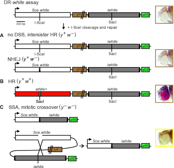
Figure 1: DR-white DSB reporter assay and repair outcomes. The Direct Repeat of white assay contains two nonfunctional copies of the white gene: Sce.white (with a 23-bp insertion containing the I-SceI recognition site and introducing a premature stop codon) and iwhite (5′- and 3′-truncated donor sequence). Crossing flies containing DR-white and an I-SceI transgene produces a site-specific DSB that can be analyzed phenotypically or molecularly. The resulting phenotypes are (A) white-eyed progeny (y+ w−), indicating no DSB, intersister HR, or NHEJ; (B) red-eyed progeny (y+ w+), indicating intrachromosomal HR that restores the wild-type SacI sequence from the iwhite donor; and (C) yellow-bodied, white-eyed progeny (y− w−), indicating SSA or mitotic CO. Figure adapted from Do et al.14. Abbreviations: CO = crossover; DR-white = Direct Repeat of white; DSB = double-strand break; HR = homologous recombination; NHEJ = nonhomologous end joining; SSA = single-strand annealing. Please click here to view a larger version of this figure.
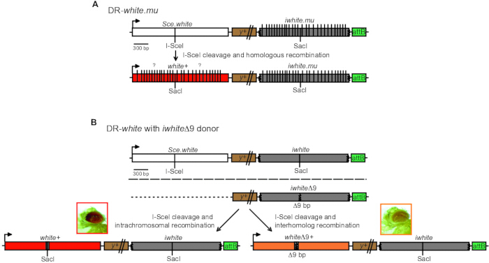
Figure 2: Derivatives of the DR-white assay. (A) The DR-white.mu assay contains 28 silent SNPs in the iwhite.mu donor sequence, producing 1.4% sequence divergence between the direct repeats. Gene conversion of individual polymorphisms varies among HR events and can be determined by sequencing. (B) DR-white with the iwhiteΔ9 donor distinguishes interhomolog from intrachromosomal recombination. HR from the intrachromosomal iwhite donor restores the wild-type SacI sequence and produces red-eyed progeny. HR from the interhomolog iwhiteΔ9 donor produces orange-eyed progeny because the 9-bp deletion decreases white expression. The figure adapted from Do et al.14 and Fernandez et al.15. Abbreviations: DSB = double-strand break; HR = homologous recombination; SNP = single-nucleotide polymorphism. Please click here to view a larger version of this figure.
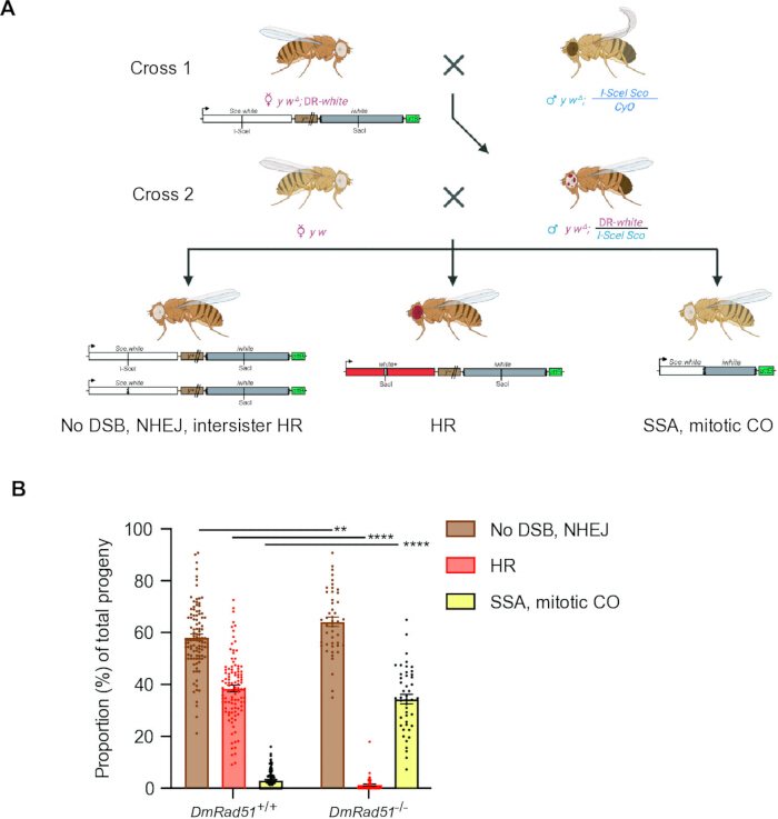
Figure 3: Experimental workflow for detecting DSB repair outcomes in the premeiotic germline. (A) Cross 1 combines the DR-white reporter (or a derivative) with an I-SceI transgene (in this example, on the second chromosome). Progeny containing both the DR-white assay and I-SceI transgene maintain repair events in their germline and somatic tissues. To analyze individual repair events in the premeiotic germline, progeny carrying both transgenes are crossed to y w tester flies (Cross 2), and the progeny are scored as described in Figure 1. (B) Individual germline DSB repair events in wild-type flies (n = 106 germlines) and DmRad51 homozygous mutants (n = 46 germlines). **P < 0.01; ****P < 0.0001 by two-way ANOVA followed by Sidak's multiple-comparisons test. Bars represent means; error bars represent SEM. Data were reanalyzed from Do et al.14. Abbreviations: ANOVA = analysis of variance; CO = crossover; DSB = double-strand break; HR = homologous recombination; NHEJ = nonhomologous end joining; SEM = standard error of the mean; SSA = single-strand annealing. Please click here to view a larger version of this figure.
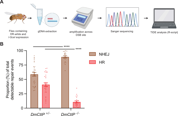
Figure 4: Experimental workflow for detecting DSB repair outcomes using Tracking of Indels by DEcomposition. (A) Flies carrying DR-white and an induced I-SceI transgene are analyzed by gDNA extraction, PCR amplification across the DSB site, Sanger sequencing, and TIDE analysis with the R script. (B) Representative data from DmCtIP-/- mutants (n = 23) and DmCtIP+/- heterozygous controls (n = 31). Detectable repair events are calculated as HR plus NHEJ with processing, excluding no DSB/NHEJ without indels. ****P < 0.0001 by two-way ANOVA followed by Sidak's multiple-comparisons test. Bars represent means; error bars represent SEM. Data were reanalyzed from Thomas et al.16. Abbreviations: ANOVA = analysis of variance; DSB = double-strand break; gDNA = genomic DNA; HR = homologous recombination; NHEJ = nonhomologous end joining; PCR = polymerase chain reaction; SEM = standard error of the mean; TIDE = Tracking of Indels by DEcomposition. Please click here to view a larger version of this figure.
Supplemental File 1: TIDE R script for identifying insertions and deletions in sequence chromatograms relative to a no-DSB control. The user-editable control filename, sample filenames, and I-SceI target sequence are marked near the end of the script. Required inputs, output files, and interpretation are described in section 2.3. The sangerseqR and Biostrings packages are required.Please click here to download this file.
Supplemental File 2: Step-by-step screenshot guide to the TIDE R script workflow. The panels illustrate the software actions described in section 2.3.Please click here to download this file.
Supplemental File 3: Example TIDE output and repair-event calculations. The 0-indel value represents no DSB or NHEJ without indels; the separate HR row represents the canonical 23-bp deletion product; and all remaining indel percentages are summed as NHEJ with processing. Total detectable repair is calculated as HR plus NHEJ with processing.Please click here to download this file.
Supplemental File 4: Raw data underlying Figure 3B and Figure 4B, organized in separate editable worksheets. These data were reanalyzed from the previously published studies cited in the corresponding figure legends.Please click here to download this file.
Discussion
The versatility of the DR-white system is a critical factor contributing to its past and future impact in the field of DNA repair. First, the attB integration sequence within the DR-white reporter construct allows investigators to target the reporter to numerous locations within the Drosophila genome21, with several targeted lines already available22. Second, the availability of different I-SceI transgenes, such as heat-shock expression via hsp70 promoter (this study), constitutive expression via ubiquitin promoter23, and premeiotic germline expression via the Bam promoter24, provides numerous developmental and tissue-specific contexts to explore DSB repair. Regardless of the I-SceI transgene used, genetic crosses must be carefully planned to ensure that the I-SceI transgene is only introduced in the final cross to ensure proper DSB induction. It is important to note that repair frequencies may vary significantly depending on the DR-white genomic location and/or the I-SceI transgene used. Future experiments could further compare genomic locations and I-SceI transgenes to determine whether genomic context or tissue specificity drives DSB repair.
A particular benefit of the DR-white assay and its derivatives is that they can identify repair of DSBs via homologous recombination, NHEJ with processing, and, in most cases, SSA. While designed to detect germline repair events phenotypically, the system also enables analysis of somatic tissues via TIDE algorithmic analysis. Despite these benefits, there are some limitations to this assay that must be considered. Of note, the DR-white assay may underestimate HR frequencies. As in other genetic reporter assays that use nuclease-induced DSBs, repair via HR from a sister chromatid (intersister HR) is indistinguishable from a no-DSB event or from NHEJ without processing. Thus, at least some of the No DSB/NHEJ class may be HR events. Considering this limitation, results comparing genetic factors (such as mutant lines) should be guarded. Other limitations of the assay include an inability to score premeiotic events that did not yield viable progeny. For example, DSB events that may not have been repaired and resulted in cell death will not be scored in the progeny. While overall fertility could be a qualitative measure of germline cell death, cell-based assays (such as TUNEL) may be required. Additionally, the TIDE R script analysis only provides the proportion of detectable events in a cell population. To determine specific indel sequence outcomes, sequencing of individual repair events using multiplex DNA sequencing is needed.
The DR-white assay has already made a profound impact in the study of DSB repair. Its application has identified factors that contribute to DSB repair in multicellular organisms, including age25, sex26, DSB location22,26, donor sequence location15, cell cycle, and tissue type26. Additionally, we have employed the assay to identify several proteins that also drive repair pathway choice and/or repair of diverged sequences in Drosophila, including Msh627, Blm28, Rif116, and CtIP16,29. Indeed, the well-established use of Drosophila as a genetic model organism with conserved DNA repair proteins provides a simple, multicellular system for investigating the genetic, cellular, and organismal factors that contribute to DSB repair in complex biological contexts.
Disclosures
The authors have no financial or non-financial conflicts to disclose.
Acknowledgements
This method was developed while J.R.L. was an AFAR Research Grant recipient from the American Federation for Aging Research. This work was also supported by the National Institute of General Medical Sciences (1R15GM110454 and 1R15GM129628) to J.R.L. Figures were developed with the assistance of BioRender (https://BioRender.com/b8sbcjp) 2026.
Materials
| Name | Company | Catalog Number | Comments |
|---|---|---|---|
| 0.5 mL RNAse/DNAse-free pestles | Fisher Scientific | 12141363 | For homogenization of samples for gDNA extraction |
| Cotton balls | Genesee Scientific | 51-101 | for standard Drosophila culturing |
| Drosophila Peltier Refrigerated Incubator | Shel Lab | SRI6PF | for standard Drosophila culturing |
| Drosophila stock: y[1] w[1] | Bloomington Drosophila Stock Center | RRID:BDSC 1495 | |
| Drosophila stock: y[2] w[Δ] cho[2] v[1] ; P{70I-SceI}2B Sco / CyO, P{GFP, w+} | LaRocque Lab | N/A | created by LaRocque lab through standard recombination of stock RRID:BDSC_6934 (y[1] w[*]; P{ry[+t7.2]=70FLP}11 P{v[+t1.8]=70I-SceI}2B sna[Sco]/CyO, S[2]) from Bloomington Drosophila Stock Center; available upon request to corresponding author |
| Drosophila stock: y[2] w[Δ] cho[2] v[1] ; DR-white (y+).3 51C,RFP+ / [CyO-GFP] | LaRocque Lab | N/A | DR-white assay integrated into stock RRID:BDSC_24482 (y[1] M{RFP[3xP3.PB] GFP[E.3xP3]=vas-int.Dm}ZH-2A w[*]; M{3xP3-RFP.attP’}ZH-51C); available upon request to corresponding author |
| Drosophila stock: y[2] w[Δ] cho[2] v[1] ; DR-white.mu (y+).3 51C,RFP+ / [CyO-GFP] | LaRocque Lab | N/A | DR-white.mu assay integrated into stock RRID:BDSC_24482 (y[1] M{RFP[3xP3.PB] GFP[E.3xP3]=vas-int.Dm}ZH-2A w[*]; M{3xP3-RFP.attP’}ZH-51C); available upon request to corresponding author |
| Drosophila stock: y[1] w[*]; iwhite2 (y+).1 51C,RFP+ / [CyO] | LaRocque Lab | N/A | iwhiteΔ9 donor integrated into stock RRID:BDSC_24482 (y[1] M{RFP[3xP3.PB] GFP[E.3xP3]=vas-int.Dm}ZH-2A w[*]; M{3xP3-RFP.attP’}ZH-51C); available upon request to corresponding author |
| Flugs Drosophila bottle closures | Genesee Scientific | 49-100 | for standard Drosophila culturing |
| Fluorescent stereomicroscope (RFP) | TriTech Research | SMT1-FL | for standard Drosophila "fly pushing" and sorting based on fluorescence |
| FlyStuff Drosophila Fly Pad | Genesee Scientific | 59-172 | for standard Drosophila "fly pushing" |
| Kimwipes | Kimberly-Clarke | 06-666A | |
| MyBath 4L Water Bath | Midwest Scientific | MB-2000-4 | for heat shock |
| NanoDrop One | Thermo Fisher Scientific | 13-400-518LY | for DNA quantitation |
| Nutri-fly Bloomington Formulation Media | Genesee Scientific | 66-121 | for standard Drosophila culturing |
| Pellet Pestle Cordless Motor homogenizer | DWK Life Sciences | K749540-0000 | For homogenization of samples for gDNA extraction |
| Polypropylene Bottles | Genesee Scientific | 32-130 | for standard Drosophila culturing |
| Polystyrene Vials | Genesee Scientific | 32-116 | for standard Drosophila culturing, including heat shock |
| Primer/oligonucleotide DR-white_8f | N/A; any oligonucleotide synthesis service is sufficient | N/A | 5'GTGGATCAGGTGATCCAGG |
| Primer/oligonucleotide DR-white_8a | N/A; any oligonucleotide synthesis service is sufficient | N/A | 5’-CTTAAGCCATCGTCAGTTGC |
| Primer/oligonucleotide DR-white_9f | N/A; any oligonucleotide synthesis service is sufficient | N/A | 5’-GAGCCCACCTCCGGACTGGAC |
| R Studio | Posit | Version 2023.06.1+524 | Free downloadable version; any version of R Studio is acceptable, although data presented was analyzed using version 2023.06.1+524 |
| R Studio packages: Biostrings | Bioconductor.org (Version 3.24) | Version 2.76.0 | R Script will prompt user to install this package |
| R Studio packages: sangerseqR | Bioconductor.org (Version 3.24) | Version 1.44.0 | R Script will prompt user to install this package |
| SapphireAMP Fast PCR | Takara | RR350B | |
| Stereomicroscope | Leica | M60 | for standard Drosophila "fly pushing" |
| Wizard SV Gel and PCR Clean up | Promega | A9281 |
References
- Khanna KK, Jackson SP. DNA double-strand breaks: signaling, repair and the cancer connection. Nat Genet. 2001;27(3):247-54.
- Cejka P, Symington LS. DNA end resection: mechanism and control. Annu Rev Genet. 2021;55:285-307.
- Oh J, Myung K. Crosstalk between different DNA repair pathways for DNA double strand break repairs. Mutat Res Genet Toxicol Environ Mutagen. 2022;873:503438.
- Figueroa-González G, Pérez-Plasencia C. Strategies for the evaluation of DNA damage and repair mechanisms in cancer. Oncol Lett. 2017;13(6):3982-8.
- Picksley SM, Attfield PV, Lloyd RG. Repair of DNA double-strand breaks in Escherichia coli K12 requires a functional recN product. Mol Gen Genet. 1984;195(1-2):267-74.
- Motamedi MR, Szigety SK, Rosenberg SM. Double-strand-break repair recombination in Escherichia coli: physical evidence for a DNA replication mechanism in vivo. Genes Dev. 1999;13(21):2889-903.
- Malkova A, Ivanov EL, Haber JE. Double-strand break repair in the absence of RAD51 in yeast: a possible role for break-induced DNA replication. Proc Natl Acad Sci U S A. 1996;93(14):7131-6.
- Frankenberg-Schwager M, Frankenberg D. DNA double-strand breaks: their repair and relationship to cell killing in yeast. Int J Radiat Biol. 1990;58(4):569-75.
- Allalunis-Turner MJ et al. Radiation-induced DNA damage and repair in cells of a radiosensitive human malignant glioma cell line. Radiat Res. 1995;144(3):288-93.
- Pierce AJ, Johnson RD, Thompson LH, Jasin M. XRCC3 promotes homology-directed repair of DNA damage in mammalian cells. Genes Dev. 1999;13(20):2633-8.
- Rong YS, Golic KG. Gene targeting by homologous recombination in Drosophila. Science. 2000;288(5473):2013-8.
- Adams MD, McVey M, Sekelsky J. Drosophila Blm in double-strand break repair by synthesis-dependent strand annealing. Science. 2003;299(5604):265-7.
- Rong YS, Golic KG. The homologous chromosome is an effective template for the repair of mitotic DNA double-strand breaks in Drosophila. Genetics. 2003;165(4):1831-42.
- Do AT, Brooks JT, Le Neveu MK, LaRocque JR. Double-strand break repair assays determine pathway choice and structure of gene conversion events in Drosophila melanogaster. G3 (Bethesda). 2014;4(3):425-32.
- Fernandez J, Bloomer H, Kellam N, LaRocque JR. Chromosome preference during homologous recombination repair of DNA double-strand breaks in Drosophila melanogaster. G3 (Bethesda). 2019;9(11):3773-80.
- Thomas MS et al. The epistatic relationship of Drosophila melanogaster CtIP and Rif1 in homology-directed repair of DNA double-strand breaks. G3 (Bethesda). 2024;14(11):jkae210.
- Greenspan RJ. Fly pushing: the theory and practice of Drosophila genetics. 2nd ed. Cold Spring Harbor Laboratory Press; Cold Spring Harbor, NY; 2004. 191 p.
- Sullivan W, Ashburner M, Hawley RS, editors. Drosophila protocols. Cold Spring Harbor Laboratory Press; Cold Spring Harbor, NY; 2000. 697 p.
- McIlwraith MJ et al. Human DNA polymerase eta promotes DNA synthesis from strand invasion intermediates of homologous recombination. Mol Cell. 2005;20(5):783-92.
- Sartori AA et al. Human CtIP promotes DNA end resection. Nature. 2007;450(7169):509-14.
- Bischof J et al. An optimized transgenesis system for Drosophila using germ-line-specific phiC31 integrases. Proc Natl Acad Sci U S A. 2007;104(9):3312-7.
- Janssen A et al. A single double-strand break system reveals repair dynamics and mechanisms in heterochromatin and euchromatin. Genes Dev. 2016;30(14):1645-57.
- Preston CR, Flores CC, Engels WR. Differential usage of alternative pathways of double-strand break repair in Drosophila. Genetics. 2006;172(2):1055-68.
- Chen H et al. An enhanced gene targeting toolkit for Drosophila: Golic+. Genetics. 2015;199(3):683-94.
- Delabaere L et al. Aging impairs double-strand break repair by homologous recombination in Drosophila germ cells. Aging Cell. 2017;16(2):320-8.
- Graham EL et al. The impact of developmental stage, tissue type, and sex on DNA double-strand break repair in Drosophila melanogaster. PLoS Genet. 2024;20(4):e1011250.
- Do AT, LaRocque JR. The role of Drosophila mismatch repair in suppressing recombination between diverged sequences. Sci Rep. 2015;5:17601.
- Ertl HA et al. The role of Blm helicase in homologous recombination, gene conversion tract length, and recombination between diverged sequences in Drosophila melanogaster. Genetics. 2017;207(3):923-33.
- Yannuzzi I, Butler MA, Fernandez J, LaRocque JR. The role of Drosophila CtIP in homology-directed repair of DNA double-strand breaks. Genes (Basel). 2021;12(9):1430.
Reprints and Permissions
This article has been published
Video Coming Soon