Here, a protocol is presented to dissect the midgut, ovaries, and salivary glands from Aedes albopictus, followed by total RNA extraction for molecular characterization of vector tissues in mosquito-borne virus transmission studies.
Method Article
Dissection of Aedes albopictus and Total RNA Extraction from Post-Dissection Tissues
* These authors contributed equally
In This Article
Summary
Abstract
In Foshan, Guangdong Province, China, in July 2025, an outbreak of dengue fever and chikungunya occurred. Ae. albopictus is a major vector for these viruses. This protocol describes a simple, standardized method for dissecting the midgut, salivary glands, and ovaries of Ae. albopictus and extracting total RNA from the dissected tissues. Three-day-old non-blood-fed female mosquitoes were anesthetized by freezing at −20 °C for 2 min, after which the target tissues were dissected. Total RNA was extracted with a tissue RNA extraction kit, quantified by a microvolume spectrophotometer, and used for quantitative real-time polymerase chain reaction (RT-qPCR) analysis. The results show that the dissection method rapidly and completely separates the tissues, and that RNA from the separated tissues can achieve relatively high purity. RT-qPCR demonstrated the expression patterns and relative stability of three reference genes (GAPDH, β-actin, and RPS6). The method is straightforward, validated, and provides standardized technical support for Ae. albopictus related research.
Introduction
Aedes albopictus (Ae. albopictus) is a vector for several pathogens, including the dengue virus (DENV) and chikungunya virus (CHIKV)1,2,3. It is widely distributed and has caused outbreaks of mosquito-borne diseases in many regions4,5. The ability of Ae. albopictus to transmit pathogens, known as vector competence, varies among populations and is linked to specific gene expression in midguts, ovaries, and salivary glands6. Understanding the structure of these tissues and extracting high-quality RNA are crucial for understanding the replication, dissemination, and transmission of vector-borne viruses7.
The detection of relative gene expression by quantitative real-time PCR (RT-qPCR) depends on the precise dissection of Ae. albopictus tissues and the extraction of high-quality RNA8,9. To date, published video protocols for the dissection of the midgut, ovaries, and salivary glands have focused primarily on Aedes aegypti10. Therefore, this protocol demonstrates the dissection of the midgut, ovaries, and salivary glands from Ae. albopictus, in which the dissected tissues are placed directly into grinding tubes containing RNA preservation solution, avoiding tissue transfer steps and minimizing transfer loss. Total RNA is extracted from the dissected tissues using a column-based method, which requires fewer handling steps and less time than the conventional TRIzol method11. This extraction typically yields 0.1–0.3 µg of total RNA from the salivary glands of five Ae. albopictus, with A260/A280 ratios of 1.8–2.0, meeting the purity requirements for downstream RT-qPCR. RT-qPCR is performed to measure the expression of three reference genes across Ae. albopictus tissues, thereby evaluating the quality of the RNA extracted from each tissue.
It should be noted that this protocol was validated only for the midgut, ovaries, and salivary glands of 3-day-old, non-blood-fed female Ae. albopictus, and is not suitable for RNA extraction from the fat body. Obtaining intact and individual tissues is the foundation for subsequent analysis. Through the systematic dissection of Ae. albopictus tissues and high-quality RNA extraction, this protocol provides technical support for subsequent gene expression detection, population-level vector competence assessment, and arbovirus transmission control.
Protocol
This study used only laboratory-reared Ae. albopictus mosquitoes. Accordingly, institutional ethical approval was not required for this study. The Ae. albopictus strain used in this experiment was provided by the Key Laboratory for Parasitic Disease Control and Prevention of the National Health Commission of China (Jiangsu). This colony was originally collected from Wuxi City in the 1980s and has been maintained in the insectary at Jiangsu Institute of Parasitic Diseases under standard conditions. The mosquito colony was housed in an artificial climate chamber at 25 °C ± 1 °C, 75% ± 5% relative humidity, with a photoperiod of 10 h light and 14 h dark. Larvae were reared in ultrapure water and fed a mixture of pig liver powder and yeast extract (Shanghai Brewery) at a 2:1 ratio. Adult mosquitoes were maintained on a 5% glucose solution. The reagents and the equipment used are listed in the Table of Materials.
1. Preparation of materials
- Preparation of solutions
- Preparation of 1x PBS buffer: Weigh 8.0 g NaCl, 0.2 g KCl, 1.44 g Na2HPO4, and 0.24 g KH2PO4. Dissolve in 800 mL ultrapure water with stirring. Adjust pH to 7.4. Bring the final volume to 1 L. Autoclave at 121 °C for 20 min.
- Preparation of DEPC solution: Add 1 mL DEPC stock solution to 1 L ultrapure water. Stir vigorously on a magnetic stirrer for 12–16 h. Autoclave at 121 °C for 20 min.
CAUTION: DEPC is highly toxic and carcinogenic. Wear gloves and safety goggles and handle in a fume hood.
NOTE: Cover the beaker with aluminum foil during vigorous stirring. Aliquot DEPC solution and store at -20 °C for up to 6 months.
- Preparation of consumables
- Sterilize dissecting tools: Place 1 mm dissecting needles and fine-point forceps in an aluminum container and autoclave at 121 °C for 20 min.
- Prepare 2 mL grinding tubes containing 0.5–1.0 mm silica beads. Place in an aluminum container and autoclave at 121 °C for 20 min.
- Add 200 µL of kit-provided RNA preservation solution to each grinding tube.
NOTE: RNA quality within tissues declines significantly within 1 min after removal from the organism. Prepare grinding tubes with preservation solution in advance.
2. Collection of Ae. albopictus
NOTE: Wear white lab coats and gloves. Do not wear dark clothing, as dark colors attract mosquitoes.
- Close all doors, windows, and ventilation systems in the room containing the mosquito cage to prevent escape of Ae. albopictus.
- Turn on the electric aspirator and insert it into the mosquito cage. Direct the aspirator toward Ae. albopictus resting on the inner walls of the cage and collect the mosquitoes.
- Keep the aspirator running while slowly withdrawing it from the cage. Seal the cage immediately and close the lid of the aspirator.
- Remove the collection chamber and place it at -20 °C for 2 min for cold immobilization of Ae. albopictus.
- Place a 10 cm glass dish on ice for pre-cooling. Transfer the immobilized Ae. albopictus from the collection chamber into the glass dish.
NOTE: Strictly control cold immobilization time to prevent tissue damage.
3. Identification of female Ae. albopictus Prior to dissection
- Select morphologically intact, cold-anesthetized Ae. albopictus from the glass dish and transfer them onto a glass slide. Examine under a stereomicroscope at 80x magnification.
- Identify the white longitudinal stripe on the mesonotum.
- Confirm the white bands at the base of the tarsal segments and at the base of abdominal tergites II-VI12.
- Examine the antennae at 80x magnification. Distinguish filiform antennae (female) from plumose antennae (male) to identify females.
4. Abdominal dissection of Ae. albopictus
- Place three drops of sterile 1x PBS from left to right on a sterilized glass slide, with each drop approximately 20 µL in volume and spaced approximately 5 mm apart.
NOTE: The PBS drops should be large enough to submerge the entire Ae. albopictus. - Use fine forceps to grasp the wings of Ae. albopictus and place the mosquito in the leftmost PBS drop.
- Orient the Ae. albopictus with the ventral side facing upward. Use the left hand to hold a dissecting needle and press on the second abdominal segment to immobilize the Ae. albopictus.
- Use the right hand to insert a 1 mm dissecting needle at the midpoint of the sixth abdominal segment. Make a transverse, semi-circular incision to open the abdominal cavity.
- Move the dissecting needle held in the right hand approximately 2 mm posteriorly and press on the mid-region of abdominal segments VI–VIII to secure the abdomen.
NOTE: The ovaries are located dorsally within abdominal segments II-VI. Do not move the needle randomly when pressing on segments VI-VIII to avoid damaging the intact morphology of the ovaries. - Use the left hand to grasp the second abdominal segment with the dissecting needle and pull gently outward to separate the abdomen at the sixth segment. During separation, the foregut, midgut, and Malpighian tubules will be fully exposed.
NOTE: Malpighian tubules are white and slender. The midgut is semi-translucent and fusiform. - Separate the midgut from the foregut and hindgut. Rinse the midgut in the central PBS drop twice.
- Transfer it into a grinding tube containing 200 µL of RNA preservation solution provided in the commercial RNA extraction kit.
- Press on the middle of the eighth abdominal segment with the dissecting needle to expose the paired ovaries on both sides. Remove the ovaries, rinse them in the central PBS drop twice, and transfer them into a grinding tube containing 200 µL RNA preservation solution.
NOTE: The paired ovaries are symmetric, rice-grain-shaped, and milky-white. Handle carefully to ensure the structural integrity of the ovaries during retrieval.
5. Salivary gland dissection of Ae. albopictus
- Increase the microscope magnification to 400x. Transfer the abdomen-dissected Ae. albopictus to the rightmost PBS drop.
- Apply gentle pressure on the mesothorax with the left-hand dissecting needle until the head is slightly elevated.
- Use the right hand to lift the head at the cervix with the dissecting needle and pull slowly outward. Observe the salivary glands (trilobed structure) as they detach from the thorax together with the head.
NOTE: The salivary glands are located in the anterior thorax and connect to the head via the salivary duct. Applying consistent pressure on the thorax prevents rupture of the salivary duct and allows the salivary glands to detach intact from the thorax with the head. Control of force at this step requires repeated practice. - Separate the salivary glands from the head. Rinse them in the central PBS drop twice and transfer them into a prepared grinding tube.
NOTE: Salivary glands are approximately 1 mm in size. Transfer them into a grinding tube containing 200 µL RNA preservation solution and observe against a white light source to confirm the presence of small white specks floating in the solution.
6. RNA extraction from Ae. albopictus and its salivary glands, ovaries, and midgut
NOTE: All RNA extraction steps were performed according to the manufacturer's instructions of the commercial tissue RNA extraction kit, without modification.
- Pre-cool the tissue grinder to 4 °C. Set the parameters to 30 Hz, 30 s grinding time, 10 cycles, and 8 s pause time.
- Place the grinding tubes containing tissue samples into the grinder and run the program.
NOTE: After grinding, check whether the tissue is fully dispersed in the RNA preservation solution. If grinding is incomplete, run an additional 5 cycles. - Transfer the homogenate to a sterile 1.5 mL centrifuge tube. Centrifuge at 12,000 x g at 4 °C for 5 min.
- Transfer the supernatant to a new sterile 1.5 mL centrifuge tube. Add 30 µL chloroform.
CAUTION: Chloroform is toxic and carcinogenic. Wear gloves and safety goggles and handle in a fume hood. - Vortex vigorously for 20 s. Incubate at room temperature for 15 min, then centrifuge at 12,000 x g at 4 °C for 15 min.
NOTE: Carefully aspirate the upper aqueous phase, avoiding the middle white layer to prevent protein contamination. - Transfer the upper aqueous phase to a new sterile 1.5 mL centrifuge tube. Add 75 µL isopropanol.
- Mix by inversion and transfer the entire mixture to a silica spin column. Centrifuge at 12,000 x g for 1 min.
- Add 600 µL wash buffer to the column. Centrifuge at 12,000 x g for 30 s. Discard the flow-through.
- Add 600 µL 80% ethanol to the column. Centrifuge at 12,000 x g for 30 s. Discard the flow-through. Repeat this wash step once, for a total of two washes.
- Add 30 µL DEPC water to the center of the column membrane. Incubate at room temperature for 1 min.
- Place the column into a sterile 1.5 mL centrifuge tube. Centrifuge at 12,000 x g for 1 min to elute the RNA.
- Take 1 µL of the eluted RNA and measure the concentration and purity using a microvolume spectrophotometer.
7. RNA reverse transcription
- Reaction system preparation
- Thaw 4x reverse transcription master mix, nuclease-free water, and RNA template on ice.
- In a 200 µL PCR tube, prepare a 20 µL reaction system by adding 5 µL 4x reverse transcription master mix, 10 µL total RNA template, and 5 µL nuclease-free water.
- Centrifuge briefly at 1,000 x g for 1 min to collect the reaction mixture at the bottom of the tube.
- Place the PCR tube in a thermal cycler and run the reverse transcription program: 50 °C for 5 min; 85 °C for 5 s.
NOTE: Dilute the reverse transcription product 20-fold before use to reduce the effect of potential inhibitors on RT-qPCR.
8. Real-time quantitative PCR (RT-qPCR)
- RT-qPCR reaction system preparation
- Thaw 2x SYBR Green qPCR master mix, cDNA template, and 10 µM forward and reverse primers on ice, protected from light throughout.
- In a 96-well PCR plate, prepare a 20 µL reaction system per well by adding 10 µL 2x SYBR Green qPCR master mix, 0.8 µL each of 10 µM forward and reverse primers (final concentration 400 nM), 2 µL of 20-fold diluted cDNA template, and 6.4 µL nuclease-free water.
- Seal the 96-well plate with an optical adhesive film, ensuring all wells are completely sealed.
- Centrifuge at 1,000 x g for 1 min to collect the reaction mixture at the bottom of the wells.
- Place the PCR plate in a real-time PCR detection system and run the thermal cycling program.
NOTE: Thermal cycling program: Pre-denaturation at 95 °C for 30 s (1 cycle); amplification: 95 °C for 30 s, 60 °C for 30 s (40 cycles); melt curve analysis: 95 °C for 15 s, 60 °C for 1 min, ramp to 95 °C at 0.15 °C/s. Each tissue RNA sample is subjected to qPCR in quadruplicate technical replicates. Each tissue sample consists of pooled tissues from 5 Ae. albopictus. Primer sequences used for RT-qPCR are listed in Table 1.
- Data analysis
- Inspect the melt curve for each primer pair to confirm a single, well-defined dissociation peak.
- Export the Ct values from the instrument software. Graphing and data analysis are performed using statistical and graphing software.
| Primers name | Primer sequences (5'-3') |
| Ae. albopictus β-actin Forward Primer | GGAGAAGATCTGGCATCACA |
| Ae. albopictus β-actin Reverse Primer | TGTCATCTTCTCGCGGTTAG |
| Ae. albopictus GAPDH Forward Primer | CTGCCAAGGCTGTCGGTAA |
| Ae. albopictus GAPDH Reverse Primer | TCATAGGAGGCAGGCTTGGA |
| Ae. albopictus RPS6 Forward Primer | ACAAGCTGCGTCACTTCTACGACA |
| Ae. albopictus RPS6 Reverse Primer | CTTGTCGTTTCCACCGGCAATCTT |
Table 1: Primer sequences of Ae. albopictus used in RT-qPCR.
Results
This study comprises two parts: dissection of Ae. albopictus and RNA extraction from post-dissection tissues. The overall workflow is shown in Figure 1, where Figure 1A illustrates the dissection of Ae. albopictus and Figure 1B illustrates RNA extraction and the subsequent detection of reference-gene expression levels.
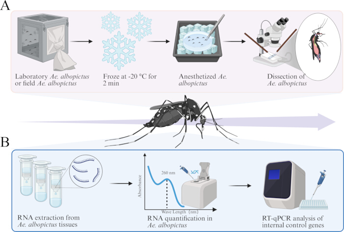
Figure 1: Schematic of the dissection of Ae. albopictus and total RNA extraction from post-dissection tissues. (A) Collection, anesthesia, and dissection of Ae. albopictus. (B) RNA extraction from post-dissection tissues, purity identification, and detection of reference gene expression levels. Please click here to view a larger version of this figure.
Five laboratory-reared Ae. albopictus were dissected to isolate the midgut, salivary glands, and ovaries. At a magnification of 80×, the transparent foregut, midgut, and hindgut of the Ae. albopictus were distinctly visible, with the grain-like ovaries positioned on either side and covered by the white Malpighian tubules (Figure 2A). When the field was adjusted to 400×, the translucent salivary glands of Ae. albopictus became clearly observable (Figure 2B). Figure 2C,D illustrate the isolated midgut and ovarian tissues. The results indicated that the tissues isolated through this dissection method preserved their structural integrity, which is essential for subsequent total RNA extraction from the post-dissection specimens.
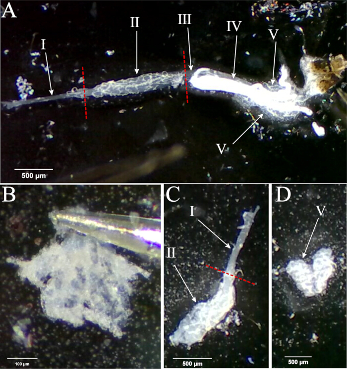
Figure 2: Post-dissection analysis of Ae. albopictus tissues. (A) Tissues are isolated from the abdomen of Ae. albopictus. (B) Translucent trilobed salivary glands of Ae. albopictus. (C) Foregut and midgut of Ae. albopictus. (D) Rice-grain-shaped paired ovaries of Ae. albopictus. I: foregut; II: midgut; III: hindgut; IV: Malpighian tubules; V: ovaries. Please click here to view a larger version of this figure.
Total RNA was extracted from dissected tissues of Ae. albopictus. Tissue samples (salivary glands, midgut, and ovaries) were fully ground prior to extraction. The RNA concentrations varied by tissue type: salivary glands were 7.8 ng/µL, while midgut, ovaries, and whole-body homogenates of female Ae. albopictus were 55.3, 45.7, and 42.7 ng/µL, respectively. All samples exhibited high purity, with A260/A280 ratios of 1.87–2.10 and A260/A230 ratios of 1.73–2.13 (Table 2). The expression levels of three candidate reference genes (GAPDH, β-actin, and RPS6) were assessed by RT-qPCR across all tissues. Although their expression varied somewhat with RNA concentration, all three genes remained relatively stable across different tissue types (Figure 3). GAPDH, β-actin, and RPS6 are suitable as stable reference genes for normalizing gene expression in different tissues of Ae. albopictus.
| Tissue | No. of samples | RNA conc. (ng/μL) | A260/A280 | A260/A230 |
| Salivary gland | 5 | 7.8 | 1.87 | 1.73 |
| Midgut | 5 | 55.3 | 1.94 | 1.98 |
| Ovary | 5 | 45.7 | 2.10 | 2.13 |
| Female | 1 | 42.7 | 2.04 | 1.92 |
Table 2: RNA concentration and purity of RNA extracted from Ae. albopictus tissue.
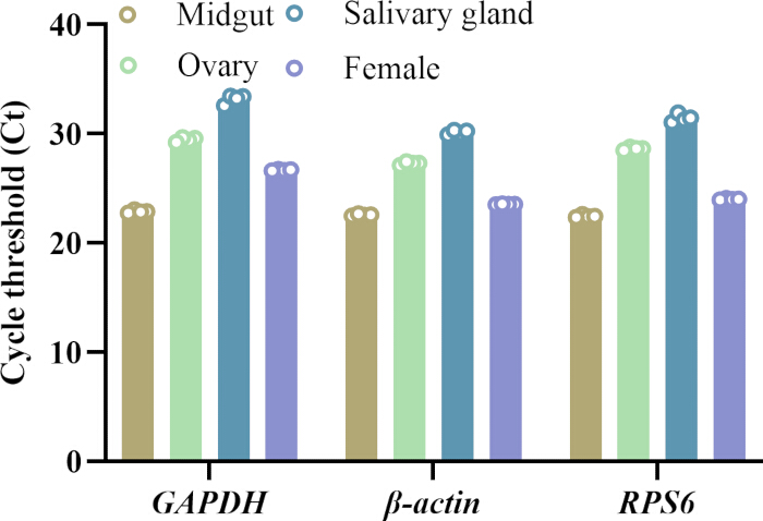
Figure 3: Ct values of reference genes (GAPDH, β-actin, and RPS6) detected by RT-qPCR. Please click here to view a larger version of this figure.
Discussion
Ae. albopictus is widely distributed in temperate, subtropical, and tropical regions across all continents except Antarctica13,14. As an important vector for arboviruses such as dengue virus (DENV), Zika virus (ZIKV), and chikungunya virus (CHIKV), it has repeatedly caused outbreaks of mosquito-borne diseases in Guangdong, China15.
To assist research on Ae. albopictus-related viruses and their transmission, we developed a straightforward method for dissecting Ae. albopictus tissues. In conventional dissection protocols, when the head is separated from the thorax during salivary gland dissection, the salivary glands may remain attached to either the thorax or the head, depending on the force applied; as the salivary glands are only approximately 1 mm in size, locating them within the thorax becomes difficult16. This method builds on standard mosquito organ isolation protocols and optimizes the separation of salivary glands located beneath the prosternal and mesosternal sternites, which should be observed under 400× magnification17. To dissect, gently press the mesothorax with one needle while using another to create pressure that separates the head, allowing the salivary glands to detach intact. This approach avoids the need to search for the salivary glands within the thorax, prevents contamination and damage to the glands, and shortens the overall dissection time. Ovaries are located near abdominal segments VI–VIII and are preserved by beginning dissection from the sixth abdominal segment and carefully extracting the midgut, Malpighian tubules, and ovaries from the abdomen18. This method produces intact salivary glands, typical ovary shapes free of adhesion, and an unbroken midgut (Figure 2). This dissection technique provides valuable guidance for studies on Ae. albopictus and the replication, dissemination, and transmission of vector-borne viruses.
RNA extraction from Ae. albopictus tissues is crucial for detecting mosquito-borne viruses. The conventional TRIzol method is time-consuming and technically demanding, but it typically provides high RNA yields and can produce high-quality RNA when performed correctly. In contrast, the column-based method offers better reproducibility and is easier to perform, but may result in lower RNA recovery and performs inefficiently when applied to the fat body19,20. In this study, dissected tissues were promptly preserved in a nucleic acid stabilizer, thoroughly ground, and processed using the spin column method to extract RNA for cDNA synthesis21. To ensure accurate normalization of RT-qPCR data amid variations in cDNA quality, stable reference genes are necessary22. While reference genes have been established for various developmental stages of Ae. albopictus, tissue-specific reference genes require further validation23,24. RT-qPCR analysis of reverse-transcribed RNA from all dissected tissues yielded consistent amplification of GAPDH, β-actin, and RPS6, confirming that the RNA quality obtained through this dissection protocol is sufficient to support downstream tissue-specific gene expression studies in Ae. albopictus.
During the dissection of Ae. albopictus tissues with this protocol, failure of the salivary glands to detach together with the head may occasionally occur. This usually results from insufficient pressure applied to the thorax combined with excessively rapid separation of the head. To resolve this, apply consistent pressure on the mesothorax and pull the head slowly outward, allowing the salivary glands to detach intact with the head. When transferring the salivary glands into a grinding tube containing RNA preservation solution, carefully examine the solution against a light source for small floating white specks. If the salivary glands cannot be located in the tube, immediately check whether they remain attached to the dissecting needle. During RNA extraction, strictly follow the kit manufacturer's protocol. If the RNA concentration obtained is low, a second elution with the elution buffer can be performed. In RT-qPCR, multiple peaks in the melt curve may occur; in this case, first verify primer specificity. If the Ct values are high, adjust the annealing temperature of the program according to the primer melting temperature. In general, the annealing temperature in the program should be set 1–2 °C below the primer melting temperature25.
This study has some limitations. The sample scope is relatively narrow, focusing only on midgut, salivary glands, and ovarian tissues, without investigating other tissues like Malpighian tubules and the fat body26. Additionally, the analysis was restricted to 3-day-old laboratory-reared female mosquitoes, with blood-fed and virus-infected individuals excluded27. Future improvements should include expanding the sample scope to include tissues like Malpighian tubules and considering Ae. albopictus individuals in different physiological states.
In summary, dissection of the midgut, ovaries, and salivary glands from Ae. albopictus, followed by total RNA extraction, provides a technical foundation for subsequent assessments of vector competence and arbovirus transmission studies.
Materials
| Name | Company | Catalog Number | Comments |
|---|---|---|---|
| 3 mL Extra-long Pasteur Pipette | WHB | WHB-S4 | 3 mL |
| ChamQ Universal SYBR qPCR Master Mix | Vazyme | Q711-02 | 500 rxns |
| HiScript IV All-in-One Ultra RT SuperMix for qPCR kit | Vazyme | R433-01 | 100 rxns |
| Micro Total RNA Extraction Kit | MedTech | MDT-CFR07 | 100T/box |
| Micropipettes 0.5-10μL | Eppendorf | 4043444 | |
| Microscope slides | CITOTEST | 80302-2104 | 75 mm×25 mm |
| MicroSpectrophotometer | KAIAO | K5600C | |
| Nucleic acid extraction buffer | MedTech | MDT-TP01 | 100 mL |
| PBS Phosphate Buffered Saline (Sterile) | Servicebio | G4202 | 500 ML |
| PCR Thermal Cycler | Eppendorf | Mastercycler X50s | |
| QuantStudioTM 7 Pro Real-Time PCR System | Applied biosystems | EN61326-1 | |
| Stemi 305 LAB Microscope Set | ZEISS | 435063-9010-000 |
References
- Hafsia S et al. Vector competence of Aedes albopictus and Aedes aegypti from the islands of the Southwestern Indian Ocean for epidemic Zika, dengue, and chikungunya viruses. Parasit Vectors. 2025;19(1):34.
- Zhang W, Wang J, Liu Q, Gong Z. A review of pathogens transmitted by the container-inhabiting mosquitoes, Aedes albopictus, a global public health threat. China CDC Wkly. 2023;5(44):984-990.
- Kraemer MUG et al. Past and future spread of the arbovirus vectors Aedes aegypti and Aedes albopictus. Nat Microbiol. 2019;4(5):854-863.
- Zhang M et al. Epidemiological characteristics and transmission dynamics of the early stage Chikungunya fever outbreak in Foshan City, Guangdong Province, China in 2025. Infect Dis Poverty. 2025;14(1):93.
- Meyer AD, Guerrero SM, Dean NE, Anderson KB, Stoddard ST, Perkins TA. Predictability of infectious disease outbreak severity: Chikungunya as a case study. Sci Adv. 2025;11(40):eadt5419.
- Wu Y et al. The making of a dangerous vector: factors shaping the vector competence of Aedes mosquitoes. Front Cell Infect Microbiol. 2025;15:1718737.
- Zhu C et al. Transcriptome analysis of Aedes aegypti midgut and salivary gland post-Zika virus infection. Curr Res Parasitol Vector Borne Dis. 2025;7:100251.
- Liu X, Yang S, Yao Y, Wu S, Wu P, Zhai Z. Opsin1 regulates light-evoked avoidance behavior in Aedes albopictus. BMC Biol. 2022;20(1):110.
- Zhang Y et al. Rapid differential detection of Japanese encephalitis virus and Getah virus in pigs or mosquitos by a duplex TaqMan real-time RT-PCR assay. Front Vet Sci. 2022;9:839443.
- Coleman J, Juhn J, James AA. Dissection of midgut and salivary glands from Ae. aegypti mosquitoes. J Vis Exp. 2007;4(5):228.
- Tesena P, Korchunjit W, Taylor J, Wongtawan T. Comparison of commercial RNA extraction kits and qPCR master mixes for studying gene expression in small biopsy tissue samples from the equine gastric epithelium. J Equine Sci. 2017;28(4):135-141.
- Ramdini C et al. First report of Aedes albopictus in Saint Barthélemy (French West Indies) confirmed by morphological, molecular and MALDI-TOF mass spectrometry approaches. Parasit Vectors. 2025;18(1):258.
- Battaglia V et al. The worldwide spread of Aedes albopictus: new insights from mitogenomes. Front Genet. 2022;13:931163.
- Laporta GZ, Potter AM, Oliveira JFA, Bourke BP, Pecor DB, Linton YM. Global distribution of Aedes aegypti and Aedes albopictus in a climate change scenario of regional rivalry. Insects. 2023;14(1):49.
- Yip SSW, Charm CYC, Ng KS, Lam SC. Chikungunya fever: a re-emerging mosquito-borne threat demanding proactive prevention strategies in Asia. Front Public Health. 2026;14:1745145.
- Tutagata J, Pocquet N, Trouche B, Reveillaud J. Dissection of mosquito ovaries, midgut, and salivary glands for microbiome analyses at the organ level. J Vis Exp. 2024;(212):67128.
- Schmid MA, Kauffman E, Payne A, Harris E, Kramer LD. Preparation of mosquito salivary gland extract and intradermal inoculation of mice. Bio Protoc. 2017;7(14):e2407.
- Airs PM, Nazarchyk MJ, Tucker BJ, Bartholomay LC. Characterizing oogenesis and programmed cell death in the eastern tree hole mosquito Aedes (Protomacleaya) triseriatus. Front Insect Sci. 2022;2:1073308.
- Chomczynski P, Sacchi N. The single-step method of RNA isolation by acid guanidinium thiocyanate-phenol-chloroform extraction: twenty-something years on. Nat Protoc. 2006;1(2):581-585.
- De Azevedo N, Lozano A, Parsons RE, Martin TC. Optimized protocols to extract total transcripts and proteins from lipid-rich tissues. Methods Protoc. 2026;9(2):65.
- García-Longoria L, Ahrén D, Berthomieu A, Kalbskopf V, Rivero A, Hellgren O. Immune gene expression in the mosquito vector Culex quinquefasciatus during an avian malaria infection. Mol Ecol. 2023;32(4):904-919.
- Lü J, Yang C, Zhang Y, Pan H. Selection of reference genes for the normalization of RT-qPCR data in gene expression studies in insects: a systematic review. Front Physiol. 2018;9:1560.
- Dzaki N, Azzam G. Assessment of Aedes albopictus reference genes for quantitative PCR at different stages of development. PLoS One. 2018;13(3):e0194664.
- Vedururu RK et al. RNASeq analysis of Aedes albopictus mosquito midguts after chikungunya virus infection. Viruses. 2019;11(6):513.
- Bustin SA et al. MIQE 2.0: revision of the Minimum Information for Publication of Quantitative Real-Time PCR Experiments guidelines. Clin Chem. 2025;71(6):634-651.
- Skowronek P, Wójcik Ł, Strachecka A. Fat body-multifunctional insect tissue. Insects. 2021;12(6):547.
- Goldman OV et al. A single-nucleus transcriptomic atlas of the adult Aedes aegypti mosquito. Cell. 2025;188(25):7267-90.e26.
Reprints and Permissions
This article has been published
Video Coming Soon