This study developed hydroxyurea-loaded nanostructured lipid carriers (NLCs) and evaluated their properties and in vitro anticancer activity. NX-ST showed high encapsulation efficiency, controlled release, and greater cytotoxicity against MCF-7 cells (IC₅₀ = 225 µg/mL) than free hydroxyurea (IC₅₀ = 722.57 µg/mL).
Research Article
Hydroxyurea-Loaded Nanostructured Lipid Carriers for Enhanced In Vitro Cytotoxicity and Drug Delivery
In This Article
Summary
Abstract
Hydroxyurea (HU) is an established anticancer drug used to treat chronic myelogenous leukemia, but its therapeutic efficacy is limited by rapid clearance, variable bioavailability, and dose-related toxicity. This study aimed to improve HU delivery by developing hydroxyurea-loaded nanostructured lipid carriers (NLCs) using different surfactant combinations. Three formulations, including NX-ST (Tween 80–SDS), NX-CT (Cremophor EL–Tween 80), and NX-CS (Cremophor EL–SDS), were prepared and evaluated for particle size, polydispersity index (PDI), zeta potential, encapsulation efficiency (EE), and cytotoxic activity. The formulations exhibited nanoscale particle sizes (67.377 to 110 nm), acceptable PDI (<0.35), high zeta potential (-30.033 to -46.433 mV), and good encapsulation efficiency (78.189 to 86.626%), indicating stable formulations. The release profile exhibited an approximately linear increase over the 0.5–6 h period, followed by a plateau. Cytotoxicity studies showed enhanced anticancer activity compared with free HU (IC₅₀ = 722.57 µg/mL). The formulation NX-ST demonstrated the highest cytotoxicity (IC₅₀ = 225 ± 21.47 µg/mL), consistent with enhanced drug delivery; the contribution of improved cellular uptake is inferred and was not directly measured in this study. The NLC formulations studied, particularly NX-ST, effectively enhanced HU's therapeutic performance by improving stability, delivery efficiency, and anticancer activity.
Introduction
Cancer remains one of the leading causes of death worldwide, with nearly 20 million new cases and 9.7 million cancer-related deaths recorded in 2022. By 2040, these numbers are projected to rise significantly, reaching 29.9 million new cases and 15.3 million cancer-related deaths annually1,2. Most current anti-cancer drug regimens rely on highly toxic compounds, but their systemic toxicity limits the therapeutic concentrations that can effectively reach cancerous tissues3. To overcome this challenge, researchers have been exploring novel drug delivery approaches to enhance tumor targeting and improve the therapeutic index of chemotherapy.
One crucial area of oncology research is the development of advanced drug delivery systems to enhance therapeutic efficacy while minimizing the side effects of anticancer drugs. Hydroxyurea (HU), a well-established chemotherapeutic and antineoplastic agent, has been a cornerstone in the management of sickle cell disease, chronic myelogenous leukemia, and certain solid tumors due to its ability to increase fetal hemoglobin production and inhibit DNA synthesis by targeting ribonucleotide reductase4,5. Despite its clinical efficacy, HU’s therapeutic potential is hampered by challenges such as rapid clearance, variable bioavailability, and a narrow therapeutic index, which often lead to variable pharmacokinetics and dose-dependent toxicities, including myelosuppression, gastrointestinal disturbances, and potential secondary malignancies6,7. To address these limitations, advanced drug delivery systems, including nanostructured lipid carriers (NLCs), have emerged as promising strategies to enhance hydroxyurea’s stability, solubility, and controlled release, thereby improving its safety and efficacy profile8.
NLCs are a type of lipid-based nanoparticle that offer several advantages over traditional drug delivery systems. They consist of a solid lipid matrix stabilized by surfactants and can encapsulate both hydrophilic and lipophilic drugs. This unique structure allows for improved drug loading capacity, controlled release, and enhanced stability9,10. The preparation of NLCs typically involves techniques such as high-pressure homogenization and ultrasonication, which facilitate the formation of nanoparticles with a narrow size distribution and high encapsulation efficiency11,12. The encapsulation of the drug in NLCs not only enhances its solubility and stability but also facilitates targeted delivery to cancer cells, thereby minimizing off-target effects. Recent studies have demonstrated the potential of drug-loaded NLCs to improve the therapeutic index and sustained-release profiles of the drug13,9. This controlled-release mechanism is crucial for maintaining therapeutic drug concentrations over extended periods, thereby reducing the frequency of administration and improving patient compliance14. Studies have demonstrated that NLCs loaded with anticancer drugs like gefitinib15, capecitabine16, and imatinib14 significantly enhance drug efficacy by improving cellular uptake and accumulation in cancer cells. For instance, gefitinib-loaded NLCs have shown a 4.5-fold increase in cytotoxicity against cancer cells compared to the free drug, highlighting the potential of NLCs to enhance the therapeutic index of tyrosine kinase inhibitors used to treat non-small cell lung cancer and colorectal cancer. Similarly, capecitabine-loaded NLCs improve drug stability and controlled release, which is crucial for maintaining therapeutic concentrations and reducing systemic toxicity. Imatinib-loaded NLCs also benefit from enhanced targeted delivery, thereby reducing off-target effects and improving patient outcomes in the treatment of chronic myeloid leukemia. These attributes make NLCs strong candidates for clinical trials and for potential applications in personalized medicine by tailoring treatments to individual patient profiles. The stability evaluation of drug-loaded NLCs is another important aspect that influences their clinical applicability. Stability studies often involve subjecting the formulations to various environmental conditions to assess their physical integrity over time.
Accelerated stability tests have shown that these NLCs maintain their structural integrity and drug content for extended periods, highlighting their potential for long-term storage and use4,17.
Despite these advantages, surfactant selection significantly influences the physicochemical properties and performance of nanostructured lipid carriers (NLCs). Lipid-surfactant systems play an important role in improving nanoparticle stability, drug encapsulation, and bioavailability18,19,20. However, limited studies have investigated how different surfactant combinations affect the physicochemical characteristics and cytotoxic activity of hydroxyurea-loaded NLCs. Therefore, this study was undertaken to develop hydroxyurea-loaded NLCs (NANOXURIA) using different surfactant systems and to systematically evaluate how these variations influence physicochemical stability, storage behavior, and anticancer activity. By establishing a clear relationship between formulation design and therapeutic performance, this work aims to provide an effective nanocarrier-based dispersed system (colloidal dispersion) to characterize hydroxyurea-loaded NLC formulations and evaluate their in vitro drug release, cytotoxicity, and short-term storage stability, generating foundational physicochemical and in vitro performance data to inform future HU delivery development, without altering the drug molecule itself.
Protocol
This study did not involve human participants, human-derived tissues, or live animals. All biological evaluations were performed in vitro using the commercially available MCF-7 human breast cancer cell line obtained from the American Type Culture Collection (ATCC). Accordingly, institutional review board (IRB) or ethics committee approval was not applicable to this work.
Materials
Hydroxyurea (HU) (assay ≥99%), sodium dodecyl sulfate (SDS) (anionic, assay 90%), Tween-80 (T80) (non-ionic, oleic acid ≥58%), Cremophor EL (non-ionic, castor oil-derived, assay ≥98%), stearic acid (assay 95–106%), and sesame oil were used as received. All other chemicals and reagents used were of analytical grade. Full supplier and catalog details for all materials and equipment are provided in the Table of Materials.
Preparation of hydroxyurea-loaded NLCs (NANOXURIA)
The preparation of NANOXURIA includes three steps, namely preparation of the lipid phase, incorporation of the drug, and preparation of the aqueous phase. The lipid phase was obtained as a melt of a mixture of stearic acid (500 mg) and sesame oil (250 µl). The melt was obtained in a beaker on a hot plate. The accurately weighed amount of HU (150 mg) was mixed with the lipid phase. The aqueous phase was prepared by dissolving surfactants (Table 1) in water (Millipore) (25 mL). The surfactant solution was heated to the same temperature as the lipid phase (70°C) to avoid premature solidification. The hot aqueous phase was gradually added to the melted lipid phase while stirring at high speed with a homogenizer. The mixture was homogenized at high speed (6,000 rpm) for 20 min to create a stable hot emulsion. The hot emulsion was cooled to room temperature under continuous stirring. During cooling, the lipid phase solidifies, forming the NLCs. The prepared HU-loaded NLCs were stored for further characterization and application.
Characterization and evaluation
Particle size, zeta potential (ZP), and polydispersity index (PDI)
The particle size analysis and polydispersity index (PDI) of NANOXURIA were determined using a Zetasizer Nano ZS90. This device operates on the principle of dynamic light scattering (DLS), which measures the Brownian motion of particles in a suspension and correlates it to their hydrodynamic diameter via the Stokes-Einstein equation. In addition to size and PDI, the surface charge of NANOXURIA was assessed by measuring the zeta potential (ZP) using the same instrument. Zeta potential was determined using electrophoretic light scattering (ELS), in which the velocity of particles in an applied electric field is analyzed to calculate their surface charge. This parameter is crucial for evaluating the colloidal stability of NANOXURIA. The measurements were conducted in a controlled environment, with the diluted samples placed in cuvettes to ensure compatibility with the instrument’s optical and electrical requirements. To enhance the robustness and reproducibility of the results, all measurements were performed in triplicate.
Morphology
The morphology of NANOXURIA was studied using an electron microscope operating in both transmission and scanning modes. Transmission electron microscopy (TEM) was used to observe the internal structure and particle shape, while field-emission scanning electron microscopy (FE-SEM) was employed to examine surface morphology and particle distribution. The samples were appropriately prepared and analyzed at suitable accelerating voltages to obtain high-resolution images. For sample preparation, NANOXURIA dispersions were diluted with purified water; for TEM, a drop of the diluted dispersion was placed on a carbon-coated copper grid, the excess was blotted, and the film was air-dried at room temperature before imaging under high vacuum at an accelerating voltage of 60-80 kV. For FE-SEM, a drop of the diluted dispersion was mounted on an aluminum stub, air-dried, and sputter-coated with a thin conductive layer prior to imaging at (1–3 kV). Representative micrographs were acquired at the different magnifications.
Entrapment efficiency analysis for NANOXURIA formulations
The entrapment efficiency (EE) of NANOXURIA was assessed using the ultrafiltration-centrifugation technique with an Amicon Ultra-2 mL centrifugal filter (3 kDa MWCO). This method separated unentrapped HU (approximately 6 mg in 1 mL of NANOXURIA) from the encapsulated fraction by centrifuging at 4,000 x g for 20 min at 25 °C, ensuring effective isolation. The filtrate, containing free HU, was diluted with purified water and analyzed using a UV-visible spectrophotometer at 213 nm to quantify the unentrapped drug, with a calibration curve based on Beer-Lambert’s law. The % EE was calculated as;

In vitro release study
The in vitro release profile of HU from NANOXURIA was evaluated using the dialysis bag diffusion technique, a widely adopted method for assessing controlled drug release. Dialysis bags (12–14 kDa MWCO) were pre-soaked in distilled water for 12 h to ensure proper hydration and removal of preservatives. One end of the dialysis bag was securely sealed, and 2 mL of the NANOXURIA formulation, equivalent to 12 mg of HU, was carefully introduced into the bag. The other end was then tightly knotted to prevent leakage. The dialysis bag was immersed in a beaker containing water (50 mL) as the release medium. The setup was maintained at 37 ± 0.5 °C to simulate body temperature and agitated at 50 rpm with a magnetic stirrer to ensure uniform mixing and minimize boundary-layer effects. At predetermined time intervals (0.5, 1, 2, 4, 6, 12, and 24 h), 2 mL aliquots were withdrawn from the release medium and immediately replaced with an equal volume of fresh, pre-warmed water to maintain sink conditions. The collected samples were filtered through a 0.45 µm syringe filter to remove any particulate matter, ensuring clarity for analysis. The membrane filter retains particulates while allowing dissolved HU to pass through. As HU is freely water-soluble, the free drug remained in the filtrate, ensuring that the retained material represented particulates rather than free drug. The filtered samples were then analyzed for HU content using a UV-visible spectrophotometer at a wavelength of 213 nm, corresponding to HU’s maximum absorption (λmax). A calibration curve was established to accurately quantify HU concentrations.
Cell culture
The study was performed using the metastatic human breast cancer cell line MCF-7, obtained from the American Type Culture Collection (ATCC). The cells were maintained in RPMI 1640 medium supplemented with 10% fetal bovine serum (FBS) to support cell growth and viability, and 1% penicillin–streptomycin solution to prevent microbial contamination during culture. The cells were then incubated under standard physiological conditions at 37 °C in a humidified CO₂ incubator, with the culture medium maintained at pH 7.4. These conditions were selected to ensure optimal cell growth, proliferation, and experimental reliability throughout the cytotoxicity studies.
Cell viability assay (MTT)
The MTT assay was performed to assess the cell death-inducing capacity of different NANOXURIA formulations16,21. Briefly, MCF-7 cells were seeded into 96-well plates at a density of 1 x 10⁴ cells/mL and incubated for 24 h in a CO₂ incubator to allow proper cell attachment and growth. Untreated cells served as the negative control, while wells containing only culture medium were used as blank controls. After incubation, the culture medium was replaced with fresh medium containing different concentrations of the test formulations, up to 200 µg/mL. The treated plates were further incubated for 72 h under standard culture conditions. Following treatment, 20 µL of MTT reagent (5 mg/mL) was added to each well and incubated for an additional 4 h to allow intracellular formazan crystals to form in metabolically active cells. Subsequently, 100 µL of dimethyl sulfoxide (DMSO) was added to dissolve the crystals, yielding a violet solution. The absorbance was measured at 570 nm using a microplate reader (SpectroStar Nano). Cell viability (%) was calculated relative to the untreated control group using the absorbance values obtained.

Stability study
The stability of NANOXURIA was systematically assessed over a 3-month period under controlled storage conditions to evaluate its suitability for long-term use. Each NANOXURIA formulation was aliquoted into airtight, amber glass containers to minimize exposure to light and moisture. The containers were stored at a constant room temperature of 25 ± 2 °C and a refrigerated temperature of 4 ± 2 °C. Samples were periodically evaluated at predefined intervals (1, 2, and 3 months) for physical and physicochemical stability. A baseline measurement was performed immediately after preparation and before storage, and was chosen as 0 months (initial). Physical assessments involved visual inspection for changes in appearance, including signs of agglomeration, sedimentation, phase separation, color alteration (e.g., from off-white to yellow), changes in consistency (e.g., viscosity or texture), and odor (e.g., rancidity due to lipid oxidation). For physicochemical stability, key characterization parameters were measured, including particle size, polydispersity index (PDI), and zeta potential (ZP).
Statistical analysis
In this study, all experimental results are expressed as mean values ± standard deviation (SD) (n = 3) to reflect the central tendency and variability of the data. Statistical analyses were performed to evaluate the formulation using a paired t-test and to compare differences between formulations using one-way analysis of variance (ANOVA) followed by Tukey's multiple-comparison test. A p-value < 0.05 was considered statistically significant. Statistical analyses were performed using GraphPad Prism version 10.
Results
Physicochemical properties
The physicochemical properties of NANOXURIA formulations, including particle size, polydispersity index (PDI), zeta potential (ZP), morphology, and entrapment efficiency (EE), are shown in Table 2. The morphological assessment via TEM and SEM, supplemented by particle-size and zeta-potential distribution graphs, is presented in Figures 1, 2, and 3. A comparison of PDI for different surfactant combinations is presented in Figure 4.
Release study
The in vitro release study of NANOXURIA formulations was conducted to assess how formulation differences affect the drug-release profile over 24 h (Figure 5). NX-CS exhibited the fastest release, starting at 55.90% at 0.5 h and reaching 104.05% at 24 h (values >100% may reflect experimental variability or complete release with slight overestimation). The release profile showed a near-linear increase from 0.5 to 6 h, followed by a plateau. NX-CT exhibited the slowest, most controlled release, starting at 46.18% at 0.5 h and reaching 82.99% at 24 h, with a plateau between 2–12 h (54.51% to 59.38%) followed by a later increase. NX-ST showed an intermediate release profile, starting at 54.05% at 0.5 h, peaking at 90.63% at 12 h, and slightly decreasing to 86.23% at 24 h. Overall, cumulative release followed the order: NX-CS > NX-ST > NX-CT. NX-CS and NX-ST exhibited stronger initial burst release, while NX-CT showed delayed release. The release kinetics indicated rapid release for NX-CS, sustained release for NX-CT, and biphasic release for NX-ST.
Cytotoxicity assay
The in vitro cytotoxicity of NANOXURIA formulations was evaluated against MCF-7 breast cancer cells using the MTT assay, with Table 3 detailing the IC₅₀ values. NX-ST demonstrated the highest cytotoxicity, with an estimated IC₅₀ of 225 ± 21.47 µg/mL (extrapolated), indicating superior anticancer activity. In contrast, NX-CS and NX-CT exhibited lower cytotoxicity, with IC₅₀ values exceeding 300 µg/mL. Free hydroxyurea (HU) showed the least cytotoxicity, with an IC₅₀ of 722.57 µg/mL. Because the IC₅₀ values for NX-CS and NX-CT were reported only as exceeding 300 µg/mL, their cytotoxicity relative to each other and relative to free HU (722.57 µg/mL) cannot be established from the present data; only NX-ST could be firmly ranked as more cytotoxic than free HU. These results indicate that NLC formulations significantly enhance the anticancer activity of HU compared to the free drug.
Statistical correlation between physicochemical parameters and cytotoxicity
All studied parameters are correlated and shown in Figure 6. Statistical analyses revealed significant differences among HU-loaded NLCs (NX-ST, NX-CT, NX-CS) in zeta potential, encapsulation efficiency (EE), and cytotoxicity (IC₅₀) [F(2,12) = 6.45, p = 0.014], but not in particle size or PDI [F(2,12) = 1.27, p = 0.31]. NX-ST showed optimal cytotoxicity with moderate EE, whereas NX-CS and NX-CT exhibited higher EE but weaker cytotoxicity. IC50 values exceeding the upper limit of the tested concentration range (>300 µg/mL) were assigned a value of 300 µg/mL for the exploratory correlation analysis. Exploratory Spearman correlation analysis showed a positive association between zeta potential and IC₅₀ (ρ = 0.866, p = 0.333). Thus, formulations with less negative zeta potential tended to exhibit higher IC₅₀ values, whereas the more negatively charged NX-ST formulation showed greater cytotoxic activity.
Stability study
Particle size stability
At 25 °C, the particle size of all NANOXURIA formulations increased over the three-month storage period, showing distinct trends (Figure 7). NX-ST, 110 nm (1 month), remained relatively stable during the first two months (106.4 nm) but increased to 150.6 nm by the third month. NX-CT, starting at 91.9 nm, showed a gradual increase to 102.2 nm in two months, followed by a sharp rise to 168.3 nm at three months. NX-CS exhibited the greatest change, increasing from 67.3 nm initially to 171.06 nm by the third month. Overall, all formulations exhibited particle size growth at 25 °C, with the extent of increase following the order: NX-CS > NX-CT > NX-ST.
At 4 °C, the NANOXURIA formulations exhibited improved stability compared to 25 °C, with smaller increases in particle size over the three-month storage period (Figure 7). NX-ST maintained relative stability, with particle sizes of 110 nm at one month, 102 nm at two months, and 120.96 nm at three months. NX-CT showed a gradual increase in particle size from 91.9 nm in one month to 116.7 nm in two months and 128.5 nm at three months. NX-CS exhibited the least variation, increasing from 67.3 nm initially to 74.75 nm in two months and 89.3 nm at three months. Overall, particle size growth at 4 °C followed the trend: NX-CT > NX-ST > NX-CS, indicating improved stability under refrigerated conditions.
PDI stability
At 25 °C, the PDI of the NANOXURIA formulations showed different trends over the three-month storage period (Figure 8). NX-ST maintained relatively stable PDI values throughout the study, indicating a consistent particle size distribution. NX-CT exhibited an increase in PDI in the second month (0.342), followed by a decrease to 0.248 by the third month. NX-CS showed a progressive increase in PDI over time, indicating a gradual loss of particle size uniformity. Overall, NX-ST demonstrated the most stable distribution, NX-CT showed transient variability, and NX-CS exhibited increasing heterogeneity.
At 4 °C, the PDI of the NANOXURIA formulations showed different trends over the three-month storage period (Figure 8). NX-ST exhibited relatively stable PDI values, decreasing from 0.347 at one month to 0.269 at two months, followed by a slight increase to 0.349 at three months. NX-CT showed a progressive increase in PDI from 0.221 in one month to 0.331 in three months, indicating a gradual loss of particle uniformity. NX-CS also showed an upward trend in PDI over time, indicating reduced homogeneity. Overall, NX-ST maintained the most stable distribution, whereas NX-CT and NX-CS showed increasing heterogeneity.
Zeta potential stability
At 25 °C, the zeta potential (ZP) of the NANOXURIA formulations showed distinct trends over the three-month storage period (Figure 9). NX-ST exhibited an initial ZP of −46.43 mV at one month, which decreased to −39.37 mV at two months and increased to −52.33 mV by the third month. NX-CT showed a gradual increase in negativity, changing from −30.76 mV at one month to −35.66 mV at two months and −37.13 mV in three months. NX-CS demonstrated an increase in ZP negativity from −30.03 mV initially to −39.6 mV in two months and −52.47 mV at three months. Overall, all formulations exhibited decreasing ZP values over time, with NX-ST and NX-CS showing larger magnitudes than NX-CT.
At 4 °C, the zeta potential (ZP) of the NANOXURIA formulations showed distinct trends over the three-month storage period (Figure 9). NX-ST maintained consistently high negative ZP values throughout the study. NX-CT exhibited a gradual increase in ZP negativity, starting at −30.76 mV and becoming slightly more negative by the third month. NX-CS showed a slight decrease in ZP negativity, from −30.03 mV initially to −28.8 mV in three months.
Data Availability:
The data supporting the findings of this study are available within the article. The raw datasets generated and analyzed during the current study are available in the Supplementary File
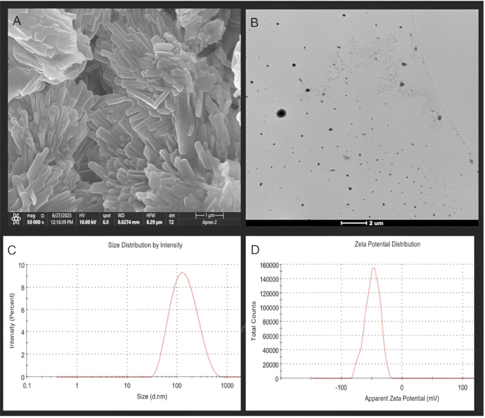
Figure 1: Morphological and physicochemical characterization of NX-ST. (A) Scanning electron microscopy (SEM) image. (B) Transmission electron microscopy (TEM) image. (C) Particle-size distribution (108.3 nm; PDI = 0.336) (n = 1). (D) Zeta-potential distribution (−48.8 mV) (n = 1). Please click here to view a larger version of this figure.
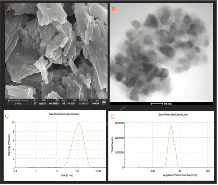
Figure 2: Morphological and physicochemical characterization of NX-CT. (A) SEM image. (B) TEM image. (C) Particle-size distribution (91.87 nm; PDI = 0.235) (n = 1). (D) Zeta-potential distribution (−31.3 mV) (n = 1). Please click here to view a larger version of this figure.
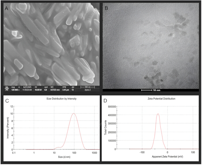
Figure 3: Morphological and physicochemical characterization of NX-CS. (A) SEM image. (B) TEM image. (C) Particle-size distribution (74.72 nm; PDI = 0.244) (n = 1). (D) Zeta-potential distribution (−36.8 mV) (n = 1). Please click here to view a larger version of this figure.
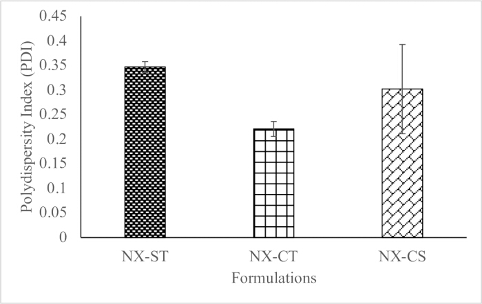
Figure 4: Comparison of PDI among NANOXURIA formulations with different surfactant combinations. PDI values are shown for NX-ST, NX-CT, and NX-CS formulations. Please click here to view a larger version of this figure.
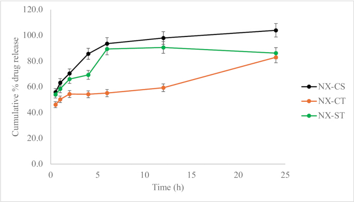
Figure 5: In vitro hydroxyurea release from NANOXURIA formulations. Cumulative hydroxyurea release from NX-CS, NX-CT, and NX-ST is shown over 24 h. Please click here to view a larger version of this figure.
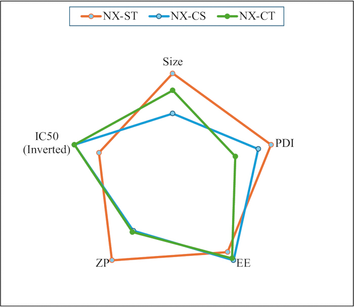
Figure 6: Comparison of physicochemical parameters and cytotoxicity among NANOXURIA formulations. Radar chart comparing particle size, PDI, zeta potential (ZP), encapsulation efficiency (EE), and IC₅₀ for NX-ST, NX-CS, and NX-CT. Please click here to view a larger version of this figure.
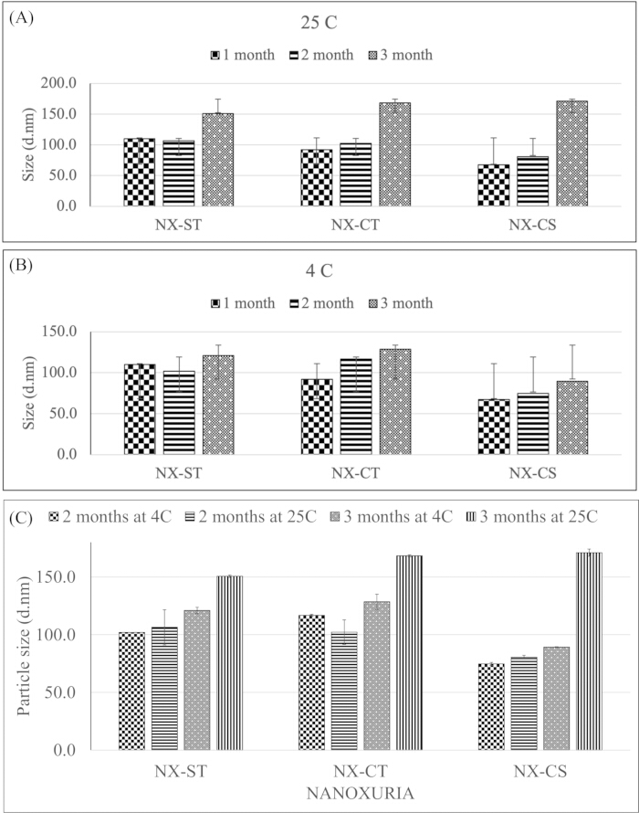
Figure 7: Effect of storage temperature and duration on NANOXURIA particle size. (A) Particle size during storage at 25 °C. (B) Particle size during storage at 4 °C. (C) Comparison of particle size between 4 °C and 25 °C at 2 and 3 months. Please click here to view a larger version of this figure.
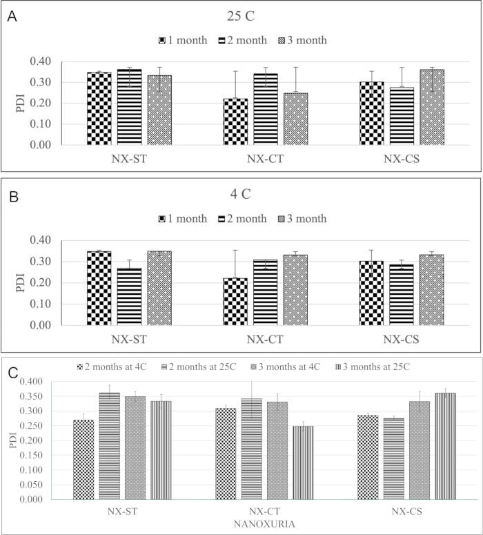
Figure 8: Effect of storage temperature and duration on NANOXURIA PDI. (A) PDI during storage at 25 °C. (B) PDI during storage at 4 °C. (C) Comparison of PDI between 4 °C and 25 °C at 2 and 3 months. Please click here to view a larger version of this figure.
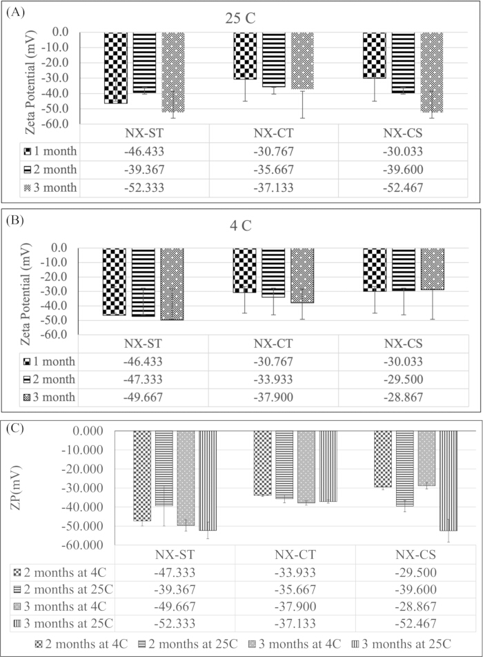
Figure 9: Effect of storage temperature and duration on NANOXURIA zeta potential. (A) Zeta potential during storage at 25 °C. (B) Zeta potential during storage at 4 °C. (C) Comparison of zeta potential between 4 °C and 25 °C at 2 and 3 months. Please click here to view a larger version of this figure.
| Formulations | Tween-80 (µL)* | Sodium dodecyl sulfate (SDS) (mg) | Cremophor EL (µL)# |
| NX-ST | 50 | 100 | - |
| NX-CT | 50 | - | 100 |
| NX-CS | - | 50 | 100 |
| Density (g/ml) = (1.07*, 1.05#) | |||
Table 1: Surfactant composition of NANOXURIA formulations. Amounts of Tween-80, sodium dodecyl sulfate (SDS), and Cremophor EL were used to prepare NX-ST, NX-CT, and NX-CS formulations.
| Formulations | Particle size (d.nm) (n = 3) (± SD) | PDI (n = 3) (± SD) | ZP (mV) (n = 3) (± SD) | EE (%) (n = 3) (± SD) |
| NX-ST | 110 ± 1.997 | 0.347 ± 0.011 | -46.433 ± 8.404 | 78.189 ± 3.49 |
| NX-CT | 91.947 ± 0.215 | 0.221 ± 0.015 | -30.767 ± 0.473 | 84.638 ± 5.0 |
| NX-CS | 67.377 ± 7.158 | 0.302 ± 0.091 | -30.033 ± 5.862 | 86.626 ± 4.676 |
Table 2: Physicochemical characterization of NANOXURIA formulations. Particle size, polydispersity index (PDI), zeta potential (ZP), and encapsulation efficiency (EE) of NX-ST, NX-CT, and NX-CS at 25 °C. Data are presented as mean ± SD (n = 3).
| Formulations | IC50 (µg/mL) | IC50 (µM) |
| NX-ST | 225 ± 21.47 | 2960 |
| NX-CS | >300 | - |
| NX-CT | >300 | - |
| HU | 722.57 | 9500 |
Table 3: In vitro cytotoxicity of NANOXURIA formulations and free hydroxyurea. IC₅₀ values of NX-ST, NX-CS, NX-CT, and free hydroxyurea (HU) against MCF-7 cells, expressed in µg/mL and µM.
Supplementary File 1: Raw data and calculations for the characterization of hydroxyurea-loaded nanostructured lipid carriers. The file contains the underlying experimental data and calculations for the NX-ST, NX-CT, and NX-CS formulations, including particle size, polydispersity index, zeta potential, encapsulation efficiency, drug-release measurements, and formulation-comparison analyses.Please click here to download this file.
Discussion
Proposed approach and key findings
The present study developed NANOXURIA using a hot homogenization approach. Stearic acid was used as the solid lipid, and the preparation temperature was maintained at approximately 70 °C, which closely matches the melting point of stearic acid (~69.3 °C), to ensure complete lipid melting and efficient homogenization while minimizing unnecessary thermal exposure of HU and the surfactants22. The surfactant systems were selected based on their hydrophilic-lipophilic balance (HLB) value, biocompatibility, and suitability for NLC preparation. Preliminary formulation trials were used to identify surfactant concentrations that produce stable nano-dispersions with small particle size and low PDI, while avoiding excessive concentrations that could adversely affect stability or cytocompatibility. The formulations were appropriately diluted with high-purity water before particle-size analysis to minimize multiple-scattering effects that can interfere with accurate determination of particle size and size distribution23.
The three formulations, NX-ST, NX-CT, and NX-CS, differed principally in their surfactant composition. NX-ST contained T80 and SDS, NX-CT contained Cremophor EL and T80, and NX-CS contained Cremophor EL and SDS. This formulation strategy enabled evaluation of the effects of non-ionic and ionic surfactants, as well as their concentrations, on the physicochemical characteristics of HU-loaded NLCs. The results demonstrated clear differences in particle size, PDI, ZP, EE, drug-release behavior, and cytotoxicity among the formulations, indicating that surfactant selection is an important determinant of NLC performance.
Particle size differed significantly among the formulations (p < 0.05), following the order NX-CS < NX-CT < NX-ST. Particle size is an important determinant of nanocarrier performance because smaller particles may provide greater surface area and influence dispersion stability, cellular interactions, biodistribution, and drug-release behavior24,25. NX-CS produced the smallest particles, which may be associated with the high emulsification efficiency of Cremophor EL combined with the moderate concentration of SDS. Cremophor EL can facilitate the formation of smaller droplets by reducing interfacial tension, whereas SDS contributes to electrostatic stabilization. The lower SDS concentration used in NX-CS compared with NX-ST may have provided sufficient interfacial stabilization without producing the excessive interfacial effects associated with the higher SDS concentration26,27,28.
NX-CT produced an intermediate particle size. The combination of Cremophor EL and T80 provides two non-ionic surfactants with complementary interfacial and steric stabilization properties. Cremophor EL contributes strong emulsification, while T80 can form a steric barrier around the particles and reduce coalescence29,30,31. The absence of an ionic surfactant also avoids strong charge-mediated interactions during particle formation. In contrast, NX-ST exhibited the largest particle size. Although SDS is a strong emulsifying surfactant, its relatively high concentration in NX-ST may have altered the interfacial environment and particle-growth behavior. T80 provides steric stabilization, but the combined effect of T80 and the higher SDS concentration may have produced a less favorable balance between emulsification and stabilization than that achieved in NX-CS.
The morphology studies further supported the formation of nanoscale lipid carriers. SEM and TEM showed predominantly spherical to near-spherical particles, with the observed morphology broadly consistent with the particle sizes determined by DLS. Differences between microscopic and DLS measurements are expected because SEM examines dried particles, whereas DLS measures the hydrodynamic diameter of particles together with their associated hydration and surfactant layers. Thus, the combined microscopic and DLS findings support the successful formation of nanoscale NLC systems.
PDI provided additional information about the uniformity of the particle populations. NX-CT exhibited the narrowest particle-size distribution, whereas NX-ST showed the highest mean PDI. NX-CS had an intermediate mean PDI but greater variability between replicate measurements. In particular, the replicate values for NX-CS indicate that the formulation exhibited greater variability than those for NX-ST and NX-CT. The overall PDI trend was NX-CT < NX-CS < NX-ST, although the reported difference was not statistically significant (p > 0.05). The relatively low PDI of NX-CT may be related to the combined steric stabilization provided by Cremophor EL and T80. Both surfactants are non-ionic and can stabilize the particle interface without introducing strong electrostatic interactions. Cremophor EL facilitates emulsification, while T80 provides steric protection, potentially promoting a more homogeneous particle population32,33. The higher PDI of NX-ST may reflect the influence of its higher SDS concentration. SDS provides electrostatic repulsion, but changes in surfactant concentration can also alter micellization and interfacial organization, potentially contributing to particle-size heterogeneity34,35,36. NX-CS showed intermediate PDI behavior, consistent with the combined effects of Cremophor EL and a lower SDS concentration37.
The formulations also differed in surface charge. NX-ST exhibited the highest negative ZP, consistent with the presence of the higher SDS concentration, followed by NX-CT and NX-CS. The higher negative surface charge of SDS-containing systems is expected because SDS is an anionic surfactant. The ZP values, therefore, indicate that SDS contributed substantially to the surface charge of the NLCs, whereas the non-ionic surfactants mainly contributed to steric stabilization. The ZP trend was NX-ST > NX-CT > NX-CS (p < 0.05). However, these differences should not be attributed exclusively to surfactant composition as residual free surfactant, ionic strength, and changes in lipid-surfactant organization can also influence measured ZP38,39,40.
Encapsulation efficiency further demonstrated the influence of composition on HU retention. All formulations exhibited relatively high EE (>75%), indicating successful incorporation of HU into the NLC systems. NX-CS showed the highest EE, followed by NX-CT and NX-ST, with a significant difference among formulations (p < 0.05). Cremophor EL may have contributed to efficient interfacial stabilization and drug retention, while the moderate SDS concentration in NX-CS may have provided additional stabilization without the potentially disruptive effects associated with the higher SDS concentration in NX-ST41,42,43. Nevertheless, because HU is hydrophilic, it is unlikely to be completely dissolved within the lipid matrix. HU may instead be present as a finely dispersed drug, associated with the lipid-water interface, or accommodated within the imperfect lipid matrix and surfactant-rich interfacial region.
The release profiles demonstrated formulation-dependent behavior, with cumulative HU release following the order NX-CS > NX-ST > NX-CT at 24 h. NX-CS exhibited relatively rapid release with an initial burst followed by a plateau, consistent with first-order-like kinetics, whereas NX-CT showed the slowest and most controlled release, approximating zero-order-like kinetics; NX-ST exhibited an intermediate biphasic release profile. The release is likely governed by diffusion of HU from the particle surface and surfactant-rich interfacial region, followed by slower diffusion of entrapped drug through the lipid matrix. The faster release from NX-CS may be related to its smaller particle size, greater surface area, and the combined effects of Cremophor EL and SDS, with SDS increasing interfacial hydrophilicity and Cremophor EL facilitating drug solubilization. In contrast, the combination of Cremophor EL and T80 in NX-CT may provide a more stable interfacial barrier, limiting initial drug diffusion and producing more sustained release, while NX-ST showed an intermediate biphasic pattern attributable to the opposing influences of SDS-facilitated release and T80-mediated stabilization. These interpretations are consistent with the proposed role of surfactants in modulating interactions between the lipid matrix and release medium41,44,45,46,47,48. Water was used as the release medium to establish baseline diffusion behavior, but it does not replicate the ionic composition and physiological conditions of biological fluids. Electrolytes present in physiological buffers can alter the electrical double layer, surfactant organization, particle aggregation, and membrane permeability, potentially modifying release kinetics.
The cytotoxicity results demonstrated a clear difference between free HU and the NLC formulations, with free HU showing the highest IC₅₀ and NX-ST exhibiting the greatest cytotoxic activity. The lower IC₅₀ values observed for the NLC formulations indicate that incorporation of HU into the lipid carrier altered its in vitro activity against MCF-7 cells. The particularly strong response of NX-ST may reflect the combined contribution of its SDS/T80 surfactant system, relatively high negative ZP, and intermediate biphasic release profile, which together may influence drug availability at the cell interface and the temporal exposure of cells to released HU49,50. Interestingly, the smaller particle size and higher EE of NX-CS did not translate into greater cytotoxicity, emphasizing that particle size and drug entrapment alone do not determine biological activity; rather, cellular interactions, drug-release behavior, surfactant effects, and drug availability may collectively influence the response51,52,53. The lower cytotoxicity of NX-CT and NX-CS compared with NX-ST, therefore, should not be interpreted simply as inferior formulation performance, particularly because NX-CT demonstrated the most controlled release.
The storage study demonstrated good physical stability, as no visible agglomeration, sedimentation, phase separation, discoloration, change in viscosity or texture, or abnormal/rancid odor was observed in any formulation throughout storage. However, the physicochemical stability data provided a more differentiated picture. At 25 °C, an increase in particle size and/or PDI indicated progressive changes in the colloidal systems, with the extent and statistical significance varying among formulations. NX-ST benefited from the strong electrostatic contribution of SDS, whereas NX-CT, which relied predominantly on steric stabilization from Cremophor EL and T80, showed greater susceptibility to changes in particle characteristics. NX-CS, despite its combination of electrostatic and steric stabilization, also showed changes during storage, indicating that the presence of two stabilization mechanisms does not completely prevent time-dependent restructuring of the NLC system. Storage at 4 °C generally provided better physicochemical preservation, consistent with reduced molecular mobility and slower lipid structural changes; NX-CS showed comparatively improved particle-size stability, while NX-ST maintained relatively consistent characteristics. Nevertheless, NX-CT continued to show progressive changes in PDI, indicating that steric stabilization alone may not be sufficient for prolonged storage. Changes in ZP during storage further suggest time-dependent reorganization of the lipid-surfactant interface, potentially related to surfactant desorption, lipid recrystallization, and HU-associated structural changes, which were not directly established47,48,54.
Advancement of the field
NLC formulations of other anticancer agents, including gefitinib, capecitabine, and imatinib, have previously demonstrated improved delivery relative to the free drug14,15,16, but comparative data on how different surfactant combinations shape the physicochemical and cytotoxic profile of hydroxyurea-loaded NLCs specifically have been limited43,44,55. By systematically comparing three surfactant systems within a single NLC platform, this study provides a direct, internally controlled comparison clarifying how surfactant ionic character and concentration jointly influence particle size, colloidal stability, drug retention, and in vitro anticancer activity for a small, highly hydrophilic drug - a combination less well represented in the NLC literature than lipophilic payloads51,54,56,57. These results extend prior reports of HU encapsulation in lipid and albumin-based nanocarriers53,58 by identifying a surfactant pairing (T80–SDS) associated with the greatest in vitro cytotoxic enhancement.
Study limitations
This study has several limitations that should be considered when interpreting the findings. First, the biological evaluation was restricted to a single in vitro MTT assay in MCF-7 breast cancer cells; although HU is used clinically in chronic myeloid leukemia and sickle cell disease, the present data do not support conclusions specific to these conditions, and confirmation in a relevant leukemia model (e.g., K562 cells) and in vivo is warranted. Second, the mechanistic explanations offered for the observed physicochemical and cytotoxic differences (e.g., surfactant desorption, lipid recrystallization, altered cellular uptake) were not directly demonstrated and should be regarded as hypotheses; techniques such as DSC, XRD, and Cryo-TEM23,41, together with cellular-uptake and release-kinetics studies, would be required to substantiate them. Third, in vitro release was assessed in water rather than a physiological buffer, which is considered an essential step for predicting in vivo performance59. Fourth, the chemical stability of HU during preparation was not directly quantified, and a dedicated assessment of drug leakage into the aqueous phase during storage was not conducted; given HU's high aqueous solubility, such leakage is possible and should be assessed. Finally, the formulation is intended for parenteral administration, and the mechanistic interpretations offered here should be viewed as exploratory pending further characterization, consistent with the broader NLC literature emphasizing the need for rigorous physicochemical validation32.
Alternative approaches
Several alternative characterization and validation strategies could strengthen future work in this area. Solid-state techniques (e.g., DSC, XRD) and cryo-TEM would help clarify the physical state of HU within the lipid matrix and directly test the surfactant-desorption and lipid-recrystallization mechanisms proposed to explain the observed stability trends36,60,61. Release testing in physiological buffer or simulated biological fluids, rather than water alone, would better predict in vivo release behavior, and direct quantification of cellular uptake (e.g., by fluorescent labeling and flow cytometry) would test the uptake-based explanation proposed for NX-ST's enhanced cytotoxicity rather than inferring it from zeta potential alone62,63,64. Confirmatory testing in a second, clinically relevant cell line (e.g., K562 for chronic myeloid leukemia) and evaluation under formal accelerated stability conditions would further help translate the present in vitro findings toward a clinically applicable formulation.
Importance and potential applications
These findings are relevant to the broader effort to improve hydroxyurea delivery, whose clinical use is currently constrained by rapid clearance, variable bioavailability, and dose-related toxicity. By showing that surfactant composition can be tuned to favor either smaller, more stable particles with higher encapsulation efficiency (NX-CS, NX-CT) or greater in vitro cytotoxic potency (NX-ST)41,65, this work provides a practical basis for selecting or further optimizing NLC composition according to the delivery attribute prioritized for a given application, without modifying the HU molecule itself. Tunable nanocarrier platforms of this type may also be applicable to other small, hydrophilic drugs that face similar delivery limitations.
Future directions
Future work should prioritize quantitative validation of the physicochemical mechanisms proposed here using solid-state and imaging techniques; drug-release testing under physiologically relevant conditions; direct measurement of cellular uptake and its relationship to cytotoxicity; testing in additional, clinically relevant cell lines and, eventually, in vivo models; formal accelerated and long-term stability studies to support shelf-life determination; and quantification of HU chemical stability and storage-related leakage. Addressing these points would help establish whether the physicochemical advantages identified here translate into meaningful therapeutic benefit.
Conclusions
This study addresses key limitations of HU therapy, including poor delivery efficiency due to rapid clearance, variable bioavailability, and dose-related toxicity, by developing nanostructured lipid carriers (NANOXURIA). The findings demonstrate that surfactant composition and storage conditions critically influence physicochemical stability, cytotoxic performance, and overall delivery efficiency. Among the formulations, NX-ST exhibited superior performance, with 3.2 times the cytotoxic activity of free HU, indicating enhanced cellular uptake and drug delivery. This improved efficacy is attributed to its balanced particle characteristics, controlled release, and effective electrostatic stabilization.
In contrast, formulations relying primarily on non-ionic surfactants showed comparatively lower stability and performance, highlighting the importance of combined stabilization mechanisms. Storage conditions significantly affected formulation behavior: 25 °C promoted particle growth and heterogeneity, whereas 4 °C better preserved stability, uniformity, and surface charge.
Overall, this study addresses the identified research gap by demonstrating that rationally designed NLC systems can enhance HU delivery without altering the drug itself. The results emphasize that optimum lipid-surfactant combinations and appropriate storage conditions are essential for achieving stable, efficient, and clinically relevant drug delivery systems.
Disclosures
All authors declare that they have no conflict of interest.
Acknowledgements
The authors extend their appreciation to the Deanship of Graduate Studies and Scientific Research at Jazan University for funding this research work through the project number: JU-RSP2026. The authors also extend their sincere gratitude to the College of Pharmacy, Jazan University, Jazan, Saudi Arabia, for providing the essential resources that facilitated the successful completion of this project.
Materials
| Name | Company | Catalog Number | Comments |
|---|---|---|---|
| Amicon Ultra-2 mL centrifugal filter (3 kDa MWCO) | Millipore, Ireland | UFC200324 | |
| Carbon-coated copper grid (TEM sample support) | N/A | ||
| Cremophor EL (non-ionic surfactant, castor oil derivative, assay ≥98%) | MedChemExpress, NJ, USA | HY-Y1890 | |
| Dialysis bags (12-14 kDa MWCO) | Spectrum Laboratories, CA, USA | 08-670-3BB | |
| Dimethyl sulfoxide (DMSO) | Fisher Chemicals, UK | 10162090 | |
| Fetal bovine serum (FBS) | Gibco, Paisley, UK | 10270-106 | |
| Field emission scanning electron microscope (FE-SEM), Apreo 2 | Thermo Fisher-FEI, USA | 1229000 | |
| Homogenizer, HG 15D | WiseTis, Germany | DH.WHG02020 | |
| Hydroxyurea (HU) (assay ≥99%) | Sudair Pharma, Riyadh, Saudi Arabia | 21L001 | |
| MCF-7 human breast cancer cell line | American Type Culture Collection (ATCC), USA | HTB-22 | |
| Microplate reader, SpectroStar Nano | BMG LABTECH, Ortenberg, Germany | 601-101 | |
| MTT reagent (5 mg/mL) | Invitrogen Corporation, San Diego, CA, USA | M6494 | |
| Penicillin-streptomycin solution | Sigma-Aldrich, St. Louis, MO, USA | P4333 | |
| RPMI 1640 medium | Gibco, Grand Island, NY, USA | 11875093 | |
| Sesame oil | Local merchant, Jazan, Saudi Arabia | N/A | |
| Sodium dodecyl sulfate (SDS) (anionic surfactant, assay 90%) | PanReac AppliChem, Darmstadt, Germany | 28312 | |
| Stearic acid (assay 95-106%) | Himedia, Mumbai, India | GRM1425 | |
| Syringe filter (0.45 µm) | Merck, SA | SLHV033RS | |
| Transmission electron microscope (TEM), Talos | Thermo Fisher, USA | N/A | |
| Tween-80 (T80) (non-ionic surfactant, oleic acid ≥58%) | Loba Chemie, Mumbai, India | Art. No. 06420 | |
| UV-visible spectrophotometer | Shimadzu, Kyoto, Japan | 220-92961-01 | |
| Zetasizer Nano ZS90 | Malvern Panalytical, Malvern, UK | ZEN3690 |
References
- Kushwaha AK, et al. A descriptive cross-sectional study on clinical epidemiology of different types of cancer in a tertiary care centre: insights from Eastern India. Cureus. 2024;16(6):e62529.
- National Cancer Institute. Cancer statistics. 2024. Available from: https://www.cancer.gov/about-cancer/understanding/statistics
- Bai X, et al. Sustained drug release from smart nanoparticles in cancer therapy: a comprehensive review. Micromachines. 2022;13(10):1623.
- Rajendra PKM, Balan AP, Selvaraj J, Jawahar N. Formulation and optimization of hydroxyurea loaded nanostructured lipid carriers using design of experiment for the effective treatment of ovarian cancer. Int J Appl Pharm. 2022;14(5):137-43.
- Bell V, Varzakas T, Psaltopoulou T, Fernandes T. Sickle cell disease update: new treatments and challenging nutritional interventions. Nutrients. 2024;16(2):258.
- Ware RE. How I use hydroxyurea to treat young patients with sickle cell anemia. Blood. 2010;115(26):5300-11.
- Lanzkron S, et al. Systematic review: hydroxyurea for the treatment of adults with sickle cell disease. Ann Intern Med. 2008;148(12):939-55.
- Pouton CW, Porter CJH. Formulation of lipid-based delivery systems for oral administration: materials, methods and strategies. Adv Drug Deliv Rev. 2008;60(6):625-37.
- Khan MS, et al. Potential of lipid-based nanocarriers against two major barriers to drug delivery-skin and blood-brain barrier. Membranes. 2023;13(3):343.
- Alam MI, et al. Nanostructured lipid carrier containing CNS acting drug: formulation, optimization and evaluation. Curr Nanosci. 2011;7(6):1014-27.
- Azhar SNAS, Ashari SE, Zainuddin N, Hassan M. Nanostructured lipid carriers-hydrogels system for drug delivery: nanohybrid technology perspective. Molecules. 2022;27(1):289.
- Haider M, Abdin SM, Kamal L, Orive G. Nanostructured lipid carriers for delivery of chemotherapeutics: a review. Pharmaceutics. 2020;12(3):288.
- Müller RH, Radtke M, Wissing SA. Nanostructured lipid matrices for improved microencapsulation of drugs. Int J Pharm. 2002;242(1-2):121-8.
- Makeen HA, et al. Preparation, characterization, and anti-cancer activity of nanostructured lipid carriers containing imatinib. Pharmaceutics. 2021;13(7):1086.
- Makeen HA, et al. Gefitinib loaded nanostructured lipid carriers: characterization, evaluation and anti-human colon cancer activity in vitro. Drug Deliv. 2020;27(1):622-31.
- Sultan MH, et al. Capecitabine-loaded NLC for breast cancer treatment: preparation, characterization, and in vitro evaluation. Curr Drug Deliv. 2025;22(7):968-82.
- Coache D, et al. Stability evaluation of compounded hydroxyurea 100 mg/mL oral liquids using a novel analytical method involving chemical derivatization. PLoS One. 2022;17(6):e0270206.
- Yasir MS, et al. Microemulsified gel formulations for topical delivery of clotrimazole: structural and in vitro evaluation. Langmuir. 2021;37(46):13767-77.
- Siddique MY, et al. Formulation of gelled microemulsion for effective permeation of celecoxib across the skin barrier. ChemistrySelect. 2024;9(3):e202302841.
- Naseem K, et al. Enzymes encapsulated smart polymer micro assemblies and their tuned multi-functionalities: a critical review. Int J Polym Mater Polym Biomater. 2024;73(9):785-816.
- Syam S, Abdelwahab SI, Al-Mamary MA, Mohan S. Synthesis of chalcones with anticancer activities. Molecules. 2012;17(6):6179-95.
- Lide DR. CRC handbook of chemistry and physics. CRC Press; Boca Raton, FL; 2009.
- Pei Y, Hinchliffe BA, Minelli C. Measurement of the size distribution of multimodal colloidal systems by laser diffraction. ACS Omega. 2021;6(22):14049-58.
- Mok ZH. The effect of particle size on drug bioavailability in various parts of the body. Pharm Sci Adv. 2024;2:100031.
- Zhang M, et al. Influencing factors and strategies of enhancing nanoparticles into tumors in vivo. Acta Pharm Sin B. 2021;11(8):2265-85. doi:10.1016/j.apsb.2021.03.033.
- Zhang Y, et al. Sodium dodecyl sulfate improved stability and transdermal delivery of salidroside-encapsulated niosomes via effects on zeta potential. Int J Pharm. 2020;580:119183.
- Westesen K, Bunjes H, Koch MHJ. Physicochemical characterization of lipid nanoparticles and evaluation of their drug loading capacity and sustained release potential. J Control Release. 1997;48(2-3):223-36.
- Bjørnestad VA, Lund R. Pathways of membrane solubilization: a structural study of model lipid vesicles exposed to classical detergents. Langmuir. 2023;39(11):3914-33.
- Rao VRS, Shao J. Self-nanoemulsifying drug delivery systems (SNEDDS) for oral delivery of protein drugs. Int J Pharm. 2008;362(1-2):2-9.
- Weiszhár Z, et al. Complement activation by polyethoxylated pharmaceutical surfactants: Cremophor-EL, Tween-80 and Tween-20. Eur J Pharm Sci. 2012;45(4):492-8.
- Khan ZU, et al. Physicochemical characterizations and pharmacokinetic evaluation of pentazocine solid lipid nanoparticles against inflammatory pain model. Pharmaceutics. 2022;14(2):409.
- Beloqui A, et al. Nanostructured lipid carriers: promising drug delivery systems for future clinics. Nanomedicine. 2016;12(1):143-61.
- Samari-Kermani M, Jafari S, Rahnama M, Raoof A. Ionic strength and zeta potential effects on colloid transport and retention processes. Colloid Interface Sci Commun. 2021;42:100389.
- Duplâtre G, Ferreira Marques MF, Da Graça Miguel M. Size of sodium dodecyl sulfate micelles in aqueous solutions as studied by positron annihilation lifetime spectroscopy. J Phys Chem. 1996;100(41):16608-12.
- Hanafy AS, Farid RM, ElGamal SS. Complexation as an approach to entrap cationic drugs into cationic nanoparticles administered intranasally for Alzheimer’s disease management: preparation and detection in rat brain. Drug Dev Ind Pharm. 2015;41(12):2055-68.
- Witayaudom P, Klinkesorn U. Effect of surfactant concentration and solidification temperature on the characteristics and stability of nanostructured lipid carrier prepared from rambutan kernel fat. J Colloid Interface Sci. 2017;505:1082-92.
- Öztürk K, Kaplan M, Çalış S. Effects of nanoparticle size, shape, and zeta potential on drug delivery. Int J Pharm. 2024;666:124799.
- Lavagna, E., Bochicchio, D., De Marco, A.L., Güven, Z.P., Stellacci, F. and Rossi, G., 2022. Ion-bridges and lipids drive aggregation of same-charge nanoparticles on lipid membranes. Nanoscale, 14.(18), pp.6912-6921.
- Ye J, et al. Comparative colloidal stability of commercial amphotericin B nanoformulations using dynamic and static multiple light scattering techniques. Int J Nanomedicine. 2022;17:6047-64.
- Honary S, Zahir F. Effect of zeta potential on the properties of nano-drug delivery systems-a review (Part 2). Trop J Pharm Res. 2013;12(2):265-73.
- Doktorovova S, Souto EB, Silva AM. Nanotoxicology applied to solid lipid nanoparticles and nanostructured lipid carriers-a systematic review of in vitro data. Eur J Pharm Biopharm. 2014;87(1):1-18.
- Bnyan R, et al. Surfactant effects on lipid-based vesicles properties. J Pharm Sci. 2018;107(5):1237-46.
- Adepu S, Ramakrishna S. Controlled drug delivery systems: current status and future directions. Molecules. 2021;26(19):5905.
- Müller RH, Mäder K, Gohla S. Solid lipid nanoparticles for controlled drug delivery-a review of the state of the art. Eur J Pharm Biopharm. 2000;50(1):161-77.
- Das S, et al. Formulation design, preparation and physicochemical characterizations of solid lipid nanoparticles containing a hydrophobic drug: effects of process variables. Colloids Surf B Biointerfaces. 2011;88(1):483-9.
- Naseri N, Valizadeh H, Zakeri-Milani P. Solid lipid nanoparticles and nanostructured lipid carriers: structure, preparation and application. Adv Pharm Bull. 2015;5(3):305-13.
- Pardeike J, Hommoss A, Müller RH. Lipid nanoparticles in cosmetic and pharmaceutical dermal products. Int J Pharm. 2009;366(1-2):170-84.
- Chauhan I, et al. Nanostructured lipid carriers: a groundbreaking approach for transdermal drug delivery. Adv Pharm Bull. 2020;10(2):150-65.
- Ali A, et al. Solid lipid-based nanoparticular system for sustained release and enhanced in vitro cytotoxic effect of 5-fluorouracil on skin melanoma and squamous cell carcinoma. PLoS One. 2023;18(2):e0281004.
- Miyazawa T, et al. A critical review of the use of surfactant-coated nanoparticles in nanomedicine and food nanotechnology. Int J Nanomedicine. 2021;16:3937-99.
- Garg J, Pathania K, Sah SP, Pawar SV. Nanostructured lipid carriers: a promising drug carrier for targeting brain tumors. Future J Pharm Sci. 2022;8(1):25.
- Raza F, et al. Recent advances in the targeted delivery of paclitaxel nanomedicine for cancer therapy. Mater Adv. 2022;3(5):2268-90.
- Alam MI, et al. Determining the anticancer effect of gefitinib-loaded NLCs through investigating the inflammatory cytokine (JAK-STAT) pathway. Res J Pharm Technol. 2024;17(11):4968-74.
- Akbari A, et al. Development and characterization of nanoliposomal hydroxyurea against BT-474 breast cancer cells. Adv Pharm Bull. 2020;10(1):39-45.
- Iqbal MA, et al. Nanostructured lipid carriers system: recent advances in drug delivery. J Drug Target. 2012;20(10):813-30.
- Heeney MM, et al. Chemical and functional analysis of hydroxyurea oral solutions. J Pediatr Hematol Oncol. 2004;26(3):179-84.
- Santos SS, Dos Santos IG, Da Silva CV, Cazedey ECL. Qualitative analysis of hydroxyurea. Drug Anal Res. 2020;4(1):18-21.
- Tazhbayev Y, Mukashev O, Burkeev M, Kreuter J. Hydroxyurea-loaded albumin nanoparticles: preparation, characterization, and in vitro studies. Pharmaceutics. 2019;11(8):410.
- Sato H, Yamada K, Miyake M, Onoue S. Recent advancements in the development of nanocarriers for mucosal drug delivery systems to control oral absorption. Pharmaceutics. 2023;15(12):2708.
- Mehnert W, Mäder K. Solid lipid nanoparticles: production, characterization and applications. Adv Drug Deliv Rev. 2001;47(2-3):165-96.
- Danaei M, et al. Impact of particle size and polydispersity index on the clinical applications of lipidic nanocarrier systems. Pharmaceutics. 2018;10(2):57.
- Fröhlich E. The role of surface charge in cellular uptake and cytotoxicity of medical nanoparticles. Int J Nanomedicine. 2012;7:5577-91.
- Zhang W, et al. Effects of morphology and size of nanoscale drug carriers on cellular uptake and internalization process: a review. RSC Adv. 2023;13(1):80-114.
- Wang HX, et al. Surface charge critically affects tumor penetration and therapeutic efficacy of cancer nanomedicines. Nano Today. 2016;11(2):133-44.
- Musielak E, Feliczak-Guzik A, Nowak I. Optimization of the conditions of solid lipid nanoparticles synthesis. Molecules. 2022;27(7):2202.