This study presents a reproducible retrospective workflow for integrating pre-grafting inflammatory biomarkers, endotoxin, β-D-glucan, and wound culture into a composite score associated with autologous skin graft take in adults with severe burns.
Research Article
Pre-Grafting Infection Biomarker Burden and Autologous Skin Graft Take in Adults with Severe Burns: A Single-Center Retrospective Cohort Study
52 views
⸱
September 11th, 2026
In This Article
Summary
Abstract
Autologous split-thickness skin grafting is a principal method of definitive wound closure in patients with severe burns, but graft take may be compromised by systemic inflammation, local microbial burden, and wound-bed instability. This retrospective cohort study evaluated whether a composite pre-grafting infection biomarker burden score was associated with graft take after definitive autologous grafting. Adult patients with severe or extremely severe burns treated at Ganzhou People’s Hospital between January 1, 2022, and December 31, 2025, were screened. The score assigned one point each for C-reactive protein ≥ 150 mg/L, procalcitonin ≥ 1.5 ng/mL, endotoxin ≥ 0.25 EU/mL, fungal β-D-glucan ≥ 100 pg/mL, and a positive pre-grafting wound culture. The primary outcome was graft take rate at the first formal assessment within postoperative days 5–10, using postoperative day 7 when available. Among 84 included patients, the mean age was 46.2 ± 15.3 years, the mean total body surface area burned was 50.6% ± 10.5%, and the mean graft take rate was 87.5% ± 9.5%. Thirty-four patients achieved graft take ≥ 90%. Mean graft take declined from 92.5% ± 6.0% in patients with scores of 0–1 to 75.7% ± 11.8% in those with scores of 4–5. After adjustment for prespecified clinical covariates, each one-point increase in the score was associated with a 3.84-percentage-point decrease in graft take rate. Higher pre-grafting infection biomarker burden was therefore associated with poorer graft take and may support structured perioperative risk assessment in adults with severe burns.
Introduction
Burn injury remains a major cause of preventable mortality, disability, disfigurement, and long-term functional impairment worldwide, with the greatest clinical burden occurring among patients with extensive or deep burns requiring specialized surgical treatment1. Severe burns simultaneously produce tissue destruction, fluid and protein loss, systemic inflammation, metabolic stress, immune dysfunction, and increased susceptibility to infection2. Timely wound closure is therefore essential not only for reconstruction but also for reducing exposed wound area, limiting ongoing fluid and heat loss, controlling microbial invasion, and supporting recovery. Autologous split-thickness skin grafting remains a principal method of definitive wound closure because it permits coverage of relatively large wound surfaces while preserving donor-site regeneration3.
Successful graft take requires close contact between the graft and a viable, adequately perfused recipient bed during plasma imbibition, inosculation, and revascularization. Graft adherence and survival are influenced by burn extent and depth, wound-bed vascularity, timing of excision and grafting, nutritional status, systemic illness, hematoma, seroma, edema, mechanical shear, and fixation quality4. In clinical practice, graft readiness is judged from wound appearance, punctate bleeding after debridement, granulation quality, exudate, surrounding inflammation, microbiological findings, and overall physiological condition. These assessments remain partly subjective. Patients with apparently acceptable wound beds may still experience partial graft loss. Infection is especially relevant because local contamination and systemic infection-related responses can disrupt perfusion, amplify tissue injury, increase exudation, and impair wound repair5.
Accordingly, graft outcome should be interpreted within a broader clinical context that includes age, diabetes, nutritional status, total body surface area and burn depth, inhalation injury, timing of excision and grafting, anemia, wound-bed granulation and perfusion, hemodynamic stability, vasopressor exposure when relevant, and postoperative fixation and dressing technique. These factors may confound or modify associations between infection-related biomarkers and graft take.
Burn wounds provide a protein-rich and immunologically vulnerable environment in which microbial colonization may develop rapidly. Repeated operations, prolonged hospitalization, invasive devices, broad-spectrum antimicrobial exposure, and delayed wound closure further alter the wound microbiome and increase the risk of resistant organisms6. Clinically important burn-wound pathogens include Pseudomonas aeruginosa, Acinetobacter baumannii, Staphylococcus aureus, and Klebsiella pneumoniae, which may persist through biofilm formation, antimicrobial tolerance, and repeated wound contamination6. Conventional wound culture remains clinically useful, but its interpretation is limited. A positive culture may represent colonization rather than invasive infection, whereas a negative culture may reflect recent antimicrobial therapy, superficial sampling, wound cleansing, or low organism recovery. Microbiological findings should therefore be interpreted together with systemic and local clinical evidence rather than used as an isolated measure of graft readiness.
Inflammatory and infection-related biomarkers provide additional information, although no individual marker can fully distinguish burn-induced inflammation from infection. C-reactive protein is sensitive to inflammatory activation but may remain markedly elevated after extensive tissue injury, debridement, or surgery. Procalcitonin is considered more closely related to bacterial infection and sepsis, but its diagnostic performance varies according to burn severity, sampling time, infection definition, organ dysfunction, and antimicrobial treatment7. Systematic evidence suggests that procalcitonin can contribute to infection assessment in burn populations, but its interpretation is most reliable when combined with clinical findings and other laboratory indicators8. Endotoxin testing may reflect exposure to gram-negative bacterial components or translocation, whereas fungal β-D-glucan may support investigation of invasive fungal disease in selected high-risk patients. Both tests are influenced by patient selection, assay availability, treatment exposure, and noninfectious causes of abnormal results9.
The five score components were selected to represent complementary infection-related domains rather than interchangeable laboratory tests: systemic inflammatory activity (C-reactive protein), bacterial systemic response (procalcitonin), gram-negative bacterial product exposure (endotoxin), fungal cell-wall antigen burden (β-D-glucan), and local wound microbiology (wound culture). White blood cell count and neutrophil percentage were retained as secondary descriptive variables because they are highly responsive to burn injury and physiological stress and were not intended to duplicate the systemic inflammatory component of the score. Interleukin-6 was not included because it was not part of the prespecified routinely available pre-grafting dataset used for complete-case score construction in this retrospective cohort.
Evidence directly connecting pre-grafting microbial status with graft survival remains comparatively limited. Previous burn research has shown that bacterial colonization of the recipient wound can be associated with subsequent skin-graft loss, supporting the biological relevance of local microbial burden before definitive coverage10. However, studies have generally evaluated wound culture, individual inflammatory markers, or broad clinical risk factors separately. This approach may not adequately represent the cumulative infection-related condition of a severely burned patient. Perioperative stabilization measures, including secure fixation and negative-pressure wound therapy, can improve graft contact and fluid control in selected wounds, but they cannot fully compensate for uncontrolled microbial burden or an unstable recipient bed11. A composite measure integrating systemic inflammatory markers, organism-related biomarkers, and wound microbiology may therefore provide a more clinically interpretable estimate of pre-grafting infection burden.
The present study developed a five-component infection biomarker burden score based on pre-grafting C-reactive protein, procalcitonin, endotoxin, fungal β-D-glucan, and wound culture results. The primary objective was to determine whether increasing score burden was associated with lower autologous split-thickness skin graft take in adults with severe or extremely severe burns. Individual score components and clinically interpretable graft-take categories were also examined to characterize the pattern and stability of the association. It was hypothesized that patients with greater cumulative pre-grafting infection biomarker burden would have lower graft take rates after definitive autologous skin grafting.
The primary endpoint was graft take rate, defined as the percentage of the grafted wound surface showing viable adherence to the recipient bed at the first formal postoperative assessment within days 5–10; postoperative day 7 was used when an assessment was available on that day. This score was newly developed for the present study and was intended as an exploratory burden index rather than a previously validated clinical prediction tool.
Protocol
The study protocol was reviewed and approved by the Ethics Committee of Ganzhou People’s Hospital (Approval No. SZITCM-EC-2025-017; approval date: 2025.04.12). The requirement for individual written informed consent was waived because the study involved retrospective analysis of routinely collected clinical data without additional sampling, intervention, treatment allocation, or patient contact.
Study design, setting, ethics, and data protection
This single-center retrospective cohort study was conducted in the Department of Burns, Plastic Surgery, and Wound Repair, Ganzhou People’s Hospital, Ganzhou, Jiangxi Province, China. Consecutive inpatient records of adults with severe or extremely severe burns treated between January 1, 2022, and December 31, 2025, were screened using the electronic medical record system, laboratory information system, microbiology database, operative and anesthesia records, pharmacy records, and inpatient wound-care documentation.
Patients were managed according to routine institutional burn-care pathways during the study period; however, graft timing, wound-bed preparation, antimicrobial treatment, and fixation decisions were individualized by the treating burn-surgery team rather than mandated by a study-specific intervention protocol.
The primary exposure was the pre-grafting infection biomarker burden score derived from laboratory and microbiological findings obtained before the index grafting procedure. The primary outcome was graft take rate at the first formal postoperative assessment between postoperative days 5 and 10, with the postoperative day 7 assessment used whenever available. The study was reported in accordance with the Strengthening the Reporting of Observational Studies in Epidemiology recommendations for observational cohort studies12.
Each eligible patient was assigned a unique study code before analysis. Direct identifiers were removed from the analytical dataset, and the linkage file connecting study codes with original medical records was stored separately on a password-protected institutional computer accessible only to authorized investigators. Results were reported in aggregate form.
Patient eligibility and index procedure
Patients were eligible if they were aged ≥ 18 years, were admitted during the study period with a diagnosis of severe or extremely severe burn, underwent definitive autologous split-thickness skin grafting during the index hospitalization, had complete documentation of the index operation, had complete data for the five infection biomarker burden score components, and underwent formal graft take assessment between postoperative days 5 and 10.
Burn severity was determined from the admission diagnosis and verified using total body surface area burned, full-thickness burn percentage, inhalation injury, burn shock, organ dysfunction, and requirement for intensive burn care. An extremely severe burn was defined as an injury involving ≥50% total body surface area, a full-thickness injury involving ≥20% total body surface area, or a burn injury accompanied by severe inhalation injury, early organ dysfunction, burn shock, or intensive care requirement. Severe burn was defined as injury involving ≥30% total body surface area, full-thickness injury involving ≥10% total body surface area, or a burn of lower extent accompanied by inhalation injury, burn shock, major organ dysfunction, or functionally critical anatomical involvement requiring specialized burn management13.
This classification was used as a prespecified operational eligibility definition and did not rely on total body surface area alone. It incorporated burn extent, full-thickness involvement, inhalation injury, shock, organ dysfunction, and intensive-care requirement. The abbreviated burn severity index (ABSI), which is principally a prognostic mortality-risk score, was not used as the eligibility definition in this retrospective cohort; this difference from ABSI-based classification is acknowledged when interpreting generalizability.
Patients were excluded if they were younger than 18 years, underwent grafting for a non-burn wound, were readmitted for reconstruction of a previous burn injury, underwent isolated grafting for scar revision or chronic wound reconstruction, or received only allogeneic skin, artificial dermis, or temporary wound coverage without definitive autologous grafting. Patients were also excluded if any infection biomarker burden score component was unavailable, if operative documentation was insufficient, if graft take was not assessed within the predefined postoperative window, or if death or transfer occurred before outcome assessment.
The index procedure was defined as the first definitive autologous split-thickness skin grafting operation performed for wound closure during the index hospitalization. When multiple grafting procedures were performed, only the first definitive autologous grafting procedure was included in the primary patient-level analysis. All preoperative biomarkers, microbiological findings, operative characteristics, and postoperative outcomes were linked to this procedure. The patient-screening process is shown in Figure 1.
The screening denominator before sequential exclusions was 286 burn-related inpatient records. Of the 104 patients who reached definitive autologous split-thickness skin grafting in the screening flow, 9 (8.7%) were excluded specifically because one or more of the five score components were unavailable; these 9 patients accounted for 3.1% of the 286 screened records.
Clinical and perioperative variables
Demographic and comorbidity variables included age, sex, body mass index, smoking history, alcohol use, diabetes mellitus, hypertension, cardiovascular disease, chronic kidney disease, chronic liver disease, and other major conditions documented in the inpatient record. Diabetes mellitus was defined by a previous diagnosis, use of antidiabetic medication, or confirmation during the index hospitalization.
Burn-related variables included burn mechanism, total body surface area burned, full-thickness burn percentage, anatomical distribution, time from injury to admission, inhalation injury, burn shock, intensive care unit admission, mechanical ventilation, and the number of debridement or wound-bed preparation procedures performed before the index operation. When more than one estimate of burn extent was documented, the value recorded after formal burn reassessment and before grafting was used. Burn area was assessed by the treating burn team using the Lund–Browder chart or the rule-of-nines method, as appropriate14. Inhalation injury was defined by bronchoscopic evidence of airway injury, documented airway burn, or an explicit diagnosis by the treating burn team based on compatible exposure and respiratory findings.
Perioperative variables included the operation date, time from admission to grafting, grafted anatomical region, grafted area, graft thickness, meshing ratio, wound-bed preparation method, perioperative blood transfusion, postoperative fixation method, and use of negative-pressure wound therapy. Systemic antibacterial and antifungal therapy administered within 72 h before and 72 h after grafting was recorded. Time to grafting was calculated as the number of calendar days from hospital admission to the index operation. Serum albumin and hemoglobin were obtained from the 48 h pre-grafting window and treated as perioperative nutritional and hematological covariates. The operational definitions, extraction windows, and data sources are summarized in Table 1.
Pre-grafting infection assessment and biomarker burden score
The primary exposure window for blood-based infection-related biomarkers was the 48 h immediately preceding the recorded start time of the index grafting procedure. When multiple results were available, the measurement obtained closest to surgery was selected. Results obtained more than 48 h before grafting were not included in the primary analysis. The closest result obtained within 72 h before grafting was used only in the expanded-window sensitivity analysis.
All blood-based score components used in the primary analysis were selected from the prespecified 48 h immediately preceding grafting, with the measurement closest to the recorded start time of the index operation used for each component. For descriptive timing, the interval between the most recent selected blood-based score-component measurement and the recorded start time of grafting was calculated for each patient.
The blood-based score components were C-reactive protein, procalcitonin, endotoxin, and fungal β-D-glucan. C-reactive protein was recorded in micrograms per liter (mg/L), procalcitonin in nanograms per milliliter (ng/mL), endotoxin in endotoxin units per milliliter (EU/mL), and fungal β-D-glucan in picograms per milliliter (pg/mL). Additional laboratory variables extracted within the 48 h window included white blood cell count, neutrophil percentage, lymphocyte count, hemoglobin, platelet count, coagulation indices, electrolytes, albumin, prealbumin, renal and liver function variables, and blood glucose.
Laboratory results were obtained directly from the central laboratory information system. Values outside clinically plausible ranges were verified against the original reports, and confirmed transcription or unit-conversion errors were corrected. Values that could not be verified were coded as missing. C-reactive protein and procalcitonin were interpreted as infection-related rather than infection-specific indicators because burn-related tissue injury and systemic inflammation may independently increase their concentrations15.
Wound culture results were obtained from the microbiology database. An eligible culture was defined as a wound specimen collected within 7 days before the index operation. When multiple cultures were available, the result obtained closest to grafting was selected. Wound specimens were collected during routine clinical care when infection, persistent exudation, delayed wound-bed preparation, tissue deterioration, or abnormal infection-related biomarkers were suspected. Superficial exudate was removed, and the wound surface was cleansed before sampling.
Culture results were classified as negative, single-organism positive, or polymicrobial positive. Positive cultures were further categorized as gram-positive bacterial, gram-negative bacterial, mixed bacterial, or fungal growth. Recorded organisms included Pseudomonas aeruginosa, Acinetobacter baumannii, Staphylococcus aureus, Klebsiella pneumoniae, Escherichia coli, Enterococcus species, Candida species, and other clinically relevant isolates. Cultures containing more than one organism were classified as polymicrobial. Systemic antibacterial and antifungal exposure within 72 h before grafting was recorded because recent treatment could affect microbial recovery and interpretation16.
The infection biomarker burden score assigned 1 point for each of the following findings: C-reactive protein ≥ 150 mg/L, procalcitonin ≥ 1.5 ng/mL, endotoxin ≥ 0.25 EU/mL, fungal β-D-glucan ≥ 100 pg/mL, and a positive wound culture within 7 days before grafting. Results below the specified thresholds and negative wound cultures were assigned 0 points. The five components were summed to produce a score ranging from 0 to 5, with higher values indicating greater infection-related biomarker burden.
The five-component score was developed a priori for the present study as an exploratory, parsimonious burden index and has not previously been validated as a clinical prediction rule. The components were selected to represent complementary domains of the pre-grafting infection-related state: systemic inflammation (C-reactive protein), bacterial systemic response (procalcitonin), gram-negative bacterial product exposure (endotoxin), fungal cell-wall antigen burden (β-D-glucan), and local wound microbiology (wound culture). The procalcitonin threshold of 1.5 ng/mL is consistent with values reported in the burn-sepsis literature17. The remaining cut points were prespecified pragmatic thresholds selected for clinical interpretability and were not optimized against graft-take outcomes in this cohort. Each abnormal component was assigned one point to preserve transparency and to avoid unstable data-derived weighting in a modest single-center sample without an independent validation cohort. Differential or regression-derived weighting was therefore not used in the primary score and should be evaluated in larger external validation datasets. Equal weighting was therefore a deliberate simplification for interpretability and does not imply that the five components have identical biological significance, diagnostic performance, or prognostic contribution.
Complete information for all five components was required. Unmeasured components were coded as missing and were not treated as negative or imputed. The score was analyzed as a continuous variable in the primary regression model and categorized as 0–1, 2–3, and 4–5 for descriptive comparisons and graphical presentation. Individual biomarkers and wound culture positivity were examined separately in secondary analyses.
Outcome definitions
The primary outcome was graft take rate after the index autologous split-thickness skin grafting procedure, defined as the percentage of the grafted wound surface showing viable adherence to the recipient bed at the first formal postoperative assessment. The postoperative day 7 assessment was used whenever available. When no assessment was recorded on postoperative day 7, the evaluation closest to that time point between postoperative days 5 and 10 was selected. For patients treated with negative-pressure wound therapy or delayed initial dressing removal, the first formal assessment within the same postoperative window was used.
Postoperative day 7 was prioritized because it corresponds to the center’s routine early graft inspection after initial adherence and revascularization, and is consistent with burn-grafting literature that has reported graft survival as an early postoperative day-7 outcome18.
Outcome data were obtained from burn-surgeon assessments, wound-care records, dressing-change documentation, operative follow-up notes, and standardized wound photographs when available. When photographs were available, a second investigator reviewed the image together with the documented graft take percentage.
The graft-take estimate used for analysis was the percentage documented during routine postoperative care by the treating burn-surgery team; the study did not use a prospectively designated single assessor or a centralized adjudication panel. Because these assessments occurred during routine care before retrospective calculation of the five-component score, treating clinicians could have access to individual laboratory and microbiology results in the medical record, but they were not evaluating grafts with knowledge of a calculated burden score. For study verification, the documented graft-take estimate was reviewed together with standardized wound photographs when available, and outcome verification was performed separately from score calculation. Thus, graft-take assessment reflected routine institutional practice rather than a study-specific blinded planimetric protocol.
Graft take rate was recorded as a continuous value from 0% to 100%. When several anatomical regions were grafted during the same operation, the documented overall graft take rate was used. If only site-specific percentages were recorded, a graft-area-weighted mean was calculated. Patients without an overall estimate or sufficient information to calculate a weighted mean were excluded during cohort construction.
Successful graft take was defined a priori as a graft take rate ≥ 90%, a clinically interpretable high-take category consistent with prior burn-grafting literature reporting >90% graft take as successful coverage17. Poor graft take was defined as <75% to identify substantial partial graft loss for descriptive analyses; this cut point is consistent with severe-burn literature that has used graft survival <75% to define poor graft survival19, but it was not treated as a validated treatment-decision threshold in the present study. Additional secondary outcomes were postoperative wound infection within 14 days, repeat grafting during the same hospitalization, and postoperative length of stay from the index operation to discharge.
Postoperative wound infection was defined by a documented clinical diagnosis supported by purulent discharge, progressive wound inflammation, a newly positive or persistently positive wound culture with compatible clinical findings, graft loss attributed to infection by the treating burn surgeon, or escalation of systemic antimicrobial therapy because of suspected wound infection. Fever or isolated elevation of inflammatory biomarkers without compatible wound findings or microbiological evidence was not sufficient to define infection.
Repeat grafting was defined as an additional autologous skin grafting procedure performed during the same hospitalization for partial or complete loss of the index graft in the same wound region. Dressing reinforcement, bedside wound care, minor graft trimming, or debridement without formal graft placement was not classified as repeat grafting.
Data extraction, quality control, and bias reduction
A standardized data-extraction form was used for all eligible records. Two trained investigators from the study team independently extracted demographic characteristics, burn severity variables, operative information, laboratory measurements, microbiological findings, perioperative treatment, graft take rate, postoperative wound infection, repeat grafting, and postoperative length of stay from the electronic medical record system, laboratory information system, microbiology database, operative and anesthesia records, pharmacy records, and wound-care documentation.
Key variables, including total body surface area burned, full-thickness burn percentage, inhalation injury, index grafting date, infection biomarker burden score components, wound culture status, systemic antimicrobial exposure, negative-pressure wound therapy use, graft take rate, postoperative wound infection, and repeat grafting, were verified against the original source records. Discrepancies were resolved through joint review of the source documentation. Cases that remained unclear were reviewed by a senior burn surgeon, whose determination was used in the analytical dataset.
Laboratory values outside clinically plausible ranges were checked against the original laboratory reports. Confirmed transcription or unit-conversion errors were corrected, whereas values that could not be verified were coded as missing. Laboratory and microbiological exposures were extracted separately from graft outcome assessment. Investigators responsible for verifying graft take outcomes did not calculate the infection biomarker burden score during outcome review.
Consecutive potentially eligible patients were screened throughout the study period. The index operation, exposure windows, score components, and postoperative assessment window were defined before analysis. Culture timing, antimicrobial exposure, burn severity, serum albumin, time to grafting, inhalation injury, diabetes mellitus, and negative-pressure wound therapy use were recorded because they could influence biomarker interpretation or graft outcome. The first formal graft assessment within postoperative days 5–10 was used consistently, and prespecified clinical covariates were included in the multivariable models to reduce measured confounding.
Sample size, missing data, and statistical analysis
The study included all patients who met the eligibility criteria during the predefined study period; therefore, no a priori sample-size calculation was used to determine cohort enrollment. The primary linear model included 84 patients and seven regression predictors (the score plus six prespecified covariates), corresponding to 12 observations per predictor. Graft take rate was retained as the continuous primary outcome to preserve statistical information, and the number of predictors was restricted according to sample size and clinical relevance. The logistic model was explicitly exploratory because only 34 patients achieved graft take ≥ 90%; it therefore used a reduced five-predictor covariate set and was interpreted cautiously rather than as a definitive prediction model.
Continuous variables were summarized as mean ± standard deviation when approximately normally distributed and as median with interquartile range when skewed. Categorical variables were summarized as numbers and percentages. Variable distributions were assessed using histograms, Q–Q plots, and the Shapiro-Wilk test. Comparisons between patients with graft take ≥ 90% and those with graft take < 90% were performed using the independent-samples t test or Mann-Whitney U test for continuous variables and the χ2 test or Fisher exact test for categorical variables, as appropriate.
Unadjusted associations between continuous infection-related biomarkers and graft take rate were examined using Spearman correlation coefficients. Graft take rate was also compared according to wound culture status and across infection biomarker burden score categories of 0–1, 2–3, and 4–5.
The primary multivariable linear regression model used graft take rate as the dependent variable and the infection biomarker burden score as the main independent variable. The model adjusted for age, total body surface area burned, inhalation injury, diabetes mellitus, pre-grafting serum albumin, and time from admission to grafting. Regression coefficients represented the percentage-point change in graft take rate associated with a one-unit increase in each predictor.
Multicollinearity was formally assessed using variance inflation factors before model interpretation. Total body surface area burned was retained as the principal anatomical severity variable, and full-thickness burn percentage was not entered simultaneously when substantial collinearity was present. Assumptions of the multivariable linear regression model were evaluated using residual diagnostics. Residual-versus-fitted plots were used to assess linearity and homoscedasticity, and normal Q–Q plots were used to examine the distribution of residuals. A reduced stability model including the infection biomarker burden score, total body surface area burned, serum albumin, and inhalation injury was also fitted to assess the stability of the principal score coefficient.
An exploratory multivariable logistic regression model used successful graft take, defined as graft take rate ≥ 90%, as the dependent variable. The model included the infection biomarker burden score, total body surface area burned, serum albumin, diabetes mellitus, and inhalation injury. Results were reported as odds ratios with 95% confidence intervals, and discrimination was assessed using the area under the receiver operating characteristic curve. The analytical models are summarized in Table 2. Because this logistic model was exploratory and only 34 patients achieved graft take ≥ 90%, no split-sample validation was performed. Calibration analysis and bootstrap optimism correction were not prespecified; accordingly, the reported area under the curve was interpreted as apparent in-sample discrimination rather than validated predictive performance.
Sensitivity analyses examined C-reactive protein, procalcitonin, endotoxin, fungal β-D-glucan, and wound culture positivity as separate exposures. An expanded-window analysis used the closest blood biomarker result obtained within 72 h before grafting when no measurement was available within the primary 48 h window. A culture-timing analysis was restricted to patients whose selected wound culture was obtained within 48 h before grafting. The primary model was also repeated after excluding graft take values below the 5th percentile or above the 95th percentile and after adding negative-pressure wound therapy as an additional covariate. Robustness was evaluated by comparing the direction, magnitude, and confidence interval of the infection biomarker burden score coefficient across the primary, reduced, and sensitivity models.
Missingness was assessed before model construction and summarized for each variable. Variables with more than 20% missing data were not included in the primary multivariable model. Calculation of the infection biomarker burden score required complete information for all five components. Missing score components were not coded as negative and were not imputed, and patients lacking any component were excluded from the primary score-based cohort. Complete-case analysis was used for covariates included in the primary and exploratory regression models.
To examine potential complete-case selection, baseline characteristics of the final analytical cohort (n = 84) were compared with those of patients excluded specifically because one or more biomarker-score components were unavailable (n = 9). Welch’s t test was used for continuous variables and Fisher’s exact test for categorical variables because of the small size of the biomarker-incomplete group.
All statistical analyses were performed using R software, version 4.3.2. All tests were two-sided, and a P value < 0.05 was considered statistically significant. Regression coefficients, odds ratios, and other effect estimates were reported with 95% confidence intervals. The infection biomarker burden score was the prespecified primary exposure. Analyses of individual biomarkers and microbiological findings were secondary and exploratory; therefore, no formal adjustment for multiple comparisons was applied.
Results
Patient selection
A total of 286 burn-related inpatient records were screened between January 1, 2022, and December 31, 2025. After excluding patients younger than 18 years, patients treated for non-burn wounds, and repeated admissions for previous burn episodes, 242 records underwent burn-severity assessment. Among these, 155 patients met the operational criteria for severe or extremely severe burns. Patients who did not undergo definitive autologous split-thickness skin grafting, received only temporary or non-autologous wound coverage, or lacked complete exposure or outcome data were subsequently excluded. The final analytical cohort comprised 84 patients with complete infection biomarker burden score components and postoperative graft take assessments (Figure 1).
Among the 104 patients who underwent definitive autologous grafting in the screening sequence, 20 were excluded before the final analysis; 9 exclusions were specifically attributable to incomplete infection-biomarker score data, with the remaining exclusions due to missing postoperative graft assessment, death before assessment, transfer before outcome evaluation, or incomplete operative records (Figure 1). Incomplete biomarker data, therefore, accounted for 9 of 104 (8.7%) patients who had reached definitive autologous grafting in the screening sequence and 9 of 286 (3.1%) of all screened records.
A baseline comparison was also performed between the 84 included patients and the 9 patients excluded specifically because of incomplete biomarker-score components. No statistically significant differences were observed in age (46.2 ± 15.3 years versus 48.9 ± 14.6 years, P = 0.611), male sex (73.8% versus 77.8%, P = 1.000), total body surface area burned (50.6% ± 10.5% versus 53.2% ± 10.9%, P = 0.511), full-thickness burn area (19.2% ± 8.8% versus 21.0% ± 9.3%, P = 0.592), inhalation injury (33.3% versus 44.4%, P = 0.489), diabetes mellitus (16.7% versus 22.2%, P = 0.650), pre-grafting serum albumin (32.5 ± 3.9 g/L versus 31.6 ± 4.2 g/L, P = 0.553), or time from admission to grafting (13.9 ± 4.3 days versus 14.7 ± 4.6 days, P = 0.629). Given the small number of biomarker-incomplete patients, these comparisons were interpreted cautiously. These results are summarized in Supplementary Table 1.
Clinical and perioperative characteristics
The cohort included 84 adults with a mean age of 46.2 ± 15.3 years, of whom 62 (73.8%) were male. Sixty patients had severe burns, and 24 had extremely severe burns. The mean total body surface area burned was 50.6% ± 10.5%, and the mean full-thickness burn area was 19.2% ± 8.8% of total body surface area. Inhalation injury was documented in 28 patients, and 14 had diabetes mellitus.
The mean interval from admission to the index grafting procedure was 13.9 ± 4.3 days. Mean pre-grafting serum albumin and hemoglobin concentrations were 32.5 ± 3.9 g/L and 115.2 ± 14.7 g/L, respectively. The overall mean graft take rate was 87.5% ± 9.5%. Thirty-four patients achieved graft take ≥ 90%, whereas 50 had graft take < 90%.
Patients with graft take < 90% had a numerically greater total body surface area burned than those with graft take ≥ 90% (52.3% ± 10.9% versus 48.1% ± 9.7%, P = 0.067). Age, sex, burn-severity category, full-thickness burn area, inhalation injury, diabetes mellitus, time to grafting, serum albumin, and hemoglobin did not differ significantly between the two groups (Table 3).
Pre-grafting infection assessment and biomarker burden score
The median interval between the most recent selected blood-based score-component measurement and the recorded start of grafting was 20.6 h (interquartile range, 12.8–30.9 h), indicating that the biomarker measurements used in the primary score generally reflected the immediate preoperative period.
Pre-grafting C-reactive protein, procalcitonin, and endotoxin concentrations were higher among patients with graft take < 90%. Median C-reactive protein was 132.05 mg/L in the graft take < 90% group and 98.50 mg/L in the graft take ≥ 90% group (P = 0.004). Corresponding median procalcitonin concentrations were 1.31 ng/mL and 0.47 ng/mL (P < 0.001), and median endotoxin concentrations were 0.26 EU/mL and 0.13 EU/mL (P = 0.001). White blood cell count, neutrophil percentage, platelet count, and fungal β-D-glucan did not differ significantly between groups.
Pre-grafting wound cultures were positive in 36 of 84 patients. Culture positivity was more frequent among patients with graft take < 90% than among those with graft take ≥ 90% (60.0% versus 17.6%, P < 0.001). Frequently identified organisms included Pseudomonas aeruginosa, Acinetobacter baumannii, Staphylococcus aureus, and Klebsiella pneumoniae. Fungal evidence was identified in two patients.
The median infection biomarker burden score was 2.00 (interquartile range, 1.00–3.00) among patients with graft take < 90% and 0.00 (interquartile range, 0.00–1.00) among those with graft take ≥ 90% (P < 0.001; Table 4). Forty-six patients had scores of 0–1, 27 had scores of 2–3, and 11 had scores of 4–5.
Mean graft take decreased across these categories, from 92.5% ± 6.0% in the score 0–1 group to 83.7% ± 7.3% in the score 2–3 group and 75.7% ± 11.8% in the score 4–5 group. Successful graft take was achieved by 29 of 46 patients (63.0%) with scores of 0–1, 4 of 27 patients (14.8%) with scores of 2–3, and 1 of 11 patients (9.1%) with scores of 4–5. These results demonstrated a graded decrease in graft take as the pre-grafting infection biomarker burden increased (Figure 2).
Associations with graft take and multivariable models
In unadjusted analyses, the infection biomarker burden score was inversely correlated with graft take rate (Spearman r = -0.594, P < 0.001). C-reactive protein, procalcitonin, and endotoxin also showed inverse associations with graft take, whereas fungal β-D-glucan, white blood cell count, and neutrophil percentage were not significantly associated with the outcome. Patients with positive wound cultures had a lower mean graft take rate than those with negative cultures (81.5% ± 10.0% versus 92.0% ± 6.0%; Figure 3).
In the primary multivariable linear regression model, each one-point increase in the infection biomarker burden score was associated with a 3.84-percentage-point reduction in graft take rate after adjustment for age, total body surface area burned, inhalation injury, diabetes mellitus, pre-grafting serum albumin, and time from admission to grafting (β = -3.84, 95% confidence interval -4.90 to -2.79, P < 0.001). Greater total body surface area burned was also associated with lower graft take (β = -0.20 per percentage-point increase, 95% confidence interval -0.35 to -0.05, P = 0.011). Age showed a small positive coefficient (β = 0.12 per year, 95% confidence interval 0.02–0.22, P = 0.021), whereas inhalation injury, diabetes mellitus, serum albumin, and time to grafting were not statistically significant in the adjusted model.
The reduced linear model produced a similar estimate for the infection biomarker burden score (β = -3.91, 95% confidence interval -4.94 to -2.88, P < 0.001). In the exploratory logistic regression model, each one-point increase in the score was associated with lower odds of achieving graft take ≥ 90% (odds ratio = 0.35, 95% confidence interval 0.21–0.57, P < 0.001). The area under the receiver operating characteristic curve for this model was 0.85 (Table 5 and Figure 4). This AUC represents apparent in-sample discrimination; formal calibration and bootstrap optimism correction were not performed in the present cohort.
Secondary outcomes and sensitivity analyses
Across infection biomarker burden categories, postoperative wound infection occurred in 5 of 46 patients with scores of 0–1, 8 of 27 patients with scores of 2–3, and 3 of 11 patients with scores of 4–5. Repeat grafting was required in 1 of 46, 7 of 27, and 4 of 11 patients, respectively. Postoperative length of stay was also longer among patients with graft take < 90% than among those with graft take ≥ 90%. The distributions of postoperative infection, repeat grafting, and postoperative length of stay are shown in Supplementary Figure 1.
Sensitivity analyses were consistent with the primary findings. C-reactive protein, procalcitonin, endotoxin, and wound culture positivity showed directionally consistent inverse associations with graft take when evaluated separately, whereas fungal β-D-glucan remained non-significant. The association between the composite score and graft take remained negative in the reduced model, the expanded 72 h biomarker-window analysis, the analysis restricted to wound cultures collected within 48 h before grafting, the analysis excluding extreme graft take values, and the model additionally adjusted for negative-pressure wound therapy.
Overall, increasing pre-grafting infection biomarker burden was consistently associated with lower autologous skin graft take, while the strongest individual signals were observed for procalcitonin, endotoxin, C-reactive protein, and wound culture positivity.
DATA AVAILABILITY:
The datasets generated analyzed during the current study are publicly available in figshare https://doi.org/10.6084/m9.figshare.33244461.v1
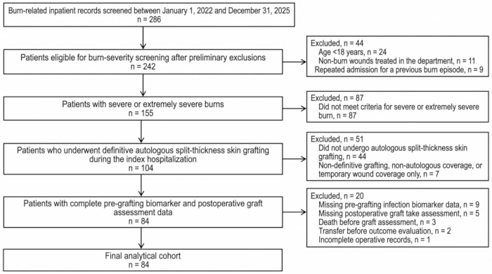
Figure 1: Patient selection flow diagram. A total of 286 burn-related inpatient records were screened. After applying the eligibility criteria, 84 adults with severe or extremely severe burns were included in the final analytical cohort. Please click here to view a larger version of this figure.
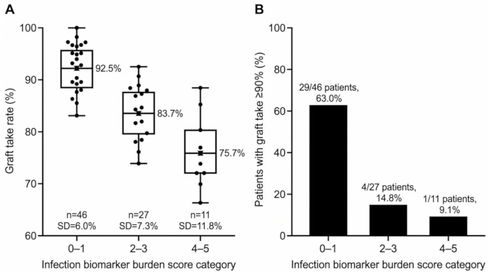
Figure 2: Graft take outcomes across infection biomarker burden score categories. (A) Graft take rate across score categories 0–1, 2–3, and 4–5. (B) Proportion of patients achieving graft take ≥ 90% in each category. Group sizes were 46, 27, and 11, respectively. Please click here to view a larger version of this figure.
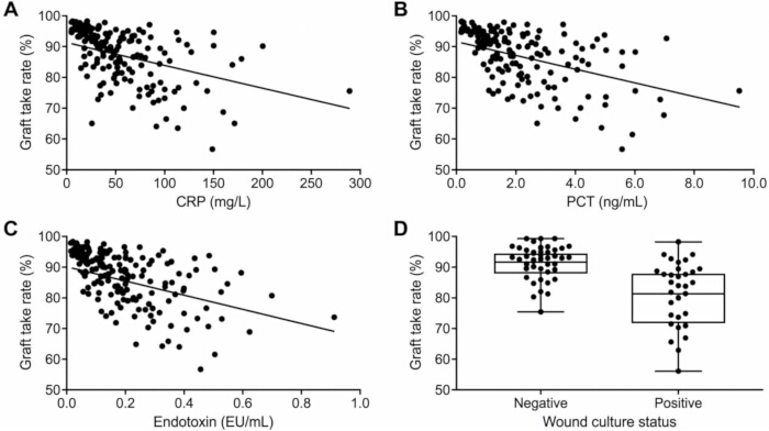
Figure 3: Associations between pre-grafting infection-related variables and graft take rate. (A) Association between C-reactive protein and graft take rate. (B) Association between procalcitonin and graft take rate. (C) Association between endotoxin and graft take rate. (D) Graft take rate according to wound culture status. CRP, C-reactive protein; PCT, procalcitonin. Please click here to view a larger version of this figure.
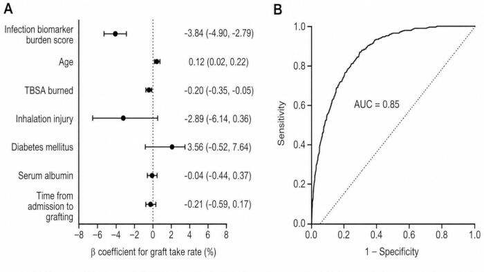
Figure 4: Adjusted model and exploratory discrimination analysis. (A) Adjusted linear regression coefficients with 95% confidence intervals for graft take rate. (B) Receiver operating characteristic curve for the exploratory model predicting graft take ≥ 90%. The area under the curve was 0.85. CI, confidence interval; ROC, receiver operating characteristic; TBSA, total body surface area. The ROC curve reflects apparent performance in the derivation cohort and was not internally or externally validated. Please click here to view a larger version of this figure.
Table 1: Operational definitions and data-extraction windows for study variables. Definitions, extraction windows, and data sources for demographic, burn-related, perioperative, laboratory, microbiological, exposure, and outcome variables. BMI, body mass index; CRP, C-reactive protein; ICU, intensive care unit; NPWT, negative-pressure wound therapy; PCT, procalcitonin; POD, postoperative day; TBSA, total body surface area. Please click here to download this Table.
Table 2: Prespecified analytical models for graft take outcomes. Outcomes, exposures, adjustment variables, and purposes of the primary, reduced, exploratory, and sensitivity analyses. NPWT, negative-pressure wound therapy; TBSA, total body surface area. Please click here to download this Table.
Table 3: Clinical and perioperative characteristics according to graft take status. Data are presented as mean ± standard deviation or number and percentage. Comparisons were made between patients with graft take ≥90% and those with graft take <90%. TBSA, total body surface area. Please click here to download this Table.
Table 4: Pre-grafting infection biomarkers and wound microbiology according to graft take status. Data are presented as mean ± standard deviation, median with interquartile range, or number and percentage. CRP, C-reactive protein; PCT, procalcitonin. Please click here to download this Table.
Table 5: Regression models for graft take outcomes. Linear regression coefficients represent percentage-point changes in graft take rate. The exploratory logistic model reports odds ratios for graft take ≥90%. β, regression coefficient; CI, confidence interval; OR, odds ratio; TBSA, total body surface area. The exploratory logistic-model AUC represents apparent in-sample discrimination and should not be interpreted as externally validated predictive performance. Please click here to download this Table. dvfrgy64e5rhopki[;pki ftrgewwee wwwsa
Supplementary Figure 1: Postoperative outcomes across infection biomarker burden score categories. (A) Postoperative wound infection within 14 days after grafting. (B) Repeat grafting during the same hospitalization. (C) Postoperative length of stay. Score categories were 0–1, 2–3, and 4–5.Please click here to download this file.
Supplementary Table 1: Baseline characteristics of included patients and patients excluded because of incomplete biomarker-score components. Data are presented as mean ± standard deviation or number (percentage). P values were calculated using Welch’s t test for continuous variables and Fisher’s exact test for categorical variables.Please click here to download this file.
Discussion
This single-center retrospective cohort study found that greater pre-grafting infection biomarker burden was associated with poorer autologous split-thickness skin graft take in adults with severe or extremely severe burns. Patients with graft take < 90% had higher pre-grafting C-reactive protein, procalcitonin, and endotoxin concentrations and were more likely to have a positive wound culture. Graft take rate decreased progressively across infection biomarker burden score categories, and the score remained inversely associated with graft take after adjustment for age, total body surface area burned, inhalation injury, diabetes mellitus, serum albumin, and time to grafting. The similar estimates obtained in the reduced and sensitivity models support the stability of this association, although the retrospective design does not establish causality.
Interpretation of infection-related biomarkers in severe burns is challenging because extensive tissue injury can produce systemic inflammatory changes that overlap with infection. Procalcitonin has been evaluated as a marker of infection and sepsis in burn populations, but its diagnostic performance varies according to burn severity, sampling time, infection definition, and clinical setting20. C-reactive protein is widely available and sensitive to inflammation, although tissue necrosis, debridement, and operative stress may reduce its specificity for infection in patients with major burns21. In the present cohort, procalcitonin, endotoxin, and C-reactive protein showed inverse associations with graft take, whereas white blood cell count and neutrophil percentage did not. These findings support interpreting infection-related laboratory results collectively rather than relying on a single nonspecific inflammatory marker.
The association between wound culture positivity and lower graft take is biologically and clinically plausible. Successful graft adherence requires a viable, adequately perfused recipient bed with controlled exudation and a low microbial burden. Previous burn research has linked bacterial wound colonization with subsequent skin-graft loss22. The organisms identified in this cohort, including Pseudomonas aeruginosa, Acinetobacter baumannii, Staphylococcus aureus, and Klebsiella pneumoniae, are common clinically relevant burn-wound isolates. Local inflammation, tissue injury, exudation, and biofilm formation may interfere with wound-bed stabilization and graft revascularization. Biofilm-associated organisms may also persist despite antimicrobial exposure and may not be fully represented by a single routine culture result23. Combining wound culture with systemic biomarkers may therefore better characterize the overall pre-grafting infection-related state than culture alone.
The present findings are broadly consistent with Wellkamp et al., who reported that bacterial wound colonization was associated with subsequent skin-graft loss in burn patients10. In contrast, Holm et al. found that a positive preoperative wound culture was not associated with graft failure in a cohort of minor burns, and many grafts achieved >90% take despite culture positivity17. The difference may reflect case mix: the present cohort comprised severe or extremely severe burns and evaluated a cumulative systemic-and-local biomarker burden, whereas the minor-burn study focused primarily on preoperative wound culture. These contrasting results reinforce that culture status alone should not determine graft timing. Because the present score was developed exclusively in adults with severe or extremely severe burns, its performance should not be extrapolated to minor or moderate burns without dedicated validation in those populations.
The graded association between the composite score and graft take suggests that cumulative infection-related abnormalities may be more informative than an isolated test result. However, the score should not be interpreted as a stand-alone criterion for delaying surgery. Total body surface area burned also remained associated with graft take, indicating that anatomical injury severity and infection-related burden contribute overlapping but distinct information. Burn extent and depth are recognized determinants of graft outcome, although wound-bed preparation, nutritional status, operative timing, and postoperative management also influence graft survival24. Similarly, split-thickness skin graft take may be affected by perfusion, hematoma, seroma, edema, shear, and fixation stability25. Negative-pressure wound therapy can improve graft stabilization in selected wounds, but its use may also reflect greater wound complexity and patient selection26. The infection biomarker burden score may therefore be most useful as a structured prompt for reassessing wound-bed readiness, antimicrobial management, nutritional status, and postoperative fixation rather than as an independent treatment rule.
The small positive age coefficient in the adjusted linear model should not be interpreted as evidence that older age improves graft survival. The between-group age difference was not significant in the unadjusted comparison, and prior burn-grafting work has not demonstrated a consistent protective effect of increasing age24. In this relatively small cohort, the adjusted coefficient may reflect residual confounding, case selection, or correlation with other clinical characteristics; its direction therefore requires replication rather than causal interpretation.
Given that the score was developed and evaluated in the same relatively small single-center cohort, the observed score categories should be regarded as hypothesis-generating risk strata rather than validated treatment thresholds. In practice, a score of 0–1 may support continuation of routine graft-readiness assessment when the wound bed and patient are otherwise suitable; a score of 2–3 should prompt closer review of wound-bed granulation and perfusion, culture results and antimicrobial response, nutritional and physiological optimization, and fixation strategy; and a score of 4–5 should trigger an especially careful multidisciplinary reassessment of reversible infection-related and wound-bed factors before proceeding. These categories should not automatically mandate postponement of grafting or escalation of antimicrobial therapy. The score is intended to complement, not replace, the burn surgeon’s examination of wound-bed quality, bleeding/perfusion, exudate, tissue viability, and overall clinical stability. Prospective multicenter validation is required before any score category is used as a formal decision threshold. Importantly, this observational study did not test whether delaying grafting, changing antimicrobial therapy, intensifying nutritional optimization, or modifying fixation solely on the basis of score category improves graft outcomes. Any score-guided management strategy therefore requires prospective evaluation before clinical adoption.
This study has several strengths. The analysis used a predefined pre-grafting exposure window, a clinically specific continuous graft-take outcome, complete availability of all five score components in the analytical cohort, and separate evaluation of individual biomarkers and the composite score. The primary analysis preserved the continuous nature of graft take, and the score association remained directionally consistent across reduced and sensitivity models. Several limitations should also be considered. The retrospective single-center design is vulnerable to residual confounding and limits generalizability. Important determinants such as detailed wound-bed granulation quality, perfusion and hemodynamic status, vasopressor exposure, nutritional status beyond albumin, renal dysfunction, timing of first excision, donor-site characteristics, topical antimicrobial therapy, fixation/dressing technique, and surgeon-related practice were not all available for complete adjustment. Graft timing, wound preparation, antimicrobial therapy, and negative-pressure wound therapy were determined during routine care rather than by a standardized study protocol. Wound cultures were obtained for clinical indications, creating possible indication bias, and recent antimicrobial exposure could reduce microbial recovery. Requiring complete data for all five score components also creates complete-case selection bias and may preferentially retain clinically complex patients who underwent broader infection-related testing. Graft take was based on clinical documentation and available photographs rather than centralized blinded planimetry. The moderate sample size limited the precision of the exploratory logistic model and the assessment of uncommon findings, particularly fungal evidence. The score is newly proposed, uses equal weighting, and has not undergone external validation. Equal weighting was a pragmatic transparency choice and should not be interpreted as evidence that the five components have identical biological or prognostic importance; in particular, wound culture positivity may convey different information from an isolated biomarker elevation, while β-D-glucan contributed limited information in this cohort because fungal evidence was uncommon. The procalcitonin threshold was informed by burn-sepsis literature, whereas the remaining cut points were prespecified pragmatic thresholds selected for clinical interpretability rather than universal guideline-defined cutoffs or outcome-optimized values. Threshold performance may vary across institutions because C-reactive protein, procalcitonin, endotoxin, and β-D-glucan measurements depend on assay platform, reference ranges, sampling conditions, and local case mix. The ≥90% and <75% graft-take categories are likewise descriptive clinical strata rather than validated treatment thresholds. Finally, the study used an operational severe/extremely severe burn definition rather than ABSI-based classification. These limitations require cautious interpretation and external validation. Although only nine patients in the definitive-grafting screening stage were excluded specifically because one or more score components were unavailable, the baseline comparison did not identify statistically significant differences in the principal demographic, burn-severity, comorbidity, nutritional, or graft-timing characteristics examined. Nevertheless, the biomarker-incomplete group was small, and the absence of statistically significant differences does not exclude modest or clinically relevant selection effects; complete-case selection bias therefore remains possible. In addition, the exploratory logistic model was developed and evaluated in the same cohort, and no formal calibration analysis, bootstrap/internal resampling validation, or independent external validation was performed. Its AUC may therefore be optimistic and should be viewed as descriptive of this dataset rather than as evidence of transportable prediction accuracy.
Future prospective multicenter studies should standardize biomarker timing, wound sampling, antimicrobial documentation, graft photography, and graft-area measurement. Repeated biomarker measurements may clarify whether temporal trends provide additional information beyond a single preoperative value. External validation should evaluate calibration, discrimination, reproducibility across assay platforms, and clinically useful decision thresholds. Within the limitations of the present study, higher pre-grafting infection biomarker burden was consistently associated with lower autologous skin graft take. Elevated C-reactive protein, procalcitonin, endotoxin, and positive wound culture contributed the clearest individual signals, whereas fungal β-D-glucan did not show an independent relationship with graft outcome. The proposed score should therefore be considered hypothesis-generating and should remain an adjunct to surgical and clinical judgment until prospectively validated in independent multicenter cohorts. These findings describe associations and should not be interpreted as evidence that the biomarker burden itself causes graft failure or that lowering the score would necessarily improve graft take.
Disclosures
The authors declare no competing financial interests or conflicts of interest.
Acknowledgements
The authors thank the medical, nursing, and wound-care teams of the Department of Burns, Plastic Surgery and Wound Repair, Ganzhou People’s Hospital, for their support in patient care and clinical data collection.
Materials
| Name | Company | Catalog Number | Comments |
|---|---|---|---|
| Cobas e 411 immunoassay analyzer | Roche Diagnostics | 4775279001 | Automated electrochemiluminescence immunoassay analyzer used for procalcitonin measurement. |
| Columbia agar with 5% sheep blood | bioMérieux | 43041 | Ready-to-use blood agar plates used for primary isolation, colony morphology, and hemolysis assessment of wound isolates. Package contains 20 plates. |
| C-reactive protein assay, Tina-quant C-Reactive Protein IV | Roche Diagnostics | 7876033190 | Immunoturbidimetric assay for quantitative determination of C-reactive protein in human serum or plasma. Compatible with cobas c 311, c 501, and c 502 systems. |
| Endotoxin assay kit for human plasma | Xiamen Bioendo Technology Co., Ltd. | KTE20 | Kinetic turbidimetric assay for quantitative determination of endotoxin in human plasma. Each kit contains materials for 20 tests, including single-test lysate vials and sample-treatment solution. |
| ESwab liquid Amies collection and transport system | COPAN Diagnostics | 480C | Regular flocked swab supplied with 1 mL liquid Amies medium for collection and transport of aerobic, anaerobic, and fastidious bacteria. |
| Fungi (1→3)-β-D-glucan detection kit | Xiamen Bioendo Technology Co., Ltd. | KCG50 | Kinetic chromogenic assay for quantitative β-D-glucan measurement. Analytical range: 25–1,000 pg/mL; 50 tests per kit. |
| Kinetic incubating microplate reader | Xiamen Bioendo Technology Co., Ltd. | ELX808IULALXH | Temperature-controlled kinetic microplate reader suitable for turbidimetric endotoxin and chromogenic β-D-glucan assays. |
| MacConkey agar with crystal violet | bioMérieux | 43141 | Selective and differential culture medium used for recovery and preliminary differentiation of gram-negative bacilli, including Enterobacterales. Package contains 20 plates. |
| Procalcitonin assay, Elecsys BRAHMS PCT | Roche Diagnostics | 9318712190 | Electrochemiluminescence immunoassay for quantitative determination of procalcitonin in human serum or plasma. One reagent pack provides 100 tests and is compatible with cobas e 411, e 601, and e 602 analyzers. |
| Pyrogen-free pipette tips, 1,000 µL | Xiamen Bioendo Technology Co., Ltd. | PT10004 | Low-endotoxin pipette tips used for sample dilution and reagent transfer during endotoxin and β-D-glucan assays. |
| Pyrogen-free pipette tips, 200 µL | Xiamen Bioendo Technology Co., Ltd. | PT2005 | Low-endotoxin pipette tips used for standards, samples, lysate reagent, and control preparation. |
| R statistical software | R Foundation for Statistical Computing | Version 4.3.2 | Used for descriptive analyses, group comparisons, correlation analyses, multivariable regression, receiver operating characteristic analysis, sensitivity analyses, and figure preparation. RRID: SCR_001905. |
| Sabouraud gentamicin chloramphenicol 2 agar | bioMérieux | 43651 | Selective culture medium used for recovery of yeasts and filamentous fungi from wound specimens. Package contains 20 plates. |
| VITEK 2 Compact identification and susceptibility system | bioMérieux | VITEK 2 Compact | Automated system used for identification of bacterial and yeast isolates and, when performed, antimicrobial susceptibility testing. |
| VITEK 2 GN identification card | bioMérieux | 21341 | Identification card for clinically relevant gram-negative bacteria, including Enterobacterales and non-fermenting gram-negative bacilli. Package contains 20 cards. |
| VITEK 2 GP identification card | bioMérieux | 21342 | Identification card for clinically relevant gram-positive bacteria. Package contains 20 cards. |
| VITEK 2 YST identification card | bioMérieux | 21343 | Identification card for clinically relevant yeasts and yeast-like organisms. Package contains 20 cards. |
References
- World Health Organization. Burns. World Health Organization; Geneva; 2023. Available from: https://www.who.int/news-room/fact-sheets/detail/burns.
- Greenhalgh DG. Management of burns. N Engl J Med. 2019;380(24):2349-59.
- Schlottmann F, Bucan V, Vogt PM, Krezdorn N. A short history of skin grafting in burns: from the gold standard of autologous skin grafting to the possibilities of allogeneic skin grafting with immunomodulatory approaches. Medicina (Kaunas). 2021;57(3):225.
- Thourani VH, Ingram WL, Feliciano DV. Factors affecting success of split-thickness skin grafts in the modern burn unit. J Trauma. 2003;54(3):562-8.
- ISBI Practice Guidelines Committee, et al. ISBI practice guidelines for burn care. Burns. 2016;42(5):953-1021.
- Roy S, et al. Microbial infections in burn patients. Acute Crit Care. 2024;39(2):214-25.
- Mokline A, et al. Procalcitonin: a diagnostic and prognostic biomarker of sepsis in burned patients. Ann Burns Fire Disasters. 2015;28(2):116-20.
- Cabral L, Afreixo V, Almeida L, Paiva JA. The use of procalcitonin (PCT) for diagnosis of sepsis in burn patients: a meta-analysis. PLoS One. 2016;11(12):e0168475.
- Palmieri TL, Heard J. Biomarkers of sepsis in burn injury: an update. Burns Trauma. 2025;13:tkae080.
- Wellkamp L, et al. Correlation between bacterial wound colonization and skin-graft loss in burn patients. J Burn Care Res. 2023;44(3):649-54.
- Scherer LA, et al. The vacuum assisted closure device: a method of securing skin grafts and improving graft survival. Arch Surg. 2002;137(8):930-4.
- von Elm E, et al. The Strengthening the Reporting of Observational Studies in Epidemiology (STROBE) statement: guidelines for reporting observational studies. Lancet. 2007;370(9596):1453-7.
- Zhang Y, et al. Epidemiological and clinical characteristics of burns in adults: a 6-year retrospective study in a major burn center in Suzhou, China. Front Public Health. 2024;12:1413986.
- Giretzlehner M, Ganitzer I, Haller HL. Technical and medical aspects of burn size assessment and documentation. Medicina (Kaunas). 2021;57(3):242.
- Egea-Guerrero JJ, et al. The utility of C-reactive protein and procalcitonin for sepsis diagnosis in critically burned patients: a preliminary study. Plast Surg (Oakv). 2015;23(4):239-43.
- Miller JM, et al. Guide to utilization of the microbiology laboratory for diagnosis of infectious diseases: 2024 update by the Infectious Diseases Society of America and the American Society for Microbiology. Clin Infect Dis. 2024;ciae104.
- Holm S, Smith MTD, Huss F, Allorto N. Is the preoperative wound culture necessary before skin grafting minor burns A pilot study in a low resource setting burn service. J Burn Care Res. 2024;45(5):1281-6.
- Di HP, et al. Effects of Meek skin grafting on patients with extensive deep burn at different age groups. Zhonghua Shao Shang Za Zhi. 2017;33(3):156-9.
- Hu G, et al. Predictive value of the prognostic nutrition index for the prognosis of patients with severe burns treated with the Meek graft. J Burn Care Res. 2021;42(3):448-53.
- Zhang P, Zou B, Liou YC, Huang C. The pathogenesis and diagnosis of sepsis post burn injury. Burns Trauma. 2021;9:tkaa047.
- Aggarwal N, et al. Serum procalcitonin and C-reactive protein as indices of early sepsis and mortality in North Indian pediatric burn injuries: a prospective evaluation and literature review. Acute Crit Care. 2024;39(3):350-8.
- Unal S, et al. Analysis of skin-graft loss due to infection: infection-related graft loss. Ann Plast Surg. 2005;55(1):102-6.
- Maslova E, Eisaiankhongi L, Sjöberg F, McCarthy RR. Burns and biofilms: priority pathogens and in vivo models. NPJ Biofilms Microbiomes. 2021;7(1):73.
- Dogan S, et al. Skin graft take rate among minor burns: a cohort study to investigate the effect of factors such as burn depth, burn size, and timing of the operation. Burns Open. 2025;9:100381.
- Braza ME, Fahrenkopf MP. Split-thickness skin grafts. StatPearls Publishing; Treasure Island, FL; 2025.
- Yin Y, et al. Negative-pressure therapy versus conventional therapy on split-thickness skin graft: a systematic review and meta-analysis. Int J Surg. 2018;50:43-8.