This study establishes a standardized protocol for isolating small extracellular vesicles (sEVs) from rat epicardial adipose tissue (EAT) through optimized enzymatic digestion and differential ultracentrifugation. The protocol yields reproducible results and complies with MISEV2023 guidelines, providing a reliable technical platform for investigating the paracrine role of EAT-derived sEVs in cardiovascular disease.
Method Article
Isolation and Characterization of Small Extracellular Vesicles Derived from Epicardial Adipose Tissue
* These authors contributed equally
In This Article
Summary
Abstract
Epicardial adipose tissue (EAT) is a distinct visceral adipose depot located between the myocardium and the visceral layer of the pericardium. Owing to the absence of an anatomical barrier between EAT and the heart, EAT can communicate with the myocardium through paracrine and vasocrine mechanisms. Small extracellular vesicles (sEVs) secreted by EAT, which are lipid bilayer-enclosed nanoparticles that mediate intercellular communication by transferring bioactive cargoes, including proteins, lipids, and nucleic acids, to recipient cells, may serve as key mediators of this crosstalk. However, the extraction of sEVs from EAT remains technically challenging. In this study, we established a reproducible protocol for isolating sEVs from rat EAT using collagenase digestion followed by differential ultracentrifugation. The isolated vesicles were characterized by transmission electron microscopy, nano-flow cytometer, and western blotting. This protocol provides a practical and reproducible experimental framework for isolating EAT-derived sEVs and may facilitate future studies exploring their roles in cardiovascular physiology, disease mechanisms, biomarker discovery, and therapeutic development.
Introduction
Cardiovascular disease (CVD) remains the leading cause of morbidity and mortality worldwide1,2. The increasing incidence of CVDs is associated with multiple risk factors, including aging, hypertension, diabetes, metabolic disorders, and obesity. With the growing global burden of obesity over recent decades, adipose tissue has become an important focus of cardiovascular and metabolic research.
Epicardial adipose tissue (EAT) is a distinct adipose depot located between the myocardium and the visceral pericardium. Compared with other fat depots, EAT has unique anatomical and physiological characteristics. It shares a common embryological origin with visceral fat and possesses a microcirculation that is closely connected with the myocardium3. Its distinct anatomy and physiology enable it to crosstalk with the myocardium through paracrine and vasocrine signaling mechanisms4. Under physiological conditions, EAT plays several protective roles. It acts as a mechanical cushion, protecting the coronary arteries and myocardium from external stress and distortion. It also provides free fatty acids to the adjacent myocardium and participates in local thermoregulation. However, under pathological conditions, the morphology and secretory profile of EAT may be altered5. Accumulating evidence suggests that dysfunctional EAT contributes to the development and progression of various CVDs, including coronary artery disease, atrial fibrillation, and heart failure6,7,8. Therefore, elucidating the molecular communication between EAT and the myocardium is important for understanding CVD mechanisms and may provide novel therapeutic targets.
Intercellular communication is essential for maintaining cellular, tissue, and organ homeostasis. In recent years, extracellular vesicles (EVs) have attracted considerable attention as important mediators of intercellular communication9. EVs, which possess a lipid bilayer membrane, can participate in intercellular communication by transferring cargoes including lipids, cytokines, RNA, DNA, and proteins, to recipient cells10. Small extracellular vesicles (sEVs), which are typically enriched in particles smaller than 200 nm, are often characterized by the expression of marker proteins such as CD9, CD63, TSG101, and Alix11. Thus, isolating and characterizing sEVs derived from EAT may improve our understanding of the role of epicardial fat in cardiac crosstalk under both physiological and pathological conditions.
Compared with sEVs isolated from cultured cell-conditioned medium, sEVs directly isolated from tissue may better reflect the in vivo microenvironment. Although protocols for isolating sEVs from cell culture medium and biological fluids, such as plasma and urine, have been widely established12, isolating sEVs from adipose tissue, particularly epicardial fat, remains challenging. Adipose tissue contains large amounts of lipids, extracellular matrix components, stromal vascular cells, blood-derived particles, lipoproteins, and soluble proteins13,14. In addition, rat EAT is limited in volume, and improper dissection may result in contamination with myocardium, mediastinal adipose tissue, paracardial adipose tissue, or circulating sEVs. These factors may reduce vesicle purity and compromise reproducibility. Therefore, a standardized and reproducible protocol is needed to obtain high-quality sEVs preparations from this specific tissue source. Compared with cerebral15, liver16 and tumor tissues17,18, EAT presents unique challenges: (1) limited tissue volume in rodents, (2) high lipid content that co-sediments with vesicles, and (3) anatomical proximity to pericardial fat and the myocardium, increasing risk of contamination. These factors necessitate a tailored isolation protocol. This study presents a detailed protocol for the isolation and characterization of sEVs derived from rat EAT. The procedures include EAT collection, tissue preprocessing, collagenase digestion, differential centrifugation-based vesicle isolation, and validation of the isolated vesicles by transmission electron microscopy (TEM), nano-flow cytometer, and sEVs marker protein detection. This protocol provides a reproducible methodological framework for studying EAT-derived sEVs and may facilitate future investigations into their roles in cardiovascular physiology and diseases. The primary objectives of this study were to: (1) establish a reproducible protocol for isolating sEVs from rat EAT, (2) characterize the isolated vesicles according to MISEV2023 guidelines19, and (3) assess the feasibility of the protocol for downstream functional studies.
Protocol
All animal experiments were performed in accordance with the Guide for the Care and Use of Laboratory Animals published by the Ministry of the People’s Republic of China (1998) and were approved by the Institutional Committee on Animal Care of Capital Medical University. The animal research was sanctioned by the Institutional Animal Care and Use Committee (IACUC) under approval number A5095F17-EA0D-4377-88F7-FED2CE3A2ED2. All methods were performed in accordance with the relevant guidelines and regulations, and followed the ARRIVE guidelines. Male SPF-class Sprague-Dawley rats (SD rats) weighing 200–250 g were used in this study. The reagents and the equipment used are listed in the Table of Materials.
1. Preparation of materials
- Sterilize all surgical tools and materials by steam sterilization, including surgical scissors and standard forceps.
- Prepare a water bath and set the temperature to 37 °C. Precool the high-speed centrifuge and ultracentrifuge to 4 °C.
- Prepare cell culture dishes (100 mm) and sterile 50 mL centrifuge tubes.
- Prepare sterile phosphate-buffered saline (PBS) and Krebs-Ringer-bicarbonate HEPES buffer in advance. According to the manufacturer’s product information, the principal components of the Krebs–Ringer–bicarbonate HEPES buffer are HEPES, sodium chloride, potassium chloride, sodium bicarbonate, potassium dihydrogen phosphate, calcium chloride, magnesium sulfate, and glucose. The buffer contains no bovine serum albumin.
- Prepare fresh digestion solution by diluting type I collagenase at a final concentration of 1 mg/mL in Krebs-Ringer-bicarbonate HEPES buffer. Sterilize the solution using a 0.22-µm filter and use it immediately or store at -20 °C for up to 1 month.
2. Anesthetization for the animals
- Anesthetize the rats by injecting a 5% pentobarbital sodium solution at a dose of 150 mg/kg into the abdominal cavity. Proceed with subsequent procedures only after confirming the absence of both the withdrawal response to a tail pinch and the corneal reflex.
- Shave or use a depilatory cream to remove the fur.
NOTE: All animal procedures must be performed in accordance with institutional animal-care regulations and approved protocols.
3. Collection of EAT from rats
NOTE: As illustrated in Figure 1, the following steps are performed:
- Spray the rats with iodophor for skin disinfection and repeat this procedure three times. Place the rats in a supine position.
- Carefully lift the skin and make a small incision. Slit the abdominal cavity and fully expose the abdominal aorta and vena cava.
- Immediately perfuse the rat with 50 mL PBS through the vena cava, then cut the abdominal aorta to flush blood from the myocardium. Pale lungs are considered a sign of adequate myocardial perfusion. Before harvesting the heart, confirm death by the absence of audible heart sounds, spontaneous respiration, and pupillary dilation. Harvest the heart.
- Rinse the heart three times in ice-cold PBS to remove residual surface blood.
- Gently tear the pericardium with forceps. Carefully remove the pericardial adipose tissue.
- Carefully tear the visceral pericardium and carefully dissect EAT from the cavity between the visceral pericardium and myocardium with forceps. Using fine-tipped forceps, gently lift the visceral pericardium at the atrioventricular groove. Carefully dissect EAT from the interventricular groove and left ventricular free wall, where EAT is most abundant. Avoid including subepicardial coronary vessels or underlying myocardial tissue.
- Avoid contamination with myocardium, blood clots, mediastinal adipose tissue, paracardial adipose tissue, or pericardial connective tissue.
- Transfer the collected EAT into a new dish containing ice-cold sterile PBS.
- Wash the tissue 3–5 times with ice-cold PBS to remove residual blood.
- Remove excess PBS with sterile filter paper and record the wet weight of the tissue.
NOTE: Rat EAT is limited in quantity. To obtain sufficient vesicle yield, EAT from multiple rats may be pooled when necessary. During dissection, the epicardial region should be carefully identified, blunt dissection should be performed gently, and the tissue should be kept moist throughout the procedure.
4. Mincing and digestion of EAT
- Mince the tissue into small pieces (1–3 mm3) with scissors in a culture dish placed on ice. Transfer the minced tissue into a 50-mL centrifuge tube.
- Add type I collagenase digestion solution (1 mg/mL) to the tissue at a ratio sufficient to fully immerse the tissue pieces (10 mL per 1 g EAT) for digestion.
- Seal the container and incubate the tissue suspension at 37 °C for 40 min with gentle shaking.
- Gently invert the tube every 10 min. Monitor the digestion progress: successful digestion yields a milky suspension with minimal visible tissue fragments. Avoid over-digestion (>60 min), which may damage vesicles.
- Terminate digestion by diluting the sample at a 1:1 ratio with PBS containing 2 mmol/L EGTA.
5. Isolation of sEVs by differential centrifugation
- Centrifuge the dissociated tissue suspension at 300 × g for 10 min at 4 °C to remove intact cells and large tissue fragments after dissociation. Carefully aspirate and discard the floating lipid-rich layer; collect only the infranatant for subsequent centrifugation steps.
- Centrifuge the supernatant from step 5.1 at 2000 × g for 20 min at 4 °C to remove cell debris, dead cells, and larger particles, and then continue to collect the supernatant.
- Filter the supernatant with a 0.22-µm membrane gently without applying pressure to minimize shear-related artifacts to remove the large particles and cell debris.
- Continue to centrifuge the filtered supernatant at 16,500 × g for 30 min at 4 °C to remove large EVs, organelle fragments, and residual particulate contaminants.
- Transfer the supernatant into the ultracentrifugation tubes and centrifuge at 120,000 × g for 70 min at 4 °C to pellet sEVs.
- Carefully discard the supernatant completely with a pipette and resuspend the pellet in PBS. Perform a second ultracentrifugation at 120,000 × g for 70 min at 4 °C to reduce soluble protein contaminants and increase sEVs purity.
- Carefully remove the supernatant and obtain the sEVs. Resuspend the pellet in 1 mL sterile PBS and transfer it into a 1.5 mL tube.
- Store the suspension at 4 °C for subsequent characterization.
NOTE: Ultracentrifuge tubes must be balanced accurately before centrifugation. If the samples are not used immediately, they should be stored at −80 °C. Repeated freeze-thaw cycles should be avoided to preserve vesicle integrity. All the above steps for the systematic isolation of sEVs are shown in Figure 2.
6. Characterization of sEVs by TEM, nano-flow cytometer, and Western blotting
- Deposit 10 µL of sEVs solution on a copper mesh, incubate at room temperature for 10 min, and rinse it with sterile distilled water. Use absorbent paper to remove any excess liquid. Next, apply 10 µL of 2% uranyl acetate onto the copper for 1 min, then remove the excess fluid with filter paper and dry the mesh under an incandescent lamp for 2 min. Observe the prepared sample under a transmission electron microscope at 80 kV.
- Use a BCA protein assay kit to measure the protein concentration of sEVs samples. Dilute the sEVs sample with PBS to 108/mL, then load the samples into the nano-flow cytometer (particle size detection range of 7–1000 nm; particle concentration was determined using 250-nm polystyrene standards, and size distribution was calculated from a side-scatter calibration curve generated using silica nanoparticles (68 nm, 91 nm, 113 nm, and 155 nm)) to measure the size distribution and particle concentration.
- Resuspend the sEVs sample in RIPA lysis buffer for protein analysis. Perform Western blotting analysis to detect the expression of EV-associated markers Alix, TSG101, HSP70, CD9, and CD63, with Calnexin assessed as a negative control.
Results
Isolation and purification of EAT-derived sEVs
To isolate sEVs from rat EAT tissue, we employed a combination of collagenase digestion and differential ultracentrifugation (Figure 2). The initial steps involved pretreating EAT to remove surface blood, followed by digestion with type I collagenase. After digestion, sequential centrifugation steps were performed to remove intact cells, cellular debris, and larger vesicles. The final ultracentrifugation step at 120,000 × g was used to pellet the sEVs. The resulting pellet was resuspended in PBS and used for subsequent characterization.
TEM examination of EAT- derived sEVs
TEM was used to assess the morphology of the isolated vesicles. The isolated particles exhibited a typical cup-shaped morphology with a clear lipid bilayer membrane, which is consistent with the characteristic ultrastructure of sEVs (Figure 3A).
Size distribution of EAT-derived sEVs
The particle size distribution and concentration of the sEVs were determined using nano-flow cytometer. The sEVs obtained from 0.536 g of EAT had a final volume of 1 mL. The protein concentration was 165.99 ng/µL. The results (Figure 3B) indicated that sEV particle size distribution ranged from approximately 40–180 nm, with a concentration of approximately 3.08 × 10¹⁰ particles/mL. This corresponded to a particle yield of approximately 5.75 × 10¹⁰ particles/g EAT, normalized to the initial tissue weight. The size measurements were consistent with previous findings on sEVs characteristics, reinforcing the isolation protocol.
Western blotting analysis
Western blotting analysis was further employed to validate the presence of sEV markers. The sEV-associated proteins TSG101, Alix, and HSP70 were detected in the isolated sEV samples, whereas Calnexin was detected in the cell lysate but not in the isolated sEV samples (Figure 3C). CD63 and CD9 were also detected in the isolated sEV samples (Figure 3D). These findings confirmed that the isolated particles were enriched in sEVs.
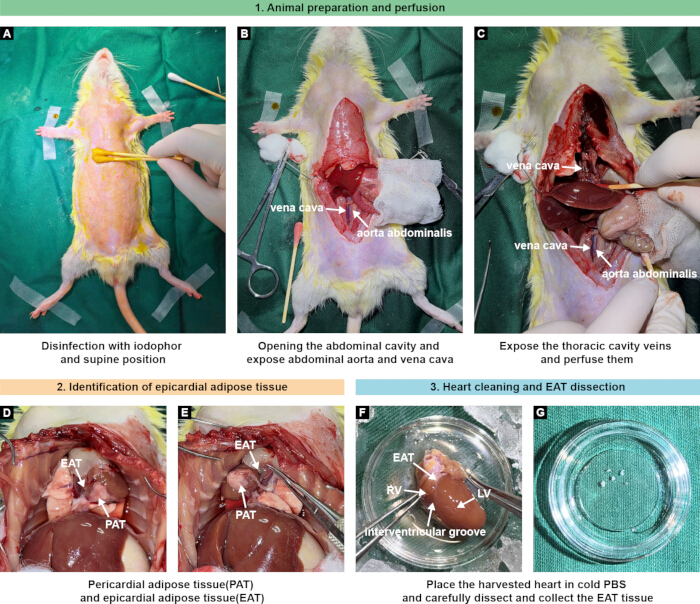
Figure 1: Dissection and collection of epicardial adipose tissue (EAT) from rats. (A) Following anesthesia and hair removal, the rat is disinfected with iodophor and positioned supine for tissue collection. (B) The abdominal cavity is opened to expose the abdominal aorta and vena cava. (C) The thoracic cavity is exposed, and PBS perfusion is performed through the vena cava to remove blood before heart collection. (D,E) Identification of pericardial adipose tissue (PAT) and epicardial adipose tissue (EAT), illustrating their anatomical locations and facilitating separation of EAT from surrounding adipose tissue. (F) The harvested heart is placed in ice-cold PBS, and EAT is carefully dissected from the epicardial surface, particularly around the interventricular groove, while avoiding contamination with the underlying myocardium and coronary vessels. RV, right ventricle; LV, left ventricle. (G) Representative EAT tissue collected following dissection and washing, prior to weighing and subsequent processing. Please click here to view a larger version of this figure.
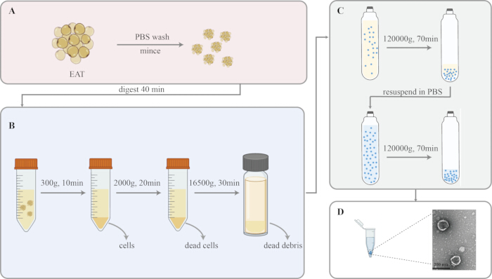
Figure 2: Workflow for isolation of small extracellular vesicles (sEVs) from rat epicardial adipose tissue (EAT). (A) EAT is washed with ice-cold PBS, minced into small pieces, and subjected to type I collagenase digestion at 37 °C for 40 min. (B) Following digestion, the tissue suspension is subjected to sequential differential centrifugation. Centrifugation at 300 × g for 10 min at 4 °C removes intact cells and large tissue fragments; the lipid-rich floating layer is discarded and the infranatant is retained. Subsequent centrifugation at 2,000 × g for 20 min at 4 °C removes dead cells and cellular debris. The supernatant is filtered through a 0.22-µm membrane and subsequently centrifuged at 16,500 × g for 30 min at 4 °C to remove large extracellular vesicles, organelle fragments, and residual particulate contaminants. (C) The resulting supernatant is ultracentrifuged at 120,000 × g for 70 min at 4 °C to pellet sEVs. The pellet is resuspended in PBS and subjected to a second ultracentrifugation at 120,000 × g for 70 min at 4 °C to reduce soluble contaminants. The final sEV pellet is resuspended in PBS for subsequent characterization. (D) Representative transmission electron microscopy image illustrating the morphology of the isolated EAT-derived sEVs; scale bar, 200 nm. Please click here to view a larger version of this figure.
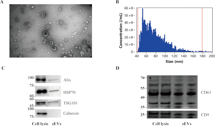
Figure 3: Characterization of small extracellular vesicles (sEVs) isolated from rat epicardial adipose tissue (EAT). (A) Representative transmission electron microscopy (TEM) image of isolated EAT-derived sEVs showing vesicles with the characteristic cup-shaped morphology and lipid bilayer appearance. Scale bar, 200 nm. (B) Particle-size distribution of the isolated sEV preparation measured using a nano-flow cytometer. The detected particles were distributed predominantly within approximately 40–180 nm. The final sEV preparation obtained from 0.536 g EAT had a volume of 1 mL and a particle concentration of approximately 3.08 × 10¹⁰ particles/mL, corresponding to approximately 5.75 × 10¹⁰ particles/g EAT. (C) Western blot analysis of the sEV-associated markers Alix, HSP70, and TSG101, with Calnexin assessed as a negative/non-EV-associated marker. Cell lysate and isolated sEV preparations are shown for comparison. (D) Western blot analysis of the sEV-associated tetraspanins CD63 and CD9 in cell lysate and isolated sEV preparations. Please click here to view a larger version of this figure.
Discussion
Recent studies suggest that EAT plays a role in the development and progression of several CVDs and may serve as a biomarker or therapeutic target4,20. EAT is increasingly recognized as an active paracrine and vasocrine tissue involved in cardiovascular pathophysiology. Therefore, establishing a reproducible and optimized method for isolating sEVs from EAT is important for elucidating the molecular mechanisms underlying epicardial fat–myocardium communication. In this work, we describe a practical workflow that combines EAT dissection, collagenase-assisted tissue dissociation, and differential ultracentrifugation, followed by complementary characterization using TEM, nano-flow cytometer, and sEVs-associated protein detection. The results showed vesicle-like morphology, a predominant particle size within the expected range, and detectable sEVs-associated proteins, supporting successful isolation of sEVs from rat EAT.
Various methods have been used to isolate EVs, including ultracentrifugation, ultrafiltration, density gradient centrifugation, tangential-flow filtration, size-exclusion chromatography, and immunoaffinity capture21. Among these techniques, ultracentrifugation remains one of the most widely used methods for EV isolation from various biological sources owing to its ability to effectively separate particles by size and density. In the present protocol, type I collagenase digestion was combined with differential ultracentrifugation to isolate sEVs derived from rat EAT. Sequential centrifugation steps were used to remove residual cells, cellular debris, and larger vesicles, thereby increasing the purity of the final sEVs.
The isolated vesicles were characterized using TEM, nano-flow cytometer, and Western blotting. The results revealed that the characteristics of the isolated sEVs were typical, exhibiting cup-shaped structures with lipid bilayer membranes ranging from 40–180 nm. Western blotting further confirmed the presence of sEVs-associated proteins, including TSG101, Alix, HSP70, CD63, and CD9. Together, these results indicate that the described protocol enables the successful isolation of sEVs derived from rat EAT.
Several steps are particularly important for preparation quality and reproducibility. First, anatomically accurate dissection is essential because rat EAT is limited in amount and lies adjacent to myocardium, pericardial fat, and blood-rich structures. Incomplete separation or rough handling can introduce non-target tissue contamination. Second, perfusion and repeated cold PBS washing before digestion are critical to reduce blood-derived particles and soluble contaminants. Third, controlled collagenase digestion (enzyme concentration, temperature, and time) is required to disperse matrix-rich adipose tissue while minimizing over-digestion-related cell damage. Fourth, strict centrifugation workflow control (duration, temperature, tube balancing) strongly influences downstream purity and yield. Finally, a second ultracentrifugation wash and gentle pellet resuspension improve consistency and reduce carryover of soluble proteins/lipids. However, this protocol has several limitations. First, the extraction and characterization procedures are time-consuming and require specialized equipment, particularly an ultracentrifuge. Second, because tissue dissociation includes collagenase treatment, the recovered particle population may include not only freely released sEVs but also matrix-associated particles; over-digestion may additionally increase the release of intracellular membrane fragments. Collagenase treatment may potentially alter EV-associated surface proteins or their accessible epitopes, which could affect surface-marker detection, antibody-based isolation, and downstream functional assays involving vesicle–cell interactions. The effects of collagenase treatment on EV surface-protein integrity were not directly assessed in this study and should be considered when interpreting downstream results. Third, although complementary characterization was performed, these assays do not fully exclude co-isolated non-vesicular material. Differential ultracentrifugation can co-enrich protein aggregates, lipoproteins, and other lipid-associated particles; thus, the absence of contamination cannot be inferred solely from EV-marker detection. Fourth, no direct comparison with mechanical mincing-only processing was performed, so the relative effects of collagenase on yield, purity, composition, and bioactivity remain unresolved. Besides, although PBS washing was included in the workflow, we did not perform a dedicated BSA spike-in assay to quantitatively evaluate soluble protein carryover before and after washing. Fifth, the protocol was established in rat EAT, where low tissue mass and dissection variability may affect reproducibility across operators and laboratories. For high-stringency applications (e.g., proteomics, RNA profiling, sensitive functional assays), additional purification and broader contaminant assessment are recommended. In conclusion, the protocol describes a reproducible and practical method for isolating sEVs from rat EAT. Tissue-derived EVs provide valuable information about the microenvironment of their tissue of origin and may offer insights into both physiological and pathological processes. By adapting and optimizing this protocol for human tissues, future studies may further explore the potential of EAT-derived sEVs as diagnostic biomarkers, mechanistic mediators, and therapeutic targets in CVDs.
Disclosures
The authors declare that they have no competing interests.
Acknowledgements
This work was supported by grants from the National Natural Science Foundation of China (Grant Nos. 82300332; 82300301), and the Beijing Natural Science Foundation (Grant No. 7252001).
Materials
| Name | Company | Catalog Number | Comments |
|---|---|---|---|
| Alix monoclonal antibody | Abcam | ab186429 | — |
| BCA protein assay kit | Thermo Fisher Scientific | 23235 | — |
| Calnexin polyclonal antibody | Proteintech | 10427-2-AP | — |
| CD63 polyclonal antibody | Abclonal | A5271 | — |
| CD9 polyclonal antibody | Proteintech | 20597-1-AP | — |
| Collagenase type I | Sigma-Aldrich | C2674 | — |
| EGTA | Absin | abs9263 | — |
| Flow NanoAnalyzer | NanoFCM Inc. | N30E | — |
| High-speed centrifuge tube (50 mL) | Beckman | 357003 | — |
| HSP70 monoclonal antibody | Abcam | ab181606 | — |
| Krebs-Ringer-bicarbonate HEPES buffer | LEAGENE | CZ0104 | — |
| Phosphate-buffered saline (PBS) | Cytiva | AF29561133 | — |
| RIPA lysis buffer | Thermo Fisher Scientific | 89900 | — |
| Transmission electron microscope | Hitachi | H-7650 | — |
| TSG101 monoclonal antibody | ABclonal | A5789 | — |
| Ultracentrifuge tube (26.3 mL) | Beckman | 355654 | — |
References
- Timmis A, et al.; ESC National Cardiac Societies. European Society of Cardiology: the 2023 Atlas of Cardiovascular Disease Statistics. Eur Heart J. 2024;45(38):4019-4062.
- Joynt Maddox KE, et al. American Heart Association. Forecasting the burden of cardiovascular disease and stroke in the United States through 2050-prevalence of risk factors and disease: a presidential advisory from the American Heart Association. Circulation. 2024;150(4):e65-e88.
- Packer M. Epicardial adipose tissue may mediate deleterious effects of obesity and inflammation on the myocardium. J Am Coll Cardiol. 2018;71(20):2360-2372.
- Iacobellis G. Epicardial adipose tissue in contemporary cardiology. Nat Rev Cardiol. 2022;19(9):593-606.
- Li C, et al. The role of epicardial adipose tissue dysfunction in cardiovascular diseases: an overview of pathophysiology, evaluation, and management. Front Endocrinol (Lausanne). 2023;14:1167952.
- Zhao J, et al. Excessive accumulation of epicardial adipose tissue promotes microvascular obstruction formation after myocardial ischemia/reperfusion through modulating macrophages polarization. Cardiovasc Diabetol. 2024;23(1):236.
- Pedersen JJ, et al. Epicardial adipose tissue and new-onset atrial fibrillation in a Danish cohort. Eur Heart J. 2025;46(47):5192-5194.
- Yang CD, et al. Epicardial adipose tissue volume and density are associated with heart failure with improved ejection fraction. Cardiovasc Diabetol. 2024;23(1):283.
- van Niel G, D'Angelo G, Raposo G. Shedding light on the cell biology of extracellular vesicles. Nat Rev Mol Cell Biol. 2018;19(4):213-228.
- Berumen Sánchez G, Bunn KE, Pua HH, Rafat M. Extracellular vesicles: mediators of intercellular communication in tissue injury and disease. Cell Commun Signal. 2021;19(1):104.
- Jia Y, et al. Small extracellular vesicles isolation and separation: current techniques, pending questions and clinical applications. Theranostics. 2022;12(15):6548-6575.
- Dong L, et al. Comprehensive evaluation of methods for small extracellular vesicles separation from human plasma, urine and cell culture medium. J Extracell Vesicles. 2021;10(2):e12044.
- Chait A, den Hartigh LJ. Adipose tissue distribution, inflammation and its metabolic consequences, including diabetes and cardiovascular disease. Front Cardiovasc Med. 2020;7:22.
- Trayhurn P, Beattie JH. Physiological role of adipose tissue: white adipose tissue as an endocrine and secretory organ. Proc Nutr Soc. 2001;60(3):329-339.
- Morgan J, Aarons T, Lace G, Mukhopadhyay A. Harnessing the power of microRNA cargoes in small extracellular vesicles released from fresh-frozen human brain sections. J Vis Exp. 2024;213:e67379.
- Ishiguro K, Yan IK, Patel T. Isolation of tissue extracellular vesicles from the liver. J Vis Exp. 2019;150:e58649.
- Yao J et al. Collection of naturally released exosomes from tumor tissue. J Vis Exp. 2025;220:e67739.
- Wang X, et al. An enrichment method for small extracellular vesicles derived from liver cancer tissue. J Vis Exp. 2023;192:64499.
- Welsh JA, et al. MISEV Consortium. Minimal information for studies of extracellular vesicles (MISEV2023): from basic to advanced approaches. J Extracell Vesicles. 2024;13(2):e12404.
- Fang W, Xie S, Deng W. Epicardial adipose tissue: a potential therapeutic target for cardiovascular diseases. J Cardiovasc Transl Res. 2024;17(2):322-333.
- Li B, Wu W, Xu W, Qian H, Ji C. Advances of extracellular vesicles isolation and detection frontier technology: from heterogeneity analysis to clinical application. J Nanobiotechnol. 2025;23(1):678.