This study aims to explore the potential molecular mechanisms underlying amygdalin's antioxidant and anti-inflammatory effects in the alopecia areata model in C3H/HeJ mice and in a human primary epidermal melanocyte injury model induced by H2O2.
Research Article
Amygdalin Regulates FOXO3a Signaling to Inhibit Oxidative Stress and Improve Alopecia Areata in Mice
* These authors contributed equally
In This Article
Summary
Abstract
This paper aims to explore the mechanism of amygdalin's (AMG) therapeutic effect on alopecia areata (AA). Hematoxylin-eosin staining was used to probe AMG's hepatorenal toxicity. After establishing the AA model of C3H/HeJ mice and H2O2-damaged human primary epidermal melanocytes (HPEMs), and following AMG intervention, the reactive oxygen species (ROS) content and the levels of forkhead box O3A (FOXO3a) protein and gene were monitored. Biochemical kits or enzyme-linked immunosorbent assay were used to detect the contents of superoxide dismutase (SOD), catalase (CAT), glutathione peroxidase 4 (GPX-4), malondialdehyde (MDA), interleukin-15 (IL-15), interferon-gamma (IFN-γ), and interleukin-6 (IL-6). A Western blot was performed to assess the levels of MHC I, IL-15, FOXO3a, SOD, CAT, GPX-4, IFN-γ, IL-6, and IL-15. ChIP-PCR was performed to assess the enrichment of AMG at the promoters of SOD, CAT, and GPX-4 in FOXO3a. Meanwhile, ROS, apoptosis, and the protein and gene levels of FOXO3a, SOD, CAT, and GPX-4 in HPEMs with low FOXO3a expression were investigated. AMG has no potential liver or kidney toxicity and can reduce the expression of MHC I and IL-15 proteins in skin, as well as the serum IL-15. AMG can enhance the expression of the FOXO3a protein and gene in the skin, as well as the levels of SOD, CAT, and GPX-4, while reducing ROS and MDA levels. AMG can inhibit MHC I, IFN-γ, IL-6, and IL-15, MDA, apoptosis, and ROS production in HPEMs, and increase SOD, CAT, and GPX-4 contents. Importantly, AMG can upregulate the expression of the FOXO3a, SOD, CAT, and GPX-4 genes and proteins, and enrich FOXO3a at the promoters of SOD, CAT, and GPX-4. These data suggest that AMG can promote FOXO3a expression and inhibit oxidative stress and inflammatory responses, thereby improving AA in C3H/HeJ mice.
Introduction
As an autoimmune disorder characterized by sudden, non-scarring hair loss, alopecia areata (AA) has a global prevalence of approximately 2–3%, and there are over 30 million patients in China1. Epidemiological data manifest that the disease can affect individuals across all age groups, but it exhibits a higher prevalence among young adults, with about 60% of patients experiencing their first onset before the age of 20, with a nearly equal male-to-female ratio1,2. Globally, the annual incidence of AA is approximately 0.2–0.3%, with 40% being single-site AA, 35% multi-site AA, 5% total baldness, and 2% universal alopecia3,4,5. In terms of complications, about 30% of patients have nail depression, longitudinal ridges, and other damages, and the co-morbidity rate with autoimmune diseases such as thyroid disorders and vitiligo exceeds the levels observed in the general public6,7. From a harmful perspective, AA not only affects appearance but is also prone to causing psychological problems such as depression and anxiety, with about 70% of new-onset patients experiencing major stress events within 3 months before the onset8,9. Untreated for a long time, patients may progress to total baldness or universal alopecia, severely affecting their social function and quality of life10.
Currently, western medicine mainly uses a combination of local drugs, oral medications, physical therapy, and hair transplantation for the treatment of AA11,12. Local treatment mainly includes minoxidil solution and corticosteroids (such as fluticasone propionate cream), which stimulate hair follicles and inhibit local immune responses to promote hair growth, but long-term usage may lead to adverse reactions such as skin atrophy13,14. For patients with progressive disease, oral immunosuppressants (such as prednisone and cyclosporine) can effectively regulate the systemic immune response, but regular monitoring of liver and kidney function is required12,15. In physical therapy, narrow-band medium-wave ultraviolet radiation and low-energy laser therapy improve microcirculation to assist hair growth, but multiple treatments are required, and the effect varies from person to person16,17. For stubborn cases, hair transplantation surgery can transplant healthy hair follicles to the balding area, but there is a risk of transplantation failure, and it requires medication maintenance18. The advantages of Western medicine treatment lie in its quick onset and high standardization, especially for early localized AA19. However, its disadvantages include being prone to rebound for hormone drugs, significant side effects for immunosuppressants, and their limited efficacy for total baldness/universal alopecia patients20,21. In addition, the treatment cycle is long (usually 3–6 months), and the cost is high (such as the annual cost of JAK inhibitors, biological agents, exceeding 100,000 yuan), which also restricts its wide application19,22. For the numerous reasons mentioned above, the authors urgently need to explore new targeted drugs.
Oxidative stress contributes substantially to the pathogenic mechanism of AA. During the process of melanocyte pigment synthesis, a large amount of hydrogen peroxide (H2O2) is produced23,24. Under normal circumstances, this can be cleared by antioxidant enzymes (such as superoxide dismutase (SOD) and catalase (CAT))25. Nevertheless, in individuals with AA, the activity of anti-oxidant enzymes is often lower, resulting in the accumulation of oxidative products and causing DNA damage and cell apoptosis26,27. This oxidative stress state will further activate the inflammatory response, releasing pro-inflammatory mediators (such as TNF-α and IFN-γ), forming a vicious cycle of oxidative stress and inflammation28. The inflammatory response attacks the hair follicles through T lymphocytes, causing the hair follicles to enter a resting phase and fall off29. Clinical trials have found that patients with AA often have other autoimmune diseases (such as thyroid diseases)30, suggesting that systemic immune dysregulation serves as a critical precipitating factor. In addition, oxidative stress and inflammation jointly promote hair follicle cell apoptosis, and the disorder of apoptotic signals will further damage the hair follicle's regenerative ability, preventing hair from regrowing31. Therefore, the treatment of AA needs to take into account anti-oxidant, anti-inflammatory, and regulation of apoptotic pathways to break this vicious cycle.
Amygdalin (AMG) is a naturally occurring cyanogenic glycoside compound, mainly contained in plant seeds belonging to the Rosaceae family (such as apricot, almond, peach kernels, and loquat)32,33,34. Previous studies have shown that AMG can improve psoriasis by enhancing the skin's barrier function35. Meanwhile, the anti-apoptotic and anti-oxidant stress effects of AMG make it possible for its use in the treatment of AA36,37. The previous evidence also confirmed that AMG can inhibit the inflammatory response to alleviate the AA symptoms in C3H/HeJ mice38. However, the molecular mechanism by which AMG inhibits inflammation and alleviates oxidative stress to mitigate AA has not been well reported. This study mainly demonstrated the forkhead box O3A (FOXO3a) signaling-mediated oxidative stress events by AMG intervention in the treatment of the AA model of C3H/HeJ mice and the human primary epidermal melanocytes (HPEMs) injury model induced by H2O2.
Protocol
Ethics approval and consent to participate
The experimental protocols involving animals were authorized by the Ethics Committee of Scientist Biotechnology Co., Ltd. (Ethical Approval Number: SYST-2024-014), following the ARRIVE 2.0, NIH Guide for the Care and Use of Laboratory Animals. And the Ethics Committee of Sichuan Provincial People’s Hospital granted approval for the ethical use of human clinical samples for in vitro experiments (Ethical Approval Number: 2024103). Informed consent was obtained from all tissue donors. All commercial details of the reagents and the equipment used in this study are provided in the Table of Materials.
Construction of the AA model in C3H/HeJ mice and AMG administration
Based on the previous literature, the AA model of C3H/HeJ mice was induced and treated with AMG38,39. In brief, thirty female C3H/HeJ mice (6–8 weeks old) underwent a one-week acclimatization period before being randomly assigned to five distinct groups: Control, AA, AMG-L, AMG-M, and AMG-H. Except for the control group, the AA model in the remaining four groups was established by applying 0.05 g imiquimod cream to the murine dorsal skin. This application was performed three times weekly, accumulating to a total of nine doses. The area of the skin that was applied was 1.5 cm × 1.5 cm. Mice in both the control and AA cohorts received 0.02 mL/g of normal saline by gavage daily. While the mice in AMG groups were given intragastric injections of 60, 90, and 120 mg/kg daily. The administration period is 21 days. For the general observation and assessment of the AA model, a well-defined circular alopecia patch ≥ 1 cm in diameter, with no scarring or severe inflammation, and with broken hair shafts, is required. If mice show diffuse whole-body depilation, severe skin ulceration, or spontaneous rapid hair regrowth, they are excluded.
HPEMs
Following the previous method40, healthy skin tissues from patients who underwent circumcision in the hospital were selected and immersed in 75% alcohol for 5 min. The skin tissues were then repeatedly washed with PBS containing ampicillin (100 U/mL) and streptomycin sulfate (100mg/mL), and incubated in dispase II solution at 4°C overnight for digestion. The separated and isolated epidermal layers were minced and filtered via a sterile membrane to yield the cell suspension. After centrifugation, the sedimentary lower-layer cells were harvested and subsequently resuspended in melanocyte 254 medium without serum and reseeded at a density of approximately 5 × 10⁵ in culture flasks and cultured in a CO2 incubator. After three passages, pure melanocytes were employed for subsequent experiments.
Establishment of H2O2-induced human melanocyte oxidative stress injury model and AMG intervention
Human melanocytes with a fusion degree of 60% to 80% were divided into a control group and groups with different concentrations of H2O2 (200, 400, 600, 800, and 1000 µM). After a 24-h incubation, cell viability was assessed by the CCK-8 method. Subsequently, the optimal concentration of H2O2 was used to establish the melanocyte oxidative stress model, and different concentrations of AMG (0.1, 0.25, 0.5, 1, 2.5, 5, and 10 mg/mL) were added for a 24-hour culture. Cell viability was then chosen to assess and screen the concentration of AMG.
HE examination
Mouse liver and kidney tissues were fixed in 4% paraformaldehyde for 24 h and embedded with paraffin, followed by a dehydration process in the following steps: 70% ethanol for 4 h, 80% ethanol for 2 h, 90% ethanol for 2 h, 95% ethanol for 2 h, anhydrous ethanol, twice, 1 h each time; xylene, twice, 30 min each time38,39,41. Then, 4-µm paraffin sections underwent H&E staining, were dehydrated, cleared, and mounted with neutral gum. A 3D HISTECH scanner was employed to digitize the tissue sections, and images at 20× and 40× magnifications were captured.
Enzyme-linked immunosorbent assay (ELISA) and biochemical detection
Skin tissues were homogenized in a buffer solution at a 1:9 (w/v) ratio, then centrifuged at 1006–1789 × g for 10 min. After protein quantification in serum, skin tissue extracts, and cell supernatants using BCA, the levels of MDA, IL-6, IL-15, and IFN-γ were assessed using the corresponding ELISA kits. And the enzyme activities of SOD, CAT, and GPX-4 were detected according to the instructions of the respective biochemical kits.
Flow cytometry for detecting reactive oxygen species (ROS)
Serum-free culture medium was used to dilute 10µM DCFH-DA at a 1:1000 ratio. And then, mouse skin tissues or human melanocytes were prepared into cell suspensions of 1–20×106 cells/mL using 10µM DCFH-DA. Cells were incubated at 37°C for 20 min. During this period, cells were shaken and mixed every 3–5 min. Following three washes with serum-free medium, intracellular ROS levels were quantified via flow cytometry, setting the excitation and emission wavelengths at 488nm and 525nm, respectively42,43.
Immunofluorescence measurement
FOXO3a (1:1000) primary antibody was incubated with 4-µm skin tissue sections or fixed HPEMs overnight at 4°C. And then, the specimens were treated with secondary antibodies labeled with Alexa Fluor fluorophores and counterstained with DAPI solution. Finally, the sections were observed and captured through fluorescence microscopy. For fluorescence imaging with ROS, a 4-µm frozen section of skin tissue or HPEMs slides were rewarmed at room temperature and dried. A circle was drawn around the tissue with a histochemical pen. The slides were treated with an autofluorescence quencher for 5 min and then rinsed under running water for 10 min. ROS staining solution was dropped and incubated in the dark at 37°C for 30 min. The sections underwent three 5-minute washes in PBS using a decolorization shaker. Then, DAPI staining solution was dropped and incubated for a duration of 10 min under dark conditions at room temperature. The slides were sealed with an anti-fluorescence quencher medium. The specimens were then observed, and images were collected via a fluorescence microscope. The excitation/emission profiles are 330–380/420 nm for DAPI and 510–560/590 nm for Cy3.
Western blot
For mouse skin tissue or human melanocytes, 50 mg of the skin tissue or 2 × 10⁵ cells and 1 mL of RIPA lysis buffer, enriched with protease inhibitors, were dispensed into a 1.5-mL microcentrifuge tube. After an ice bath for 30 min, a 5-min centrifugation was performed at 12,000 g to collect the supernatant fraction. After protein concentration was determined by the BCA method, the protein solution and 5× SDS loading buffer were mixed in a volume ratio of 4:1, and then boiled in a metal bath at 95 °C for 5 min. After the sample was subjected to 10% SDS-PAGE electrophoresis at 80 V or 120 V, proteins were electrotransferred onto a 0.22-µm PVDF membrane at a constant current of 200 mA for 1 h44,45. The membranes were then blocked at room temperature on a centrifuge shaker for 1 h using 5% skimmed milk (prepared with TBST buffer). The membranes were then exposed to primary antibodies specific to major histocompatibility complex class I (MHC I) (1:1000), IFN-γ (1:1000), IL-6 (1:1000), IL-15 (1:1000), β-Actin (1:1000), SOD (1:1000), CAT (1:1000), glutathione peroxidase 4 (GPX-4) (1:1000), and FOXO3a (1:1000) at 4 °C overnight. And the membranes were probed with the secondary antibody (1:10000) for 1 h at room temperature. ECL chemiluminescent reagent was employed to develop and capture images using ChemiScope 6100.
Quantitative reverse transcription-polymerase chain reaction (qRT-PCR)
Firstly, the sample RNA from mouse skin tissue or human melanocytes was obtained in accordance with the manufacturer’s instructions of the total RNA extraction kit. According to the operation instructions of the reverse transcription kit, the PCR instrument parameters were set as 25°C for 10 min, 42°C for 15 min, and 85°C for 5 min to obtain cDNA samples. Similarly, the parameters for setting up the PCR instrument were as follows to obtain sufficient DNA copy numbers: Pre-denaturation: 95°C for 5 min, 1 cycle; Denaturation: 95°C for 10 s, 40 cycles; Annealing and extension: 60°C for 30 s, 40 cycles. The normalized expression of the gene of interest is calculated using the following formula: 2‑ΔΔCq (ΔΔCq = Cqtarget‑Cqβ‑Actin)46,47. The information on the gene primer sequences for qRT-PCR is shown in Table 1.
ChIP-PCR analysis
ChIP-PCR analysis was carried out in accordance with the previous protocol48. First, the cells were cross-linked with 1% formaldehyde for 10 min, and glycine solution was added to terminate the cross-linking reaction. After washing with pre-cooled PBS containing PMSF, the cells were lysed with SDS lysis buffer containing PMSF. Subsequently, the chromatin DNA was sheared into fragments mainly ranging from 200 to 1,000 bp using an ultrasonic cell disruptor, and the fragment size was verified by agarose gel electrophoresis. The sheared chromatin samples were resuspended in ChIP dilution buffer containing PMSF, and 20µL of the sample was taken as the input control group. The remaining samples were pre-treated with Protein A+G Agarose/Salmon Sperm DNA. Then, 0.5µg of primary antibody was applied, followed by an overnight incubation at 4°C with slow shaking. The negative control group was treated with an equal amount of rabbit IgG. The next day, Protein A+G agarose beads were added to capture the antigen-antibody complexes, and the complexes were washed successively with low salt immune complex wash buffer, high salt immune complex wash buffer, LiCl immune complex wash buffer, and TE buffer. The immunoprecipitated complexes were eluted with elution buffer, and both the elution products and the samples in the input control group underwent a heat treatment of 65°C for 4h to break the cross-links. Subsequent to proteinase K-mediated protein digestion, DNA was isolated via phenol-chloroform extraction followed by ethanol precipitation. The purified DNA was dissolved in TE buffer and used for subsequent qRT-PCR analysis. And the gene sequence information for ChIP-PCR is shown in Table 2.
The candidate genomic sites for ChIP-PCR were selected through in silico prediction using the JASPAR database (https://jaspar.elixir.no/). Specifically, researchers retrieved the promoter sequences spanning 2000 bp upstream of the TSS of the CAT, SOD, and GPX4 genes. These sequences were scanned for FOXO3a binding motifs using the corresponding position weight matrix. To balance sensitivity and specificity, the authors set a relative profile score threshold of >80%. Among all predicted motifs meeting this criterion, the authors selected the top two highest-scoring putative sites for each gene as candidate regions for ChIP-PCR validation. For example, in the CAT gene, the selected sites were located at -733 bp and -1259 bp relative to the TSS. The anti-FOXO3a antibody used in this study is a commercially available ChIP-grade antibody. To ensure specificity, the authors applied two independent negative controls for every ChIP-PCR assay: isotype IgG and a negative genomic locus. The isotype IgG control was performed as parallel immunoprecipitations using non-immune IgG from the same species. For the negative genomic locus, the authors designed a specific primer pair targeting a non-binding region that lacks any predicted FOXO3a consensus motif. This control region allowed the authors to exclude enrichment due to random chromatin shearing or nonspecific PCR amplification. The fold enrichment was calculated using a three-step normalization strategy.
Step 1: Input normalized ΔCt, ΔCt [normalized IP] = Ct[IP]-(Ct[Input]-log₂(DF)) (1)
ΔCt [normalized IgG] = Ct[IgG] - (Ct[Input] - log₂(DF)). (2)
DF = Input Dilution Factor; in the experiment, DF = 10, as they used 10% input.
Step 2: ΔΔCt between IP and IgG:
ΔΔCt [IP/IgG] =ΔCt [normalized IP]-ΔCt [normalized IgG] (3)
Step 3: Fold enrichment = 2(-ΔΔCt [IP/IgG]). (4)
Statistical analysis
Data are expressed as mean ± standard deviation based on a minimum of three independent experiments. Statistical analyses were performed using GraphPad Prism software (version 6.0). Comparison of data among multiple groups following a normal distribution and homogeneity of variance was assessed using one-way analysis of variance (ANOVA), followed by Tukey’s post-test. Statistical significance was established at p < 0.05.
Results
AMG has no hepatorenal toxicity in normal C3H/HeJ mice
To clearly identify the potential liver and kidney toxicity of AMG from an experimental perspective, researchers administered the three doses (60, 90, and 120 mg/kg) previously studied in normal C3H/HeJ mice by gavage 38. As shown in Figure 1, after intragastric administration of AMG at three doses for 21 days, the architecture of the hepatic lobules was distinct, with hepatocytes arranged in an orderly manner. The cortical and medullary tissues of the kidney were normal, and no damage or lesions were observed in the glomeruli or renal tubules. During the AMG administration period, none of the mice exhibited abnormal behavioral manifestations. Indeed, in this paper, biochemical toxicity/body-weight data were not tested, which may affect the rationality of AMG not having liver and kidney toxicity. However, based on the histopathological examination results of the liver and kidney, these data partly suggest that AMG treatment for AA at the doses set by the authors is reasonable.
AMG inhibits the occurrence and development of AA as well as the generation of ROS
Firstly, they measured the MHC I and IL-15 indicators that are intrinsically linked to the onset and progression of AA. As shown in Figure 2A–C, under the pathological conditions of AA simulation, the amount of MHC I and IL-15 proteins in the skin tissues of C3H/HeJ mice significantly increased (p < 0.001). Whereas, the treatments with AMG-M and AMG-H significantly downregulated the expression levels of these two proteins (p < 0.01 or p < 0.001). Likewise, the three doses of AMG treatment significantly suppressed the level of IL-15 in the serum (p < 0.001, Figure 2D). Furthermore, the immunofluorescence results indicated that there was a large amount of ROS in the skin of the AA group mice, while the AMG intervention significantly reduced the expression of ROS compared to the AA group (p < 0.001, Figure 2E and F). Consistently, flow cytometry results showed that ROS levels in the skin tissues of the AA group were significantly elevated compared with the control group. However, compared with the AA group, the AMG intervention reversed the increase in ROS (p < 0.001, Figure 2G and H).
AMG stimulated the protein and gene expression of FOXO3a
To investigate the effect of AMG on FOXO3a, the authors tested its expression levels at both the protein and genetic levels. The immunofluorescence results demonstrated that the AA group exhibited a marked reduction in FOXO3a protein expression, while AMG stimulated the FOXO3a protein level in the skin tissue of C3H/HeJ mice (p < 0.001, Figure 3A and B). Further qRT-PCR data indicate that AMG significantly increased FOXO3a gene expression (p < 0.001; Figure 3C). Similarly, the Western blot experiment indicated that AMG substantially escalated the protein expression of FOXO3a (p < 0.05 or p < 0.001, Figure 3D and E).
AMG inhibited the oxidative stress events in the skin tissues of AA mice
The pathological progression of AA stimulated by oxidative stress leads to its deterioration. The biochemical test results in Figure 4 show that compared with the control group, the anti-oxidant indicators SOD, CAT, and GPX-4 in the skin tissues of mice in the AA group were significantly inhibited (p < 0.001, Figure 4A and B), while the pro-oxidative indicator MDA increased substantially (p < 0.001, Figure 4C). As expected, researchers found that after treatment with AMG, antioxidant indicators increased significantly, while pro-oxidative indicators decreased significantly (p < 0.001; Figure 4A–C). Similarly, the Western blot data also suggested that AMG intervention significantly increased the protein expression levels of the antioxidant indicators SOD, CAT, and GPX-4 (p < 0.001; Figures 4D and 4E).
AMG lowered the apoptosis and inflammatory events of HPEMs induced by H2O2
The results shown in Figure 5A indicate that increasing concentrations of H2O2 (200–1000 µM) progressively impaired the viability of HPEMs (p < 0.001). Among them, 600 µM H2O2 was the most suitable concentration for reducing the cell viability of HPEMs (p < 0.001). Furthermore, the authors found that under the condition of 600 µM H2O2 stimulating HPEM cells, 0.1–10 mg/mL of AMG could significantly increase the cell viability (p < 0.05 or p < 0.001, Figure 5B). Consistent with animal experimental results, the authors also found that AMG significantly inhibited H2O2-induced apoptosis in HPEMs, as indicated by flow cytometry for apoptosis detection in Figure 5C and D (p < 0.001). For the MHC I indicator, Western blot analysis further demonstrated that AMG markedly suppressed protein expression in HPEMs subjected to H2O2-induced injury (p < 0.05 or p < 0.01; Figures 5E and 5F). For the detection of inflammatory factors, ELISA results indicated that AMG significantly reduced the abundance of pro-inflammatory cytokines, including IFN-γ, IL-6, and IL-15, in HPEMs induced by H2O2 (p < 0.001; Figure 5G). Further, the inhibition of these three pro-inflammatory proteins by AMG was further validated via Western blot analysis, as presented in Figure 5H and I (p < 0.01 or p < 0.001).
AMG lowered the ROS levels of HPEMs induced by H2O2
Compared with HPEM cells that were not treated with H2O2 and AMG, the stimulation by 600 µM H2O2 significantly promoted the production of ROS, as evidenced by the outcomes of immunofluorescence (p < 0.001, Figure 6A and B) and flow cytometry experiments (p < 0.001, Figure 6C and D), respectively. Interestingly, AMG significantly reduced the amount of ROS (p < 0.001, Figure 6).
AMG confined the oxidative stress event of HPEMs induced by H2O2
Similarly, the authors assessed how AMG influences oxidative stress in HPEMs induced by H2O2. Compared with the control group, H2O2 stimulation led to a decline in the functions of SOD, CAT, and GPX-4 (p < 0.001, Figure 7A and B), while significantly increasing the MDA levels (p < 0.001, Figure 7C). Surprisingly, the AMG intervention significantly enhanced the activities of SOD, CAT, and GPX-4 (p < 0.001, Figure 7A and B), while reducing the level of MDA (p < 0.001, Figure 7C). Further Western blot results also substantiated that the AMG intervention significantly increased the protein expression of SOD, CAT, and GPX-4 (p < 0.05 or p < 0.01, Figure 7D and E).
AMG promoted FOXO3a level and FOXO3a-regulated SOD, CAT and GPX-4 of HPEMs induced by H2O2
Compared with the control group, H2O2 stimulation resulted in a marked reduction in FOXO3a gene expression (p < 0.001, Figure 8A), whereas AMG intervention significantly attenuated this effect (p < 0.05, Figure 8A). Further Western blot (p < 0.05, Figure 8B and C) and immunofluorescence (p < 0.05, Figure 8D and E) analyses also affirmed that the application of AMG substantially increased the protein expression of FOXO3a. The ChIP-PCR experiment confirmed that AMG can enhance the expression levels of SOD (p < 0.001), CAT (p < 0.01) and GPX-4 (p < 0.01), as shown in Figure 8F–I, as well as the enrichment level of FOXO3a at the promoters of SOD (p < 0.001), CAT (p < 0.001) and GPX-4 (p < 0.001) (Figure 8J–L).
AMG repressed FOXO3a-regulated oxidative stress and apoptotic phenomena of HPEMs induced by H2O2
Initially, researchers established an HPEM line with low expression of FOXO3a, which was characterized by a substantial decline in protein expression (p < 0.001, Figure 9A and B) and gene (p < 0.001, Figure 9C) expression levels of FOXO3a. The flow cytometry results further indicated that, in comparison to the H2O2 group, AMG markedly reduced the ROS levels and cell apoptosis in HPEMs with low FOXO3a expression (p < 0.001, Figure 9D–G). Meanwhile, AMG significantly increased the FOXO3a protein and gene expression (p < 0.01, Figure 9H–J) and the expression levels of FOXO3a-regulated SOD, CAT, and GPX-4 proteins (p < 0.05, p < 0.01, or p < 0.001, Figure 9H, K–O) and genes in HPEMs with low FOXO3a expression.
DATA AVAILABILITY:
All the raw data is available in the public data repository in the following URL https://doi.org/10.5281/zenodo.22090466.
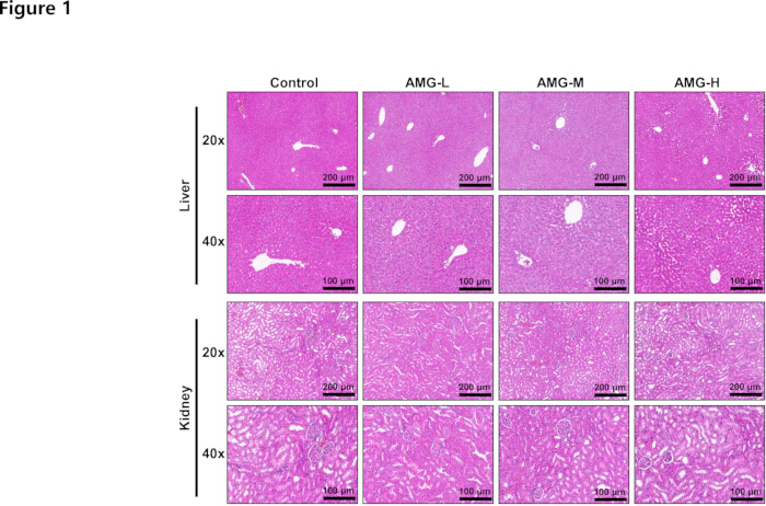
Figure 1: HE staining. HE staining was used to evaluate the dose-dependent toxicity of AMG (60, 90, and 120 mg/kg) on the liver and kidney of normal C3H/HeJ mice. No specific pathological damage to the liver and kidney was observed under the magnification of 20× (200 µm) and 40× (100 µm). n = 6. Please click here to view a larger version of this figure.
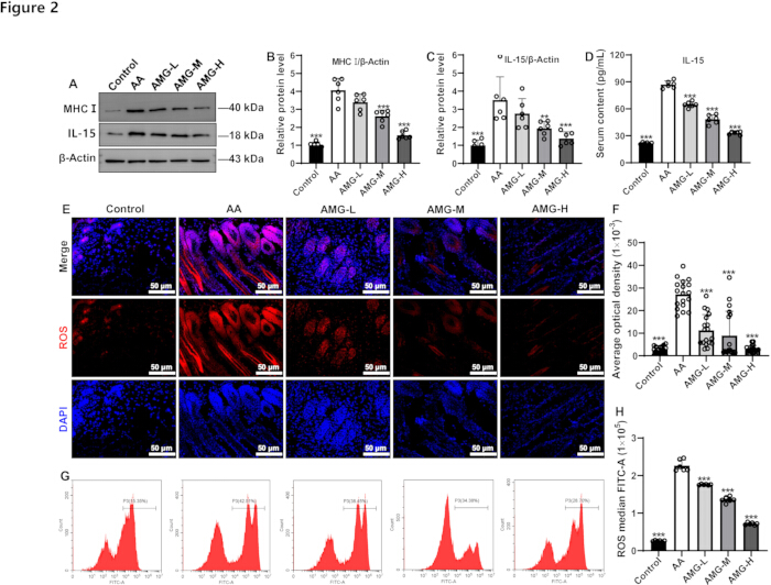
Figure 2: Effects of AMG on MHC I, IL-15, and ROS in the skin samples of AA mice. (A–C), AMG reduced the protein expression levels of MHC I and IL-15, as evaluated by Western blot. (D), AMG lowered IL-15 levels in the mice's serum. (E and F), AMG reduced ROS levels in mouse skin samples, as detected by the IF assay. (G and H), AMG reduced ROS levels in mouse skin samples, as determined by flow cytometry. Experimental results are presented in the form of mean ± SD. **p < 0.01 and ***p < 0.001 vs. the AA group; n = 6. Please click here to view a larger version of this figure.
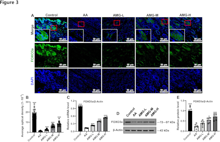
Figure 3: Effects of AMG on FOXO3a protein and gene levels in the skin samples of AA mice. (A) AMG increased FOXO3a protein levels, as evaluated by IF assay. (B) The quantitative results of IF. (C) AMG increased FOXO3a gene expression. (D) AMG increased FOXO3a protein expression, as detected by Western blot. (E) The quantitative results of the Western blot. Experimental results are presented in the form of mean ± SD. *p < 0.05 and ***p < 0.001 vs. the AA group; n = 6. Please click here to view a larger version of this figure.
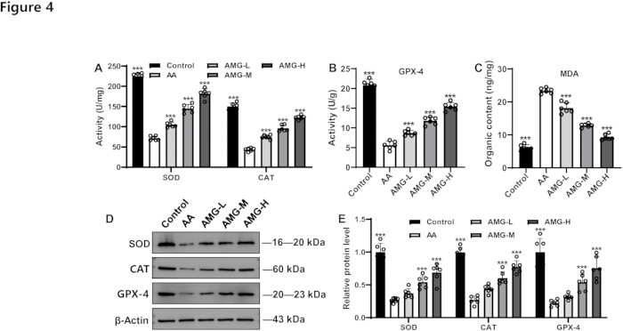
Figure 4: Effects of AMG on oxidative stress indicators in the skin samples of AA mice. (A) AMG stimulated the levels of SOD and CAT, as evaluated using a biochemical test kit. In (B) and (C), AMG increased GPX-4 levels and suppressed MDA levels. (D) representative protein bands of SOD, CAT, and GPX-4. (E) The quantitative results of protein expression for SOD, CAT, and GPX-4. Experimental results are presented in the form of mean ± SD. ***p < 0.001 vs. the AA group; n = 6. Please click here to view a larger version of this figure.
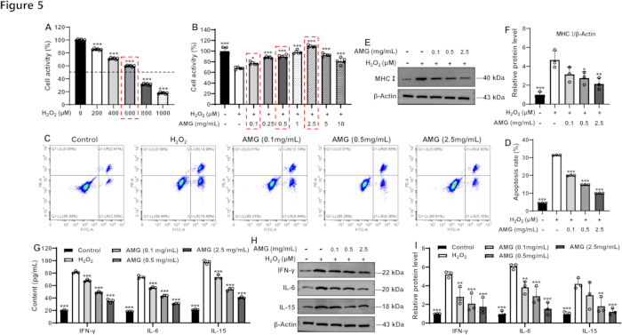
Figure 5: Effects of AMG on the apoptosis and inflammatory indicators of HPEMs induced by H2O2. (A) consequences of varying concentrations of H2O2 on HPEMs viability (n = 6). (B) Effects of varying concentrations of AMG on the survivability of HPEMs induced by H2O2 stimuli (n = 6). (C) Flow cytometric analysis was employed to assess AMG-mediated apoptosis in HPEMs induced by H2O2 stimuli. (D) The statistical results of cell apoptosis. (E) Western blot band of MHC I. (F) The quantitative result of the relative expression of protein MHC I. (G) effects of AMG on IFN-γ, IL-6, and IL-15 in HPEMs induced by H2O2 stimuli. (H) Western blot bands of IFN-γ, IL-6, and IL-15. (I) The quantitative result of the relative level of proteins IFN-γ, IL-6, and IL-15. Experimental results are presented in the form of mean ± SD. *p < 0.05, **p < 0.01 and ***p < 0.001 vs. the H2O2 group; n = 3. Please click here to view a larger version of this figure.
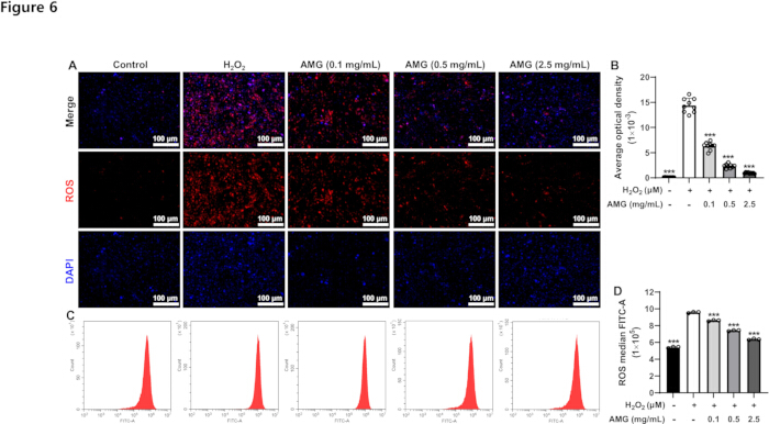
Figure 6: Effects of AMG on ROS in HPEMs induced by H2O2. (A) The fluorescence image of ROS. (B) The fluorescence quantitative statistical results of ROS. (C) The flow cytometry image of ROS. (D) The flow cytometric quantitative statistical results of ROS. Experimental results are presented in the form of mean ± SD. ***p < 0.001 vs. the H2O2 group; n = 3. Please click here to view a larger version of this figure.
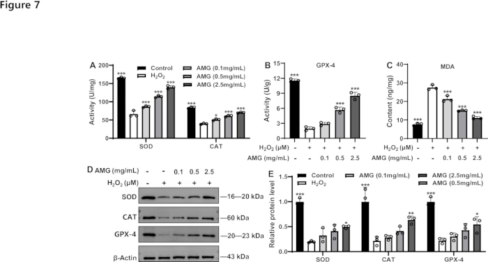
Figure 7: Effects of AMG on oxidative stress indicators in HPEMs induced by H2O2. The levels of SOD, (A) CAT, (B) GPX-4, and (C) MDA in HPEMs were measured using biochemical or ELISA kits. Western blot bands of SOD, CAT, and GPX-4 (D), and their quantitative results (E). Experimental results are presented as mean ± SD. *p < 0.05, **p < 0.01 and ***p < 0.001 vs. the H2O2 group; n = 3. Please click here to view a larger version of this figure.
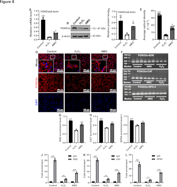
Figure 8: Effects of AMG on FOXO3a in HPEMs induced by H2O2. (A) the expression level of the FOXO3a gene. Western blot bands of FOXO3a (B) and its quantitative results (C). The fluorescence image of FOXO3a (D) and its fluorescence quantitative statistical results (E). (F) representative electrophoretic patterns of ChIP-PCR for FOXO3a-SOD, FOXO3a-CAT, and FOXO3a-GPX-4. Quantitative analysis of the gray values of the ChIP-PCR bands for SOD (G), CAT (H), and GPX-4 (I). The enrichment levels of FOXO3a at the promoters of SOD (J), CAT (K), and GPX-4 (L) were detected by ChIP-PCR. Experimental results are presented in the form of mean ± SD. *p < 0.05, **p < 0.01 and ***p < 0.001 vs. the H2O2 group, or vs. the indicated groups; n = 3. Please click here to view a larger version of this figure.
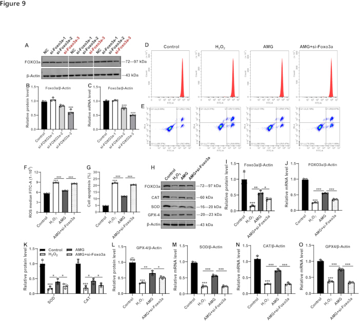
Figure 9: Effects of AMG on oxidative stress and apoptosis in HPEMs cells with low expression of FOXO3a and H2O2 stimuli. (A) Western blot bands of FOXO3a and its quantitative result (B). (C) quantitative result of FOXO3a mRNA expression. (D) flow cytometry graph of ROS and cell apoptosis (E). And the associated statistical findings (F and G). (H) western blot bands of FOXO3a, SOD, CAT, and GPX-4. (I) The quantitative result of FOXO3a protein expression. (J) quantitative result of FOXO3a mRNA expression. (K and L) quantitative results of SOD, CAT, and GPX-4 protein expression. Quantitative results of SOD (M), CAT (N), and GPX-4 (O) gene expression. Experimental results are presented in the form of mean ± SD. *p < 0.05, **p < 0.01, and ***p < 0.001 vs. the control group, or vs. the indicated groups; n = 3. Please click here to view a larger version of this figure.
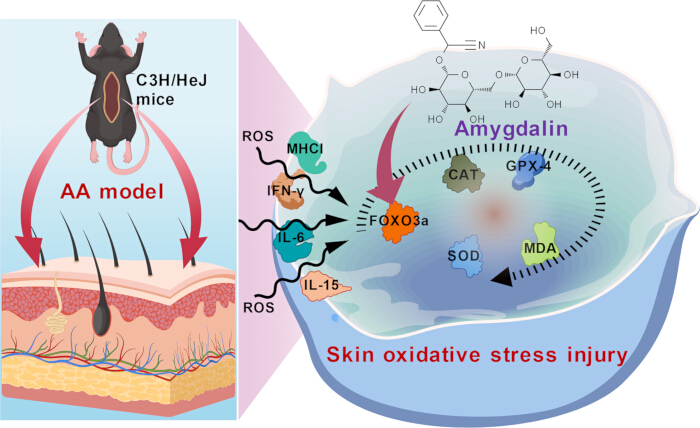
Figure 10: Schematic diagram. This diagram illustrates the mechanism by which AMG stimulates FOXO3a expression, thereby inhibiting oxidative stress and inflammation and improving AA. It is created by the authors using BioGDP.com, and no copyright concerns are involved. Please click here to view a larger version of this figure.
| Genes | Species | Sequences (5' - 3') | Length (bp) |
| FOXO3a | Mouse | Forward: GCAAGCCGTGTACTGTGGA | 116 |
| Reverse: CGGGAGCGCGATGTTATCC | |||
| FOXO3a | Human | Forward: CGGACAAACGGCTCACTCT | 150 |
| Reverse: GGACCCGCATGAATCGACTAT | |||
| SOD | Human | Forward: AAAGATGGTGTGGCCGATGT | 167 |
| Reverse: CAAGCCAAACGACTTCCAGC | |||
| CAT | Human | Forward: CTATCCTGACACTCACCGCC | 129 |
| Reverse: CCACCCTGATTGTCCTGCAT | |||
| GPX-4 | Human | Forward: GCAAGGGCATCCTGGGAAAT | 84 |
| Reverse: GTTCTTGTCGATGAGGAACTGTG | |||
| β-Actin | Human | Forward: AATCTGGCACCACACCTTCTACAA | 172 |
| Reverse: GGATAGCACAGCCTGGATAGCAA | |||
| β-Actin | Mouse | Forward: GTGCTATGTTGCTCTAGACTTCG | 174 |
| Reverse: ATGCCACAGGATTCCATACC |
Table 1: Gene sequence information for qRT-PCR. The primers used for qRT-PCR.
| Genes | Species | Sequences (5' - 3') | Length (bp) |
| SOD | Human | Forward: TGCCAGAAGAGGGACAGAACAGG | 173 |
| Reverse: ATCCGCCCACAGACACGAGAG | |||
| CAT | Human | Forward: GGGAAGCAGATTTCTCCAGTG | 141 |
| Reverse: GGGTGTTGATTTCCTCCTTTACC | |||
| GPX-4 | Human | Forward: TGGCACATTTTGGGGTTGGAAC | 103 |
| Reverse: TGAACGCTCCTCCTCCTTAAACG |
Table 2: Gene sequence information for ChIP-PCR. The primers used for ChIP-PCR.
Discussion
AMG itself has no direct toxicity. It is hydrolyzed by intestinal microbial enzymes or gastric acid, generating and releasing highly toxic hydrogen cyanide49. Hydrogen cyanide is then rapidly absorbed through the digestive tract into the bloodstream and spreads throughout the body tissues, causing hypoxia in the nervous system, cardiovascular system, and respiratory system tissues50. In terms of mechanism, hydrogen cyanide combines with the iron ions in cytochrome oxidase, blocking the cellular respiratory chain, resulting in tissue hypoxia and cellular metabolic disorders, and triggering acute poisoning51. The toxic symptoms include headache, nausea, vomiting, and breathing difficulties49,52. In severe cases, it can lead to respiratory failure and death49,52. Therefore, ensuring the safe dosage range of AMG is a prerequisite for studying its pharmacological effects. In this study, the authors first confirmed through pathological examination that the doses they used (60, 90, and 120 mg/kg) did not cause liver or kidney toxicity in normal C3H/HeJ mice, which is the foundation for further research on the treatment of alopecia areata with AMG.
IFN-γ serves as a critical driver of inflammation during the development of AA, mainly secreted by activated CD4+/CD8+ T cells53,54,55. It disrupts the immunological privilege of hair follicles (such as by inhibiting the low expression of MHC I), prompting autoreactive T cells to attack the melanocytes of hair follicles, and resulting in vacuolation of hair mother cells and abnormal keratinization of the hair bulb56,57. Animal experiments have shown that mice lacking the IFN-γ gene exhibit resistance to AA58. IL-6 and IL-15 can enhance Th1-type immune responses by activating the JAK/STAT signal transduction cascade and promoting the release of IFN-γ 59. Moreover, IL-15 can stimulate the proliferation of NK cells and T cells, exacerbating the inflammatory infiltration surrounding the hair follicle structures60. Normal hair bulbs evade immune surveillance by low expression of MHC I, while in AA patients, MHC I is abnormally upregulated, making the melanocytes of hair follicles the target of T cell attack61. In this study, the authors noted AMG treatment significantly downregulated the levels of MHC I and IL-15 proteins in the skin of C3H/HeJ mice. The previous animal experiments also confirmed that AMG can inhibit the levels of IFN-γ and IL-6 in AA mice38. In H2O2-evoked HPEMs injury, MHC I, as well as pro-inflammatory cytokines including IFN-γ, IL-6, and IL-15, were considerably restricted after AMG treatment. These data suggest that the potential of AMG in treating AA may be related to maintaining the balance between immunity and inflammation.
The imbalance between the anti-oxidant enzyme system (SOD, CAT, and GPX-4) and lipid peroxidation markers MDA and ROS plays a crucial role in the pathogenesis of AA62. SOD reduces oxidative stress by catalyzing the conversion of O2·⁻ to H2O2, while CAT and GPX-4 further decompose H2O2 into H2O, preventing the accumulation of ROS. Evidence suggests that excessive ROS can trigger lipid peroxidation, leading to elevated MDA levels and directly damaging hair follicle cells63. In patients with AA, the clearance ability of ROS in hair follicles decreases, and the accumulation of MDA exacerbates apoptotic death of hair papilla cells, resulting in the hair follicles prematurely entering the resting phase63. Moreover, oxidative stress may promote the release of pro-inflammatory factor IFN-γ, further aggravating immune attack and hair follicle degeneration64. In this study, researchers clarified that AMG intervention reduced the overexpressed ROS in the skin of the AA mice and H2O2-induced HPEM cells injury. Meanwhile, in vivo and ex vivo experimental investigations both manifested that AMG treatment significantly augmented the anti-oxidant indicators (SOD, CAT, and GPX-4), while weakening the pro-oxidative indicator MDA. These evidences indicate that maintaining the balance of oxidative stress is the contribution of AMG to AA. Considering that the compromised GPX-4 activity would lead to unchecked MDA generation, this might trigger the ferroptosis process. These molecular processes can also contribute to the aggravation of AA and serve as a potential mechanism for AMG treatment. All of these require further experimental verification in the future.
As a key transcription factor, FOXO3a may be involved in AA pathogenesis through both oxidative stress and immune regulation pathways65. It can translocate to the nucleus under oxidative stress, thereby upregulating key antioxidant enzymes such as SOD and CAT to clear excess ROS and reduce oxidative damage to scalp tissue. By mitigating local oxidative burden, FOXO3a can stabilize the immune privilege of hair follicles and restrain the autoimmune attack that drives non-scarring hair loss. Regarding oxidative stress regulation, FOXO3a activation can upregulate the expression of antioxidant enzymes such as SOD and CAT and inhibit ROS accumulation65. However, impaired FOXO3a function can trigger elevated MDA generation, a key lipid peroxidation product, thereby exacerbating cell apoptosis 66. At the immune regulation level, FOXO3a triggers autoimmune attacks by regulating the MHC I-restricted antigens67. This study reported that AMG dramatically increased FOXO3a protein and gene levels in the skin tissue of C3H/HeJ mice and in H2O2-induced HPEMs cells. Furthermore, AMG reduced ROS levels and cell apoptosis, while elevating FOXO3a expression and the levels of FOXO3a-regulated SOD, CAT, and GPX-4 in HPEM cells with low FOXO3a expression. Collectively, these findings indicate that the therapeutic effect of AMG on AA is closely linked to the FOXO3a-mediated regulatory balance between oxidative stress and cellular apoptosis. Considering the causal relationship of AMG-mediated FOXO3a to improve AA, it is necessary to conduct FOXO3a overexpression or pharmacological inhibition under the intervention of AMG in future studies. In addition, whether AMG-mediated FOXO3a can mitigate oxidative stress and modulate inflammatory responses to protect hair follicle stem cells from ROS-induced damage and immune-mediated destruction remains to be investigated. Another point worth noting is that whether AMG exerts its antioxidant effect by directly counteracting the toxicity of H₂O₂ or by regulating intracellular signal transduction remains an unsolved issue in this research.
In conclusion, this study first confirmed that AMG was not potentially toxic to the liver and kidneys at the tested dosages. Moreover, it was demonstrated at the molecular level that AMG might promote the expression of FOXO3a, thereby transcriptionally activating the expressions of SOD, CAT, and GPX-4, and inhibiting inflammatory responses and apoptotic events to treat AA (Figure 10). However, some limitations that do exist in this study still need to be explored and clarified in subsequent research. Firstly, the clarification of the long-term toxicity and pharmacokinetic properties of AMG in AA animals serves as the foundation and prerequisite for in-depth research on its pharmacological effects and molecular mechanisms. Secondly, the pharmacological effects of AMG in treating AA still require confirmation through large-scale studies in multiple animal models and clinical trials. In terms of the molecular mechanism, it is still necessary to conduct rescue experiments to further confirm whether it is FOXO3a that mediates the pharmacological effect of AMG. Whether it is the direct antioxidant activity of AMG or the antioxidant effect exerted by AMG-mediated FOXO3a requires further exploration.
Disclosures
The authors have no conflicts of interest to declare.
AUTHORS’ CONTRIBUTION:
Xun He: Investigation, Methodology, Writing – original draft. Jingsong Liu, Jiawei Wang, and Zhe Dai: Data curation. Yugang Gong, Wei Lu, and Xiaowei Sha: Software, Visualization, and Validation. Xun He: Conceptualization, Writing – Review & Editing, Funding acquisition, and Supervision.
Acknowledgements
This work was supported by the Sichuan Provincial Cadres Healthcare Committee (2024-221).
Materials
| Name | Company | Catalog Number | Comments |
|---|---|---|---|
| 5×All-In-One MasterMix (with AccuRT genomic DNA removal kit) | Applied Biological Materials Inc., Jiangsu, China | G492 | |
| β-Actin antibody | Affinity Biosciences Co., Ltd., Wuhan, China | AF7018 | |
| Amygdalin | Chengdu Herbpurify Co., Ltd., Chengdu, China | K-001-181216 | |
| Catalase antibody | Proteintech, Wuhan, China | 66765-1-Ig | |
| Catalase kit | Elabscience, Wuhan, China | E-BC-K031-M | |
| C3H/HeJ mice | Chengdu Yaokang Biotechnology Co., Ltd., Chengdu, China | N000190 | |
| ChIP assay kit | Beyotime Biotechnology, Shanghai, China | P2078 | |
| 254 culture medium | Gibco, Thermo Fisher Scientific, America | M-254-500 | |
| DAPI staining reagent | Servicebio, Wuhan, China | G1012 | |
| Dispase II | Shanghai Yuanye Bio-Technology Co., Ltd., Shanghai, China | S25046 | |
| EvaGreen Express 2×qPCR MasterMix-No Dye | Applied Biological Materials Inc., Jiangsu, China | MasterMix-ES | |
| Fast black HB | Sangon Biotech Co., Ltd., Shanghai, China | A602008-0025 | |
| Fetal bovine serum | Every Green | 13011-8611 | |
| Fluorescence microscope | SOPTOP | CX40 | |
| FOXO3a antibody | Proteintech, Wuhan, China | 10849-1-AP | |
| Freezing microtome | Thermo | Cryotome E | |
| Glutathione peroxidase 4 antibody | Proteintech, Wuhan, China | 67763-1-Ig | |
| Glutathione peroxidase 4 kit | Elabscience, Wuhan, China | E-BC-K883-M | |
| Image-pro plus 6.0 | Media Cybernetics, Inc., Rockville, MD, USA | —— | |
| Imiquimod cream | Sichuan Mingxin Pharmaceutical Co. Ltd., Chengdu, China | H20030129 | |
| Interferon gamma antibody | Proteintech, Wuhan, China | 15365-1-AP | |
| Interferon gamma kit | Elabscience, Wuhan, China | E-EL-H0108 and E-EL-M0048 | |
| Interleukin 6 antibody | Proteintech, Wuhan, China | 21865-1-AP | |
| Interleukin 6 kit | Elabscience, Wuhan, China | E-EL-H6156 and E-EL-M0044 | |
| Interleukin 15 antibody | Affinity Biosciences Ltd., Wuhan, China | DF2521 | |
| Interleukin 15 kit | Elabscience, Wuhan, China | E-EL-H0222 and E-EL-M0728 | |
| Malondialdehyde kit | Elabscience, Wuhan, China | E-EL-0060 | |
| MHC antibody | Affinity Biosciences Ltd., Wuhan, China | DF8558 | |
| Optical microscope | ZEISS | Primo Star | |
| Paraffin slicer | Thermo scientific | HM325 | |
| Pathological scanning software | 3DHISTECH (Hungary) | Slide Viewer2.5.0 | |
| Penicillin-synthroid solution | Biosharp, Beijing, China | BL505A | |
| Rabbit IgG | Beyotime Biotechnology, Shanghai, China | A7016 | |
| ROS assay kit | Beyotime Biotechnology, Shanghai, China | S0033M | |
| Superoxide dismutase 1 antibody | Proteintech, Wuhan, China | 10269-1-AP | |
| Tissue embedder | Wuhan Junjie Electronics Co., Ltd., Wuhan China | JB-P5 | |
| Tissue hydroextractor | Thermo scientific | STP420 ES | |
| Total RNA isolation kit | Foregene, Chengdu, China | RE-03014 | |
| Total superoxide dismutase kit | Elabscience, Wuhan, China | E-BC-K020-M | |
| White vasoline | Shanghai Aladdin Biochemical Technology Co., Ltd., Shanghai, China | P434227 |
References
- Li XQ, et al. Burden of alopecia areata in China, 1990-2021: Global Burden of Disease Study 2021. Chin Med J (Engl). 2025;138:318-324.
- Rodríguez-Tamez G, Maskan-Bermudez N, Tosti A. Alopecia areata incognita: Current evidence. Dermatol Ther (Heidelb). 2025;15:635-645.
- Zhou J, et al. Global burden of alopecia areata and associated diseases: A trend analysis from 1990 to 2021. J Cosmet Dermatol. 2025;24:e70076.
- Jeon JJ, Lee S, Kim MS, Kim YS, Hong SH, Yon DK, et al. The burden of alopecia areata among United States, 2000-2021: A systematic analysis for the global burden of disease study 2021. J Am Acad Dermatol. 2025;S0190-9622(25)02673-8.
- Todorova L, Kluger N. Alopecia: A Google trends analysis of the past 5 years (2018-2023). Skin Appendage Disord. 2025;11:282-286.
- Pratt CH, King LEJr, Messenger AG, Christiano AM, Sundberg JP. Alopecia areata. Nat Rev Dis Primers. 2017;3:17011.
- Mistry J, Sibbald C. Clinical presentations and comorbidities of pediatric alopecia areata. Pediatr Dermatol. 2025;42 Suppl 1:5-11.
- Kuty-Pachecka M. Psychological and psychopathological factors in alopecia areata. Psychiatr Pol. 2015;49:955-964.
- Marahatta S, Agrawal S, Adhikari BR. Psychological impact of alopecia areata. Dermatol Res Pract. 2020;2020:8879343.
- Dainichi T, Iwata M, Kaku Y. Alopecia areata: What's new in the epidemiology, comorbidities, and pathogenesis J Dermatol Sci. 2023;112:120-127.
- Lintzeri DA, Constantinou A, Hillmann K, Ghoreschi K, Vogt A, Blume-Peytavi U. Alopecia areata - Current understanding and management. J Dtsch Dermatol Ges. 2022;20:59-90.
- Singh R, Kumar P, Kumar D, Aggarwal N, Chopra H, Kumar V. Alopecia areata: Review of epidemiology, pathophysiology, current treatments and nanoparticulate delivery system. Ther Deliv. 2024;15:193-210.
- Rizzetto G, De Simoni E, Gioacchini H, Molinelli E, Offidani A, Simonetti O. Fractionated CO2 laser in combination with topical tacrolimus for chronic alopecia areata: A case series study. Life (Basel). 2024;14:1128.
- Gasques L, de Brito FF, Zobiole R, Bortolini L, Assumpção VCC, Carvalho CL. Corticosteroid atrophy plaque on the scalp treated with 5-fluourouracil and bleomycin infusion in the MMP technique. Skin Appendage Disord. 2024;10:425-428.
- Lee D, et al. Serum levels of IL-18 and sIL-2R in patients with alopecia areata receiving combined therapy with oral cyclosporine and steroids. Exp Dermatol. 2010;19:145-147.
- Mlacker S, et al. A review on laser and light-based therapies for alopecia areata. J Cosmet Laser Ther. 2017;19:93-99.
- Jafari MA, Bazgir G, Hosseini-Baharanchi FS, Jafarzadeh A, Goodarzi A. Efficacy and safety of laser therapy and phototherapy in cicatricial and noncicatricial alopecia: A systematic review study. Health Sci Rep. 2024;7:e70180.
- Civaş E, Aksoy B, Aksoy HM, Eski M, Yucel K. Hair transplantation for therapy-resistant alopecia areata of the eyebrows: is it the right choice J Dermatol. 2010;37:823-826.
- King BA, Craiglow BG. Janus kinase inhibitors for alopecia areata. J Am Acad Dermatol. 2023;89:S29-S32.
- Chim I, Ghiya R, Sinclair RD, Eisman S. Novel investigational drugs for alopecia areata and future perspectives. Expert Opin Investig Drugs. 2024;33:441-449.
- Valdez-Zertuche JA, Ramírez-Marín HA, Tosti A. Efficacy, safety and tolerability of drugs for alopecia: A comprehensive review. Expert Opin Drug Metab Toxicol. 2025;21:347-371.
- Liu M, et al. Janus kinase inhibitors for alopecia areata: A systematic review and meta-analysis. JAMA Netw Open. 2023;6:e2320351.
- Sohn KC, et al. Effect of thioredoxin reductase 1 on glucocorticoid receptor activity in human outer root sheath cells. Biochem Biophys Res Commun. 2007;356:810-815.
- Sun C, Ding HH, Zhang L, Wang JL, Su MY. Co-regulatory mechanisms and potential markers of oxidative stress-related genes in vitiligo and alopecia areata. Skin Res Technol. 2024;30:e70001.
- Perlmutter J, et al. The role of reactive oxygen species in the pathogenesis of alopecia areata: A systematic review and meta-analysis. Skin Pharmacol Physiol. 2025;38:59-67.
- Xie B, Sun JY, Song XZ. Hair follicle melanocytes initiate autoimmunity in alopecia areata: A trigger point. Clin Rev Allergy Immunol. 2022;63:417-430.
- Shin JM, et al. The crosstalk between PTEN-induced kinase 1-mediated mitophagy and the inflammasome in the pathogenesis of alopecia areata. Exp Dermatol. 2024;33:e14844.
- Mustafa AI, Khashaba RA, Fawzy E, Baghdady SMA, Rezk SM. Cross talk between oxidative stress and inflammation in alopecia areata. J Cosmet Dermatol. 2021;20:2305-2310.
- Cohen JN, et al. Regulatory T cells in skin mediate immune privilege of the hair follicle stem cell niche. Sci Immunol. 2024;9:eadh0152.
- Kinoshita-Ise M, Martinez-Cabriales SA, Alhusayen R. Chronological association between alopecia areata and autoimmune thyroid diseases: A systematic review and meta-analysis. J Dermatol. 2019;46:702-709.
- Chen DY, Yang XY, Wang HX, Fan WL, He YQ. Molecular mechanism of polygonum multiflorum in the treatment of androgenic alopecia: based on bioinformatics and molecular docking method. Diagnosis and Therapy Journal of Dermato-Venereology 2022;29:101–108.
- Iwasa K, et al. Long term administration of loquat leaves and their major component, ursolic acid, attenuated endogenous amyloid-β burden and memory impairment. Sci Rep. 2023;13:16770.
- Makovi CM, Parker CH, Zhang K. Determination of amygdalin in apricot kernels and almonds Using LC-MS/MS. J AOAC Int. 2023;106:457-463.
- Li XJ, et al. Identifying the active components and mechanisms of Persicae Semen in treating androgenetic alopecia: Insights from network pharmacology and experimental evaluations. J Ethnopharmacol. 2025;347:119755.
- Wang Q, et al. Amygdalin alleviates psoriasis-like lesions by improving skin barrier function. Arch Dermatol Res. 2025;317:115.
- Li SY, et al. Effects of amygdalin on ferroptosis and oxidative stress in diabetic retinopathy progression via the NRF2/ARE signaling pathway. Exp Eye Res. 2023;234:109569.
- Kimura K, Liu YH, Hsieh CL. Amygdalin's neuroprotective effects on acute ischemic stroke in rats. J Ethnopharmacol. 2025;345:119621.
- He X, et al. Amygdalin ameliorates alopecia areata on C3H/HeJ mice by inhibiting inflammation through JAK2/STAT3 pathway. J Ethnopharmacol. 2024;331:118317.
- He X, et al. The antiinflammatory effects of Xuefu Zhuyu decoction on C3H/HeJ mice with alopecia areata. Phytomedicine. 2021;81:153423.
- Ma LP, et al. Melatonin inhibits senescence-associated melanin pigmentation through the p53-TYR pathway in human primary melanocytes and the skin of C57BL/6 J mice after UVB irradiation. J Mol Med (Berl). 2023;101:581-593.
- Hou Y, et al. Longzhibu disease and its therapeutic effects by traditional Tibetan medicine: Ershi-wei Chenxiang pills. J Ethnopharmacol. 2020;249:112426.
- Hou Y, Fan FH, Xie N, Zhang Y, Wang XB, Meng XL. Rhodiola crenulata alleviates hypobaric hypoxia-induced brain injury by maintaining BBB integrity and balancing energy metabolism dysfunction. Phytomedicine. 2024;128:155529.
- Hou Y, et al. Salidroside intensifies mitochondrial function of CoCl2-damaged HT22 cells by stimulating PI3K-AKT-MAPK signaling pathway. Phytomedicine. 2023;109:154568.
- Fan FH, et al. Association between systemic immune-inflammation indexes and Nuodikang capsule’s protection in LPS-induced acute lung injury in mice. Phytomedicine. 2025;147:157229.
- Wang XB, et al. Mechanistic insights into salidroside's mitochondrial protection via AMPK/Sirt1/HIF-1α pathway in hypoxic HT22 cells. JoVE. 2025;218:e66923.
- Wang XB, et al. Rhodiola crenulata attenuates apoptosis and mitochondrial energy metabolism disorder in rats with hypobaric hypoxia-induced brain injury by regulating the HIF-1α/microRNA 210/ISCU1/2(COX10) signaling pathway. J Ethnopharmacol. 2019;241:111801.
- Fan SJ, et al. PRMT5 encourages cell migration and metastasis of tongue squamous cell carcinoma through methylating ΔNp63α. Cell Death Differ. 2026; 33:465-479.
- Zhao Y, et al. Integrated analysis reveals that EGR1 promotes epithelial IL33 production in T2 asthma. J Transl Med. 2025;23:203.
- He XY, Wu LJ, Wang WX, Xie PJ, Chen YH, Wang F. Amygdalin - A pharmacological and toxicological review. J Ethnopharmacol. 2020;254:112717.
- Alan-Albayrak E, Simonsen U. Cyanide beyond toxicity: A systematic review of its effects on vascular function. Basic Clin Pharmacol Toxicol. 2025;137:e70124.
- Lachowicz JI, Alexander J, Aaseth JO. Cyanide and cyanogenic compounds-toxicity, molecular targets, and therapeutic agents. Biomolecules. 2024;14:1420.
- Jaszczak-Wilke E, Polkowska Ż, Koprowski M, Owsianik K, Mitchell AE, Bałczewski P. Amygdalin: Toxicity, anticancer activity and analytical procedures for its determination in plant seeds. Molecules. 2021;26:2253.
- Deeths MJ, Endrizzi BT, Irvin ML, Steiner LP, Ericson ME, Hordinsky MK. Phenotypic analysis of T-cells in extensive alopecia areata scalp suggests partial tolerance. J Invest Dermatol. 2006;126:366-373.
- Garzorz N, et al. Dissecting susceptibility from exogenous triggers: the model of alopecia areata and associated inflammatory skin diseases. J Eur Acad Dermatol Venereol. 2015;29:2429-2435.
- Belpaire A, Demeyer A, Van Caelenberg E, van Geel N, Speeckaert R. The AhR pathway is dysregulated in alopecia areata. J Transl Autoimmun. 2025;10:100282.
- Gilhar A, Schrum AG, Etzioni A, Waldmann H, Paus R. Alopecia areata: Animal models illuminate autoimmune pathogenesis and novel immunotherapeutic strategies. Autoimmun Rev. 2016;15:726-735.
- Oelert T, Gilhar A, Paus R. T-cell "induced-self" MHC class I/peptide complexes may enable "de novo" tolerance induction to neo-antigens occurring outside of the thymus: Lessons from the hair follicle. Exp Dermatol. 2017;26:529-531.
- Freyschmidt-Paul P, et al. Interferon-γ-deficient mice are resistant to the development of alopecia areata. Br J Dermatol. 2006;155:515-521.
- Lensing M, Jabbari A. An overview of JAK/STAT pathways and JAK inhibition in alopecia areata. Front Immunol. 2022;13:955035.
- Suzuki T, et al. Interleukin-15 is a hair follicle immune privilege guardian. J Autoimmun. 2024;145:103217.
- Fitzhugh MH, Hansen JG, Jabbari A, Berrebi KG. Pathophysiology of Alopecia Areata in the Pediatric Patient. Pediatr Dermatol. 2025;42:24-30.
- Ma YQ, Sun Z, Li YM, Xu H. Oxidative stress and alopecia areata. Front Med (Lausanne). 2023;10:1181572.
- Bakry OA, Elshazly RM, Shoeib MA, Gooda A. Oxidative stress in alopecia areata: A case-control study. Am J Clin Dermatol. 2014;15:57-64.
- Peterle L, Sanfilippo S, Borgia F, Cicero N, Gangemi S. Alopecia areata: A review of the role of oxidative stress, possible biomarkers, and potential novel therapeutic approaches. Antioxidants (Basel). 2023;12:135.
- Yang X, et al. Exosomes derived from platelet-rich plasma promote hair regeneration by regulating the SIRT1/FoxO3a pathway to alleviate oxidative stress. Regen Ther. 2025;30:710-718.
- Xu SC, Huang P, Yang JH, Du HX, Wan HT, He Y. Calycosin alleviates cerebral ischemia/reperfusion injury by repressing autophagy via STAT3/FOXO3a signaling pathway. Phytomedicine. 2023;115:154845.
- Tang QY, et al. Orlistat facilitates immunotherapy via AKT-FOXO3a-FOXM1-mediated PD-L1 suppression. J Immunother Cancer. 2025;13:e008923.