This study uses 16S rRNA gene sequencing to characterize breast tissue microbiota alterations associated with plasma cell mastitis.
Research Article
Breast Microbiome Dysbiosis in Plasma Cell Mastitis: A Ribosomal RNA Gene Sequencing Study
* These authors contributed equally
In This Article
Summary
Abstract
Plasma cell mastitis (PCM) is a chronic breast condition with limited effective treatment options. Surgical intervention is a standard treatment approach but may be associated with postoperative complications. Recent studies suggest that alterations in the microbiome may be associated with the onset, progression, and recurrence of PCM. In this study, high-throughput 16S ribosomal RNA (rRNA) gene sequencing was used to compare the breast tissue microbiota of patients with PCM with that of the non-PCM control (NC) group. A total of 86 participants were enrolled. No significant differences were observed between the groups in age, body mass index (BMI), or lymphocyte count, whereas white blood cell (WBC) count, neutrophil count, prolactin level, and the prevalence of nipple inversion differed significantly between the groups. A total of 214 bacterial genera showed nominally significant differences in relative abundance between the two groups based on unadjusted P values. Staphylococcus and Bacteroides were more abundant in the PCM group, whereas Plesiomonas and Acinetobacter were more abundant in the NC group. These results indicate that the breast tissue microbiota differed between the PCM and NC groups. The findings demonstrate alterations in the breast microbiome associated with PCM and provide a basis for further investigation of their potential relevance to PCM and future treatment strategies.
Introduction
Plasma cell mastitis (PCM) is a chronic non-lactational inflammatory breast disease characterized by mammary duct ectasia, periductal inflammatory reactions, and plasma cell infiltration1,2,3. Although PCM is not a common malignant breast lesion, its prolonged course, high tendency to recur, and frequent clinical manifestations, including breast mass, pain, nipple discharge, nipple retraction, and even fistula formation, can seriously impair patients’ quality of life and psychological well-being2,3. At present, the clinical management of PCM still faces considerable challenges4. Current studies have highlighted the potential roles of immune dysregulation, endocrine abnormalities, and structural alterations of the mammary ducts in PCM development. In particular, persistent inflammatory activation and changes in the local tissue microenvironment are considered important contributors to disease initiation and progression. Previous studies have suggested that PCM is not driven by a single factor and that its onset and progression may be closely associated with immune abnormalities, endocrine imbalance, structural alterations of the mammary ducts, and a local chronic inflammatory microenvironment5,6. However, because of the lack of mature and stable cell and animal models, studies on the molecular mechanisms and pathological evolution of PCM remain relatively limited5, which, to some extent, restricts the development of more effective therapeutic strategies. Therefore, further exploration of the potential pathogenic mechanisms of PCM is important for identifying new therapeutic targets and improving clinical outcomes.
In recent years, with the rapid development of microbiome research, accumulating evidence has shown that microbial communities at different body sites play important roles in maintaining tissue homeostasis, regulating local immunity, and influencing disease onset and progression7,8. Microbial dysbiosis has been reported to be closely associated with a variety of diseases, including periodontitis9, gastric cancer10, and neurodegenerative diseases11. In PCM, the role of microbial factors is attracting increasing attention. Although PCM has traditionally been regarded as a non-infectious inflammatory disease2, clinical and pathological studies have shown that it is often accompanied by ductal obstruction and dilatation, together with infiltration of multiple inflammatory cell types, including neutrophils, macrophages, and plasma cells, suggesting persistent inflammatory stimulation and microenvironmental imbalance in local tissues3. Therefore, an important knowledge gap remains regarding whether PCM is associated with specific alterations in the local breast tissue microbiome. Although previous studies have extensively investigated the immune, endocrine, and inflammatory mechanisms involved in PCM, the microbial characteristics of breast tissue in patients with PCM have not been systematically characterized.
Based on this rationale, we hypothesized that, compared with non-PCM breast tissues, breast tissues from patients with PCM may exhibit distinct alterations in microbial composition and diversity and that these changes may reflect local microbial dysbiosis associated with the inflammatory microenvironment of PCM. To test this hypothesis, we performed high-throughput 16S rRNA gene sequencing to compare the breast tissue microbiota of patients with PCM with that of non-PCM control tissues collected adjacent to benign breast lesions, with a particular focus on community composition, microbial diversity, and differentially enriched bacterial taxa. This study aimed to provide preliminary insight into the potential association between breast tissue microbial dysbiosis and the development of PCM, thereby laying a foundation for further investigation of its role in PCM pathogenesis and the potential clinical relevance of associated microbial signatures.
Protocol
All participants provided informed consent, and the study was approved by the Ethics Committee of Jiangsu Provincial Hospital of Traditional Chinese Medicine (Number: 2018NL-163-02), in accordance with the World Medical Association Declaration of Helsinki.
Clinical Samples
This study included 41 patients diagnosed with PCM and 45 control subjects who were recruited from Jiangsu Provincial Hospital of Traditional Chinese Medicine between January 2021 and December 2022. The diagnosis of PCM was confirmed by preoperative biopsy or intraoperative rapid frozen-section examination, followed by histopathological evaluation. The diagnosis was based on characteristic pathological findings, including mammary duct ectasia, periductal inflammatory cell infiltration, and prominent plasma cell infiltration, while malignant lesions and other specific inflammatory breast diseases were excluded. The NC group comprised patients undergoing surgery for benign breast tumors. Histologically normal breast tissue adjacent to the benign lesions was collected for microbiome analysis. Breast tissue samples were collected during surgery under sterile conditions using sterile instruments and standardized handling protocols to minimize environmental contamination. After surgical excision, specimens designated for microbiome analysis were immediately placed in sterile tubes, snap-frozen in liquid nitrogen, and stored at −80°C until DNA extraction. This study compared the clinical characteristics of the two groups, focusing on factors such as age, BMI, white blood cell count, neutrophil count, lymphocyte count, prolactin levels, and nipple abnormalities. Exclusion criteria included malignancies, other specific inflammatory breast diseases, and active infectious diseases unrelated to PCM. Active infectious diseases were defined as clinically diagnosed infections at enrollment, including systemic infectious symptoms (e.g., fever or chills), local signs of acute infection (e.g., progressive erythema, warmth, fluctuance, or purulent drainage), documented positive microbiological results when available, or ongoing antimicrobial treatment. These conditions were evaluated based on medical history, physical examination, and clinical records. PCM-related inflammatory manifestations, such as breast pain, mass, nipple discharge, or nipple retraction, were not considered independent infectious diseases in the absence of additional evidence of infection. Participants who had used antibiotics, probiotics, or prebiotics within 1 month before sample collection were also excluded.
High-throughput 16S ribosomal RNA gene sequencing
To assess the microbial composition of the samples, we performed 16S ribosomal RNA gene amplicon sequencing. DNA was extracted from approximately 150–200 mg of breast tissue using a DNA isolation kit according to the manufacturer's protocol. PCR negative controls were included during amplification. The PCR products were examined by agarose gel electrophoresis, and no visible amplification bands were observed in the negative controls. Accordingly, the negative controls were not subjected to library preparation or sequencing. The V3–V4 regions of the bacterial 16S rRNA gene were amplified using the 341F/806R primer pair (341F: 5′-CCTACGGGNGGCWGCAG-3′; 806R: 5′-GGACTACHVGGGTATCTAAT-3′). PCR amplification was performed in a total reaction volume of 50 μL containing 10 μL of 5× Q5 Reaction Buffer, 10 μL of 5× Q5 High GC Enhancer, 1.5 μL of 2.5 mM dNTPs, 1.5 μL each of the forward and reverse primers (10 μM), 0.2 μL of Q5 High-Fidelity DNA Polymerase, approximately 50 ng of template DNA, and nuclease-free water to a final volume of 50 μL. The PCR program consisted of an initial denaturation at 95°C for 5 min, followed by 30 cycles of denaturation at 95°C for 1 min, annealing at 60°C for 1 min, and extension at 72°C for 1 min, with a final extension at 72°C for 7 min. The amplicon libraries were sequenced on a NovaSeq 6000 platform using paired-end 250-bp sequencing. Raw paired-end sequencing reads were quality-filtered using fastp (v0.18.0). Reads containing ≥10% ambiguous bases (N), reads in which ≥50% of bases had Phred quality scores ≤20, and reads containing adapter sequences were removed. The resulting clean reads were merged into tags using FLASH (v1.2.11), with a minimum overlap of 10 bp and a maximum mismatch rate of 2%. The merged tags were further quality-filtered by truncating sequences at the first position where three consecutive bases had quality scores ≤3; tags in which the remaining high-quality sequence was shorter than 75% of the original tag length were discarded. Clean tags were clustered into operational taxonomic units (OTUs) at 97% sequence similarity using the UPARSE12 algorithm implemented in USEARCH (v9.2.64). Chimeric sequences were identified and removed using UCHIME, and the most abundant sequence within each OTU was selected as the representative sequence. Representative OTU sequences were taxonomically classified using the RDP Classifier (v2.2) against the SILVA database (v132). Krona charts were used to visualize the taxonomy summary data13.
Assessment of biological diversity
Alpha diversity was assessed using the ACE and Chao1 richness indices and the Shannon and Simpson diversity indices. Alpha-diversity analyses were performed using QIIME v1.9.1.14. Rarefaction analysis was performed to assess sequencing depth and sampling sufficiency. Rarefaction curves were evaluated at a sequencing depth of 30,000 tags per sample, at which Good’s coverage exceeded 0.99 for all samples, indicating adequate sequencing coverage. Microbiota community structures were visualized using principal coordinate analysis (PCoA) and tested using permutational multivariate analysis of variance (PERMANOVA) with 999 permutations. Heat maps were generated based on the nonparametric Wilcoxon test at the genus level, and differential taxa were identified using linear discriminant analysis effect size (LEfSe, v1.0), with P < 0.05 and an LDA score >2.0 used as the significance and effect-size thresholds, respectively.
Statistical Analysis
Clinical characteristics were analyzed using SPSS version 25.0. The distribution of each continuous variable was assessed using the Shapiro–Wilk test separately in the PCM and NC groups. Variables that were normally distributed in both groups were compared using the independent-samples t-test, whereas variables that did not meet the normality assumption in either group were compared using the two-sided Mann–Whitney U test. Categorical variables were compared using Pearson's chi-square test or Fisher's exact test, as appropriate. All clinical statistical tests were two-sided, and P < 0.05 was considered statistically significant. Microbiome data from the two groups were analyzed for microbial composition and diversity in breast tissue samples. R version 3.4.3 was used to process the data. Between-group differences in alpha-diversity indices were assessed using the two-sided Wilcoxon rank-sum test. The reported P values were unadjusted, and a two-sided P < 0.05 was considered statistically significant. Genus-level differences in microbial abundance between the PCM and NC groups were assessed using the two-sided Wilcoxon rank-sum test. No correction for multiple comparisons was applied to the genus-level differential abundance analysis. The reported differentially abundant genera were identified using unadjusted (raw) P values, with a two-sided P < 0.05 considered statistically significant.
Results
Demographic and clinical characteristics of the PCM and NC groups
The demographic and clinical characteristics of the study participants are presented in Table 1. A total of 86 individuals were enrolled, including 41 patients with PCM and 45 subjects in the NC group. No significant differences were found between the two groups in age, BMI, or lymphocyte count (P = 0.060, P = 0.188, and P = 0.179, respectively). In contrast, white blood cell count, neutrophil count, and prolactin level were significantly higher in the PCM group than in the NC group (all P < 0.001).
| Parameter | PCM (Mean ± SD) | NC (Mean ± SD) | Total (Mean ± SD) | P value |
| Age, years | 32.73 ± 5.78 | 37.40 ± 10.69 | 35.17 ± 9.06 | 0.06 |
| BMI, kg/m2 | 23.83 ± 2.84 | 22.93 ± 3.40 | 23.36 ± 3.20 | 0.188 |
| White blood cell count, ×109/L | 9.49 ± 2.64 | 6.13 ± 1.35 | 7.73 ± 2.68 | <0.001 |
| Neutrophil count, ×109/L | 6.78 ± 2.74 | 3.59 ± 1.18 | 5.11 ± 2.61 | <0.001 |
| Lymphocyte count, ×109/L | 1.71 ± 0.60 | 1.89 ± 0.61 | 1.81 ± 0.62 | 0.179 |
| Prolactin, ng/mL* | 39.01 ± 22.06 | 16.72 ± 9.98 | 27.35 ± 20.43 | <0.001 |
Table 1: Demographic and clinical characteristics of the PCM and NC groups. Values for continuous variables are presented as mean ± SD. P values represent comparisons between the PCM and NC groups. BMI, body mass index; NC, non-PCM control; PCM, plasma cell mastitis; SD, standard deviation.
Comparison of nipple inversion between the PCM and NC groups
The comparison of nipple inversion between the two groups is summarized in Table 2. Nipple inversion was detected in 16 of 41 patients with PCM but in none of the NC subjects, representing a statistically significant difference between the two groups (P < 0.001).
| Nipple inversion | PCM (n = 41) | NC (n = 45) | P value |
| Yes | 16 | 0 | <0.001 |
| No | 25 | 45 | — |
Table 2: Comparison of nipple inversion between the PCM and NC groups. Data are presented as numbers of participants. The P value represents the comparison of nipple inversion between the PCM and NC groups. NC, non-PCM control; PCM, plasma cell mastitis.
Community structure, richness, and diversity of the breast microbiota
The structure of the microbial community was assessed using principal coordinate analysis (Figure 1A). Community richness was evaluated using both the Chao1 (P = 0.004) and ACE (P = 0.002) estimators across all samples. Both measures indicated lower richness in the PCM group than in the NC group (Figure 1B,C). Diversity was measured using the Shannon and Simpson indices, with no significant differences observed between the two groups (P > 0.05). Beta-diversity analysis revealed a significant difference in microbial community composition between the PCM and NC groups (Figure 1D), as confirmed by PERMANOVA based on Bray–Curtis distances (F = 2.8113, R2 = 0.0324, P = 0.007; 999 permutations). Overall, the findings suggest alterations in the community structure, richness, and diversity of the breast microbiota in patients with PCM compared with the NC group.
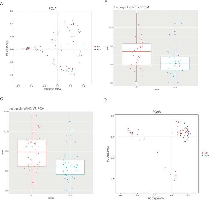
Figure 1: Community structure, richness, and diversity of the breast microbiota in the PCM and NC groups. (A) Principal coordinate analysis (PCoA) showing microbial community structure in the PCM and NC groups. (B, C) Community richness assessed using the Chao1 (B) and ACE (C) indices. Between-group differences in alpha-diversity indices were assessed using the two-sided Wilcoxon rank-sum test. Chao1 and ACE richness were significantly lower in the PCM group than in the NC group (Chao1, P = 0.004; ACE, P = 0.002). The reported P values are unadjusted. (D) PCoA showing differences in beta diversity between the PCM and NC groups. PERMANOVA based on Bray–Curtis distances confirmed a significant difference in microbial community composition between the two groups (F = 2.8113, R2 = 0.0324, P = 0.007; 999 permutations). Please click here to view a larger version of this figure.
Comparison of overall microbiome composition between the PCM and NC groups
Rarefaction curves indicated that the sequencing depth was sufficient, with Good’s coverage exceeding 0.99 for all samples at a sequencing depth of 30,000 tags (Supplementary Figure 1). Further comparison of the breast tissue microbial communities between the PCM and NC groups revealed compositional differences at the phylum, class, order, and genus levels (Figure 2A–D). Overall, the community composition was relatively similar at the phylum level, whereas differences became more pronounced at the order and genus levels, suggesting substantial remodeling of the PCM-associated breast microbiota at lower taxonomic ranks. The Circos plot illustrated the genus-level distribution patterns of the two groups (Figure 2E). The Venn diagram showed that the NC and PCM groups contained 113 and 101 unique genera, respectively, with 282 genera shared between them (Figure 2F).

Figure 2: Overall breast microbiome composition in the PCM and NC groups. Relative abundance of bacterial taxa at the (A) phylum, (B) class, (C) order, and (D) genus levels in the PCM and NC groups. (E) Circos plot showing the genus-level distribution of bacterial taxa between the PCM and NC groups. (F) Venn diagram showing 282 genera shared between the groups, with 113 genera unique to the NC group and 101 genera unique to the PCM group. Please click here to view a larger version of this figure.
Comparison of differential bacterial taxa between the PCM and NC groups
Using the Wilcoxon rank-sum test with an unadjusted P < 0.05 as the significance criterion, a total of 214 genera showed nominally significant differences in relative abundance between the PCM and NC groups. Among these, Staphylococcus and Bacteroides were more abundant in the PCM group, whereas Plesiomonas and Acinetobacter were more abundant in the NC group (Figure 3A). In addition, LEfSe analysis identified discriminative taxa between the two groups, which were visualized in a phylogenetic cladogram (Figure 3B).
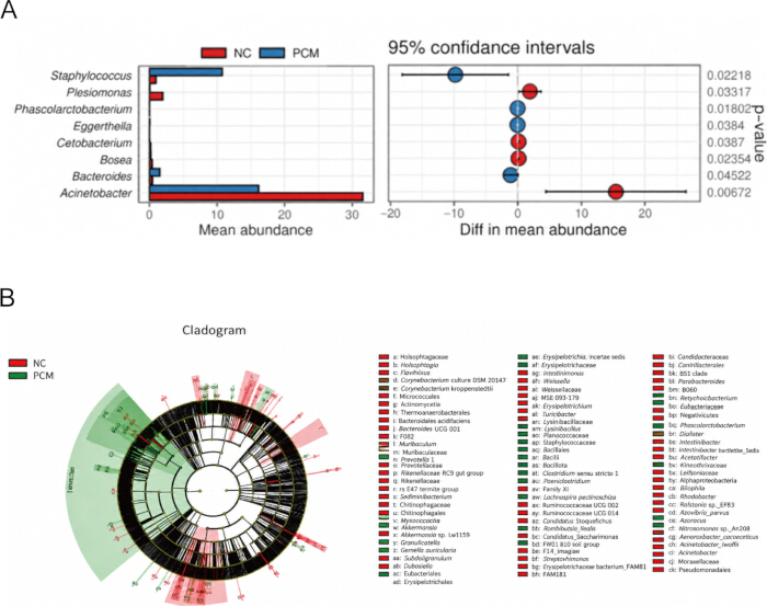
Figure 3: Differentially abundant bacterial taxa in the PCM and NC groups. (A) Comparison of the relative abundance of selected bacterial genera between the PCM and NC groups. Between-group differences were assessed using the two-sided Wilcoxon rank-sum test. The displayed P values are unadjusted (raw) P values, and P < 0.05 was considered statistically significant. (B) Linear discriminant analysis effect size (LEfSe) cladogram showing differentially abundant bacterial taxa between the PCM and NC groups. A significance threshold of P < 0.05 and an LDA score >2.0 were used for LEfSe analysis. Red and green indicate taxa enriched in the NC and PCM groups, respectively. Please click here to view a larger version of this figure.
Overall, the results demonstrate differences in breast tissue microbial community structure, richness, composition, and the relative abundance of specific bacterial taxa between the PCM and NC groups. These findings support an association between PCM and alterations in the breast tissue microbiota without establishing a causal relationship.
Data Availability:
The data supporting the findings of this study are available from the corresponding authors upon reasonable request. Available data include the raw paired-end 16S rRNA gene sequencing reads and the processed operational taxonomic unit (OTU) abundance and taxonomic data used for the microbiome analyses.
Supplementary Figure 1: Rarefaction analysis and sequencing coverage of breast tissue microbiota samples. (A) Rarefaction curves based on the observed species (Sobs) index for individual breast tissue samples from the PCM and NC groups. The x-axis represents the number of sequencing tags sampled, and the y-axis represents observed microbial richness. (B) Good’s coverage curves for individual samples. Good’s coverage exceeded 0.99 for all samples at 30,000 tags per sample, indicating adequate sequencing coverage. Please click here to download this file.
Discussion
In this study, we first compared the clinical characteristics between the PCM and NC groups. The results showed no significant differences between the two groups in age, BMI, or lymphocyte count. In contrast, white blood cell and neutrophil counts and prolactin levels were significantly higher in the PCM group, and the prevalence of nipple inversion was also significantly higher than that in the NC group. The elevated white blood cell and neutrophil counts may reflect greater inflammatory activity in patients with PCM. Previous studies have indicated that although PCM is usually classified as a non-infectious inflammatory disease, its histopathological features are often accompanied by infiltration of neutrophils, macrophages, and plasma cells, suggesting that the local inflammatory microenvironment plays an important role in disease progression15. In addition, hyperprolactinemia has been proposed to contribute to the occurrence of PCM by promoting mammary duct secretion retention and ductal dilation, thereby inducing local immune responses16. Nipple inversion may lead to obstruction of mammary duct drainage, creating conditions for bacterial colonization and subsequently contributing to a chronic inflammatory microenvironment. Together, these findings suggest that endocrine disturbance, ductal obstruction, and sustained inflammatory activation may create a permissive microenvironment for microbial dysbiosis in PCM.
From the perspective of microbial diversity, further analysis revealed that the richness of the breast microbiota in the PCM group, as reflected by the Chao1 and ACE indices, was significantly lower than that in the NC group, whereas the Shannon and Simpson indices showed no significant differences. Reduced richness may reflect a less complex breast microbial community in patients with PCM. However, the ecological and functional significance of this finding remains uncertain. Previous studies have shown that decreased microbial richness is commonly associated with chronic inflammatory diseases, such as inflammatory bowel disease and metabolic disorders17. Although the changes in α-diversity indices were limited, β-diversity analysis and PCoA results showed clear separation between the two groups in community structure, indicating that the microbial community composition differed substantially between the groups. These findings suggest that PCM may be associated more closely with the abnormal enrichment of specific bacterial taxa and an imbalance in community structure than simply with an overall reduction in microbial diversity.
The key finding of this study is that the microbial community composition differed between the breast tissues of patients with PCM and those of the NC group. A total of 214 bacterial genera showed nominally significant differences in relative abundance between the two groups based on unadjusted P values. Staphylococcus and Bacteroides were enriched in the PCM group, whereas Plesiomonas and Acinetobacter were enriched in the NC group. Staphylococcus, particularly Staphylococcus aureus, has been widely recognized as one of the most common pathogens in mastitis and breast abscesses18,19 and can promote persistent infection and tissue damage through virulence factor secretion, biofilm formation, and activation of inflammatory signaling pathways20,21. However, the present 16S rRNA gene sequencing data did not provide sufficient species-level resolution to determine whether the enriched Staphylococcus sequences represented S. aureus. Clinically, staphylococcal infection often presents with abscess formation, wound infection, and marked pain22, which are partly consistent with the clinical manifestations of PCM. Compared with lactational breast abscesses, non-lactational cases tend to have a more complex pathogen spectrum and are more frequently associated with anaerobic bacteria23, including Bacteroides24, and are often related to subareolar lesions, recurrent disease, and ductal abnormalities25,26. The enrichment of Bacteroides may be compatible with local conditions favoring anaerobic taxa, such as secretion stasis and reduced oxygen availability. However, these microenvironmental conditions were not directly measured in the present study27. As a Gram-negative bacterium, Bacteroides contains outer membrane components, particularly lipopolysaccharide (LPS), which can participate in host innate immune recognition and trigger inflammatory signaling, thereby potentially contributing to the persistence of local inflammation28. Taken together, these findings support a potential dysbiosis-associated inflammatory mechanism rather than a direct pathogenic role for any single detected genus. Alterations in the breast tissue microbiota may modify local exposure to microbial-associated molecular patterns and thereby influence innate immune signaling and inflammatory mediator production. Previous studies of nonpuerperal mastitis have suggested interactions between the breast tissue microbiota, host gene regulation, and the local immune microenvironment29. In addition, experimental evidence has implicated TLR-related inflammatory signaling in mastitis and demonstrated that activation of the IL-6/JAK2/STAT3 pathway can promote PCM in a mouse model30. Persistent inflammatory activation may facilitate immune-cell recruitment and sustain the plasma cell-rich periductal inflammation characteristic of PCM. In turn, ductal ectasia and secretion stasis may favor persistent microbial imbalance and inflammatory stimulation, suggesting a potential dysbiosis–inflammation–ductal injury cycle in PCM.
Reports of Plesiomonas in breast tissue are limited. Plesiomonas shigelloides, a representative species of this genus, has been described as an opportunistic Gram-negative bacterium31,32. However, the biological significance of the higher relative abundance of Plesiomonas in the NC group remains unclear. Although Acinetobacter was more abundant in the NC group, the biological significance of this finding also remains uncertain. Because breast tissue is a low-biomass specimen and no extraction blank or negative-control sequencing was available, potential low-level contamination cannot be excluded. Therefore, this finding should be interpreted cautiously and should be validated in future studies incorporating appropriate extraction blanks and sequenced negative controls.
Although these clinical differences coexisted with the observed alterations in breast tissue microbial composition, our study did not directly assess correlations between individual bacterial genera and white blood cell count, neutrophil count, prolactin level, nipple inversion, pain, nipple discharge, or other clinical manifestations. Recurrence and treatment outcomes were also not evaluated. Therefore, no direct genus–symptom or genus–outcome relationship can be inferred from the present data. Overall, our findings indicate that PCM is associated with alterations in breast tissue microbial composition in the context of endocrine abnormalities, ductal structural changes, and chronic inflammation. The enrichment of Staphylococcus and Bacteroides, together with differences in microbial community structure, provides preliminary evidence of PCM-associated local microbial dysbiosis. These findings warrant further prospective and mechanistic studies to determine whether specific microbial signatures have diagnostic, prognostic, or therapeutic relevance. However, the present study did not evaluate treatment response or clinical efficacy and therefore does not support any specific microbiome-targeted intervention in current clinical practice.
Several limitations should be considered when interpreting the present findings. First, breast tissue is a low-biomass specimen and is therefore susceptible to reagent- and laboratory-derived contamination. Although PCR no-template controls showed no visible amplification, no DNA extraction blank was included, and the negative controls were not subjected to sequencing. Consequently, potential low-level contamination could not be formally assessed or excluded. Accordingly, findings involving potentially contaminant taxa, particularly Acinetobacter, should be interpreted with caution and should not be considered evidence of a specific biological role. Second, the cross-sectional 16S rRNA gene sequencing design identifies associations in microbial composition but does not establish a causal relationship between microbial alterations and PCM or directly assess microbial function. Third, genus-level differential abundance testing was based on unadjusted P values without correction for multiple comparisons; therefore, the identified taxa should be considered exploratory and require independent validation. Fourth, the NC samples were histologically normal breast tissues adjacent to benign breast lesions rather than tissues obtained from healthy individuals, which may limit the generalizability of the findings. In addition, potential confounding by clinical characteristics that differed between the groups was not addressed using multivariable analyses. Direct correlations between individual bacterial genera and clinical features were also not analyzed, and recurrence or treatment outcomes were not evaluated. Further studies incorporating appropriate negative controls, longitudinal designs, and complementary functional analyses are needed to validate these findings and clarify their biological relevance.
Disclosures
Conflict of Interest:
The authors have no conflicts of interest to disclose.
Acknowledgements
The authors thank all participating patients and control subjects for supporting this study by donating biomaterials for research. This work was supported by the National Youth Science Foundation Project of China (82002788); the Natural Science Foundation for Young Scientists of Jiangsu Province (BK20191089); the Peak Academic Talent Training Project of Jiangsu Province Hospital of TCM (y2021rc34); and Jiangsu Provincial Science and Technology Development Program for Traditional Chinese Medicine, General Project (MS2022022).
Materials
| Name | Company | Catalog Number | Comments |
|---|---|---|---|
| 16S rRNA V3–V4 forward primer 341F | N/A | N/A (custom oligonucleotide; catalog number not documented) | Sequence: 5′-CCTACGGGNGGCWGCAG-3′. Used with 806R for bacterial 16S rRNA V3–V4 amplification. |
| 16S rRNA V3–V4 reverse primer 806R | N/A | N/A (custom oligonucleotide; catalog number not documented) | Sequence: 5′-GGACTACHVGGGTATCTAAT-3′. Used with 341F for bacterial 16S rRNA V3–V4 amplification. |
| 5× Q5 High GC Enhancer | New England Biolabs (NEB) | Exact catalog/pack not documented | 10 μL per 50 μL PCR reaction. |
| 5× Q5 Reaction Buffer | New England Biolabs (NEB) | Exact catalog/pack not documented | 10 μL per 50 μL PCR reaction. |
| Biowest Agarose | Beijing Mengyimei Trading Center | BY-R0100 | Used for agarose gel electrophoresis of PCR products and PCR negative controls. |
| Circos | Open-source software | v0.69-3 | Used to generate the genus-level Circos plot. |
| dNTPs (2.5 mM) | Supplier not documented | N/A | 1.5 μL per 50 μL PCR reaction. |
| fastp | Open-source software | v0.18.0; RRID: SCR_016962 | Used for quality filtering of raw Illumina paired-end reads. |
| FLASH | Open-source software | v1.2.11; RRID: SCR_005531 | Used to merge paired-end clean reads into tags (minimum overlap, 10 bp; maximum mismatch rate, 2%). |
| GoldView I nucleic acid stain | Beijing Mengyimei Trading Center | D0125 | Used for visualization of nucleic acids in agarose gels. |
| HiPure Stool DNA Kit | Magen (Guangzhou, China) | D3141 | Used for DNA extraction from approximately 150–200 mg of breast tissue, following the manufacturer's protocol. |
| Illumina DNA Prep Kit | Illumina | 20060059 | Used for sequencing-library preparation. |
| Krona | Krona development team | v2.6; RRID: SCR_012785 | Used to visualize taxonomic summary data. |
| LEfSe | Open-source software | v1.0; RRID: SCR_014609 | Used to identify discriminative taxa; significance threshold P < 0.05 and LDA score >2.0. |
| Microbial community amplicon sequencing analysis service | Guangzhou Genedenovo Biotechnology Co., Ltd. | Service; project GHD22050460-2_std_1 | 16S rRNA V3–V4 amplicon sequencing and project-specific bioinformatics analysis for NC and PCM breast tissue samples. |
| NovaSeq 6000 S2 Reagent Kit | Illumina | 20042038 | Used for sequencing of the prepared 16S rRNA V3–V4 amplicon libraries on the NovaSeq 6000 platform. |
| NovaSeq 6000 sequencing platform | Illumina | N/A | Used for paired-end 250-bp (PE250) sequencing of 16S rRNA V3–V4 amplicon libraries. |
| Q5 High-Fidelity DNA Polymerase | New England Biolabs (NEB) | Exact catalog/pack not documented | 0.2 μL per 50 μL PCR reaction. |
| QIIME | QIIME development team | v1.9.1; RRID: SCR_008249 | Used for alpha-diversity analysis. |
| R statistical software | R Foundation for Statistical Computing | v3.4.3; RRID: SCR_001905 | Used for microbiome data processing and statistical analysis. |
| RDP Classifier | Ribosomal Database Project | v2.2; RRID: SCR_022773 | Used for taxonomic assignment of representative OTU sequences. |
| SILVA database | SILVA | v132; RRID: SCR_006423 | Reference database used for bacterial/archaeal taxonomic annotation. |
| SPSS Statistics | IBM | v25.0; RRID: SCR_002865 | Used for analysis of demographic and clinical characteristics. |
| UCHIME | Drive5 / Robert C. Edgar | RRID: SCR_008057; separate version not specified | Used for detection and removal of chimeric sequences. |
| UPARSE | Drive5 / Robert C. Edgar | RRID: SCR_005020; separate version not specified | Used to cluster clean tags into OTUs at 97% sequence similarity and select representative sequences. |
| USEARCH | Drive5 / Robert C. Edgar | v9.2.64; RRID: SCR_027438 | Used in the OTU analysis workflow; UPARSE was used for OTU clustering and UCHIME for chimera detection. |
| vegan R package | Open-source R package | v2.5.3; RRID: SCR_011950 | Project-report software used for Bray–Curtis, PCoA, and Adonis/PERMANOVA-related analyses. |
References
- Chen R, et al. Differential diagnosis of plasma cell mastitis and invasive ductal carcinoma using multiparametric MRI. Gland Surg. 2020;9:278-90.
- Liang Z, Zhang L. Research progress of plasma cell mastitis. Immun Inflamm Dis. 2025;13:e70199.
- Xing M, et al. Current understanding and management of plasma cell mastitis: can we benefit from what we know? Breast Care (Basel). 2022;17:321-9.
- Sun Y, et al. Risk factor analysis and development of a nomogram prediction model for plasma cell mastitis. PLoS One. 2025;20:e0338711.
- Zhang HJ, et al. MAC mediates mammary duct epithelial cell injury in plasma cell mastitis and granulomatous mastitis. Int Immunopharmacol. 2022;113:109303.
- Wang X, et al. Exosomes play an important role in the progression of plasma cell mastitis via the PI3K-Akt-mTOR signaling pathway. Mediators Inflamm. 2019;2019:4312016.
- Hou K, et al. Microbiota in health and diseases. Signal Transduct Target Ther. 2022;7:135.
- Zheng D, Liwinski T, Elinav E. Interaction between microbiota and immunity in health and disease. Cell Res. 2020;30:492-506.
- Deng ZL, et al. Dysbiosis in chronic periodontitis: key microbial players and interactions with the human host. Sci Rep. 2017;7:3703.
- Liu C, et al. Meta-analysis of mucosal microbiota reveals universal microbial signatures and dysbiosis in gastric carcinogenesis. Oncogene. 2022;41:3599-610.
- Loh JS, et al. Microbiota-gut-brain axis and its therapeutic applications in neurodegenerative diseases. Signal Transduct Target Ther. 2024;9:37.
- Edgar RC. UPARSE: highly accurate OTU sequences from microbial amplicon reads. Nat Methods. 2013;10:996-8.
- Ondov BD, Bergman NH, Phillippy AM. Interactive metagenomic visualization in a web browser. BMC Bioinformatics. 2011;12:385.
- Kuczynski J, et al. Using QIIME to analyze 16S rRNA gene sequences from microbial communities. Curr Protoc Microbiol. 2012;Chapter 1:Unit 1E.5.
- Xue B, et al. Immune imbalance and autoimmune mechanisms in plasma cell mastitis: current perspectives. Front Cell Dev Biol. 2025;13:1727675.
- Liu H, et al. Risk factors for non-puerperal mastitis: a meta-analysis. BMC Womens Health. 2025;25:562.
- Hu Y, et al. Disturbances of the gut microbiota and microbiota-derived metabolites in inflammatory bowel disease. Nutrients. 2022;14:5061.
- Hu X, et al. The gut microbiota contributes to the development of Staphylococcus aureus-induced mastitis in mice. ISME J. 2020;14:1897-910.
- Rimoldi SG, et al. The role of Staphylococcus aureus in mastitis: a multidisciplinary working group experience. J Hum Lact. 2020;36:503-9.
- Gong Z, et al. An underlying mechanism of bovine mastitis: PGE2 regulates Staphylococcus aureus-induced inflammatory response through TLR2, TLR4, and NLRP3 in macrophages. Vet Q. 2026;46:2615759.
- Wang H, et al. Characterization of TLR2, NOD2, and related cytokines in mammary glands infected by Staphylococcus aureus in a rat model. Acta Vet Scand. 2015;57:25.
- Tong SY, et al. Staphylococcus aureus infections: epidemiology, pathophysiology, clinical manifestations, and management. Clin Microbiol Rev. 2015;28:603-61.
- Boakes E, et al. Breast infection: a review of diagnosis and management practices. Eur J Breast Health. 2018;14:136-43.
- Costa Morais Oliveira V, et al. Non-lactational infectious mastitis in the Americas: a systematic review. Front Med (Lausanne). 2021;8:672513.
- Jiao Y, et al. Identification of periductal mastitis and granulomatous lobular mastitis: a literature review. Ann Transl Med. 2023;11:158.
- Zhang Y, et al. Clinical characteristics, classification and surgical treatment of periductal mastitis. J Thorac Dis. 2018;10:2420-7.
- Bah YR, et al. Bacteroides- and Prevotella-enriched gut microbial clusters associate with metabolic risks. Gut Pathog. 2025;17:55.
- Mancuso G, et al. Bacteroides fragilis-derived lipopolysaccharide produces cell activation and lethal toxicity via toll-like receptor 4. Infect Immun. 2005;73:5620-7.
- Zhu J, et al. Interactions between the breast tissue microbiota and host gene regulation in nonpuerperal mastitis. Microbes Infect. 2022;24:104904.
- Liu Y, et al. Activation of the IL-6/JAK2/STAT3 pathway induces plasma cell mastitis in mice. Cytokine. 2018;110:150-8.
- Puah SM, Puthucheary SDA, Chua KH. Virulence profiles among gastrointestinal and extraintestinal clinical isolates of Plesiomonas shigelloides. Jpn J Infect Dis. 2022;75:407-10.
- Romli NIA, et al. Plesiomonas shigelloides bacteremia: a scoping review of epidemiology, clinical characteristics, outcomes, and implications of antimicrobial stewardship. Pathogens. 2026;15:33.