This protocol describes electrical impedance tomography-guided pulmonary rehabilitation in patients with respiratory failure. Regional ventilation feedback guides positioning, vibration-assisted sputum clearance, and early mobilization. Ventilation and physiological outcomes are assessed at baseline and on three subsequent days. ICU stay and delirium are evaluated during hospitalization.
Research Article
Application of Electrical Impedance Tomography-Guided Pulmonary Rehabilitation Strategy in Patients with Respiratory Failure
In This Article
Summary
Abstract
This study investigated pulmonary rehabilitation strategies guided by electrical impedance tomography (EIT) in patients with respiratory failure. A total of 92 patients with respiratory failure admitted to the Intensive Care Unit (ICU) of a tertiary Grade A hospital in Chongqing from July 2024 to January 2025 were selected and randomly divided into a control group and an experimental group, with 46 cases in each group, using a random number table. The control group received conventional pulmonary physical therapy nursing strategies. The experimental group additionally received EIT-guided positioning, vibratory sputum clearance, and early mobilization. Relative regional tidal-ventilation distribution, PaO₂/FiO₂, respiratory rate, and pulse oxygen saturation were assessed at baseline and on the following three days; ICU stay, and delirium incidence were also compared. Finally, 44 cases in the control group and 44 cases in the experimental group completed the study. Repeated-measures ANOVA showed significant time effects across the four ventral-to-dorsal layers and significant group and interaction effects for the dorsal layer. Dorsal ventilation share was higher in the experimental group on days 2 and 3, but not significantly different on day 1. No significant group, time, or interaction effects were found for the four lung quadrants. The experimental group had higher PaO₂/FiO₂, shorter ICU stay (6.55 vs. 7.75 days), and lower delirium incidence (25.0% vs. 45.5%). Respiratory rate decreased, and pulse oxygen saturation increased over time in both groups, without significant group or interaction effects. The EIT-guided strategy may improve regional ventilation distribution and oxygenation, shorten ICU stay, and reduce delirium in patients with respiratory failure.
Introduction
Respiratory failure is caused by a variety of factors that seriously impair lung ventilation and/or gas exchange; that is, normal gas exchange cannot be maintained at rest, resulting in decreased blood oxygen content, with or without carbon dioxide retention1. The most common nursing problem for patients with respiratory failure is impaired gas exchange, so airway management and pulmonary physical therapy are important nursing measures for these patients. At present, commonly used lung imaging methods have their own clinical significance and limitations. Although X-ray, computed tomography (CT), and positron emission tomography (PET) can provide imaging and disease information, they cannot provide continuous real-time bedside monitoring. Although ultrasound can dynamically and non-radiatively evaluate the lungs, it cannot accurately assess ventilation and is highly operator-dependent.
As a non-invasive, non-radiative bedside monitoring technology, EIT can continuously and dynamically monitor regional lung ventilation, provide dynamic images, identify ventilation loss2,3, and support targeted pulmonary physical therapy and dynamic adjustment of interventions. Conventional pulmonary rehabilitation is often guided by global physiological measurements, which provide limited immediate feedback on how individual lung regions respond to positioning, sputum clearance, and mobilization. Therefore, EIT may provide useful regional feedback for adjusting these interventions. However, evidence remains limited on the use of EIT to guide an integrated pulmonary rehabilitation strategy that combines these interventions in patients with respiratory failure. Therefore, this study evaluated an EIT-guided pulmonary rehabilitation strategy incorporating individualized position management, vibration-assisted sputum clearance, early mobilization, and repeated assessment–intervention–reassessment. The study compared regional ventilation distribution, oxygenation, respiratory rate, pulse oxygen saturation, ICU length of stay, and delirium incidence between patients receiving conventional pulmonary rehabilitation and those receiving additional EIT guidance. It was hypothesized that EIT-guided rehabilitation would improve regional ventilation distribution and oxygenation compared with conventional rehabilitation.
Protocol
This study was approved by the Hospital Ethics Committee (No. KY S2024-016-01). The study was registered with the Chinese Clinical Trial Registry (ChiCTR2600132334).
Research object
A total of 92 patients with respiratory failure admitted to the ICU of a tertiary hospital in Chongqing from July 2024 to January 2025 were selected for the study. An independent investigator assigned random-number-table values to 92 enrollment slots and ranked them; the 46 lowest-ranked slots were assigned to the control group, and the remaining 46 to the experimental group. Study-team members enrolled participants, and assignments were concealed in sequentially numbered, opaque, sealed envelopes until allocation. Bedside outcome assessors and the statistician were not blinded. No formal prospective sample-size calculation was performed; the sample size was based on eligible admissions during the recruitment period. Inclusion criteria: In accordance with the diagnostic criteria of respiratory failure in the "Clinical Diagnosis and Treatment Guidelines · Respiratory Diseases Branch " of the Respiratory Diseases Branch of the Chinese Medical Association, and confirmed by the ICU attending physician. Exclusion criteria: age <18 or >80 years; pregnancy; EIT contraindications ( such as monitoring site skin damage ); severe intracranial hypertension; Pneumothorax, mediastinal emphysema, subcutaneous emphysema and other barotrauma manifestations, or accompanied by pulmonary bullae, emphysema, severe pulmonary interstitial disease and other high-risk pneumothorax; The clinician judged that the hemodynamics was unstable and could not tolerate pulmonary rehabilitation treatment; end-stage disease; family members did not sign the informed consent; Participate in other studies. Dropout criteria: One patient or their family voluntarily withdrew from treatment and was discharged during the study period; death due to disease progression; study termination criteria included force majeure factors such as transfer. During the study, one case in each group was terminated due to the family's abandonment of treatment and automatic discharge, and one case in each group was terminated due to death. Finally, 44 cases were completed in each group.
Research methods
A research team was set up. The team comprised all members of the respiratory intensive care group: 8 nurses, 1 doctor, and 1 rehabilitation therapist. Before the study's implementation, the team members received two days of EIT-specific training (covering basic knowledge, regional ventilation monitoring, operational procedures, image acquisition and interpretation, and clinical case analysis) and one month of clinical practice, and all passed the theoretical and operational assessments. Both groups followed the same usual-care principles, with ventilatory/oxygen support and sedative/analgesic treatment adjusted according to clinical need. Support settings were reviewed at each assessment.
Control group
The control group was treated with a routine comprehensive pulmonary physiotherapy nursing strategy: within 6 hours of the patient's diagnosis of respiratory failure in the ICU, according to the vital signs, pulmonary auscultation, arterial blood gas, respiratory mechanics, and imaging results, the early rehabilitation activities such as turning over, manual chest percussion, mechanical vibration expectoration, and position management were carried out. Specific measures: turning over every 2 hours; mechanical expectoration twice a day, 15 minutes each time. When the patient's Richmond Agitation-Sedation Scale (RASS) score was -5 to -2 points: the head of the bed was elevated to 30°–40°, supplemented by supine or left and right lateral position, passive activity 2 times a day, 10 minutes each time; RASS score -1 to 0 points: supported upright sitting in bed for 30 min twice a day, passive joint activity training twice a day; For patients with a RASS score of 0, activities progressed from sitting in bed for 30 min to sitting at the bedside for 30 min, with balloon-blowing exercises twice daily, starting with five repetitions per session and gradually increasing to ten. During the operation, if the fluctuation in vital signs exceeds 20% or the fluctuation in pulse oxygen saturation exceeds 5%, the operation was terminated immediately.
Experimental group
Based on the treatment in the control group, the members of the study group used the EIT instrument to monitor lung ventilation, communicated the EIT evaluation results to the competent doctor in a timely manner, and guided the lung physical therapy nursing strategy according to the lung ventilation information obtained via EIT. The 16-electrode band was placed at the 4th to 5th intercostal level of the patient’s chest4, connected to the EIT monitoring host, and the real-time state image was obtained after start-up. According to the need, it was divided into 'hierarchical' distribution and 'quadrant' distribution2. The 'hierarchical' distribution was divided from the ventral side to the dorsal side in parallel, and was set to a four-layer mode, followed by ventral (region of interest [ROI] 1), mid-ventral (ROI 2), mid-dorsal (ROI 3), and dorsal (ROI 4) layers. The ' hierarchical ' ventilation distribution of the healthy lung is usually dominated by ROI 2 and ROI 3 regions5, in which the reference range of ROI 1 and ROI 4 regions is 10%–15%, and the reference range of ROI 2 and ROI 3 regions is 30%–40%6. The 'quadrant' distribution was divided into four quadrants with the image center as the origin, followed by the right ventral region (ROI 1), the left ventral region (ROI 2), the right dorsal region (ROI 3), and the left dorsal region (ROI 4). The diagnostic criteria for insufficient ventilation in the four regions of ROI 1, ROI 2, ROI 3, and ROI 4 were <18%, <15%, <16%, and < 3%, respectively7. Whether it is 'level' distribution or 'quadrant' distribution, ROI 1 + ROI 2 + ROI 3 + ROI 4 = 100%8. An end-expiratory reference was used. The regional tidal-ventilation share was defined as the change in tidal impedance in an ROI, divided by the sum of the tidal impedance changes across the four ROIs, multiplied by 100%. The arithmetic mean of 10 artifact-free breaths was used at each assessment. Breaths affected by movement, electrode contact loss, or coughing were rejected; acquisition was repeated after correcting for movement or belt contact loss. These values describe relative regional ventilation distribution and do not represent absolute lung aeration or perfusion.
(1) Individualized guidance position management: The position management of critically ill patients is dynamic and complex, and its core lies in the continuous cycle of ' assessment-decision-implementation-reassessment '. EIT can clearly show the dynamic distribution images of patients ' lung ventilation in different positions9, and realize accurate position management supported by objective data. Position management and early activity in ICU patients require continuous assessment and adjustment according to the patient's physiological response10. If the ' hierarchical ' distribution of the patient's supine position showed ROI 3 <25% and ROI 4 <10%, the patient was positioned in a prone, sitting, bedside sitting, or prone position of 30°–45 ° according to the actual situation of the patient. After 15 minutes, the EIT was evaluated again. If the lung ventilation became more uniform, the selected position was continued. If the lung ventilation became less uniform, the selected position was discontinued. In the ' quadrant ' distribution, each ROI was independently judged: ROI 1 <18%, ROI 2 <15%, ROI 3 <16%, ROI 4 <13%, which was considered a lack of ventilation function. When the EIT image showed a lack of ventilation function in ROI 1 and ROI 3, the left 45° supine lateral or left 45° prone lateral position was used, as appropriate. EIT evaluation was repeated 4 hours after prone positioning. The position was continued if beneficial and stopped if no benefit was observed. If ventilation loss was confined to ROI 3, a reverse impedance change was observed in this region (decreased impedance during inspiration and increased impedance during expiration), and a purple image was observed in the enhanced image; pleural effusion was suspected. At this time, the right semi-recumbent position was used to facilitate gas exchange in the left lung and reduce the pressure of the effusion on the right lung tissue. The treating physician was assisted with ultrasonographic evaluation. After drainage, EIT was used to monitor increases in ventilation and end-expiratory lung impedance (EELI) in this area.
(2) To guide the precise positioning of vibration sputum excretion: Vibration sputum excretion is one of the common airway clearance techniques in clinical practice. By loosening bronchial secretions, reducing sputum adhesion, promoting its movement, and improving ciliary movement, thereby improving the efficiency of sputum excretion11, the manual-assisted sputum excretion method and the mechanical vibration sputum excretion method are often used. When airway obstruction leads to poor local ventilation, EIT can evaluate ventilation distribution in the lung area in real time, quickly identify abnormalities through the ventilation distribution view, provide continuous objective monitoring, and guide medical staff to dynamically adjust the physical therapy strategy based on real-time imaging data12. EIT showed that the ventilation of ROI 4 was significantly reduced when the ' quadrant ' distribution was used, and the dynamic image suggested that the local ventilation was delayed or missing. Considering the possibility of sputum retention in this area, the vibration sputum drainage treatment was intensified at the corresponding surface projection of ROI 4 to assist sputum drainage. After treatment, EIT was used for re-evaluation. Through the repeated 'evaluation-treatment-evaluation ' model of EIT, the operational method is optimized, costs are reduced, and nurses' workload is reduced13.
Early activity safety assessment
Early activity in the ICU is an important means of promoting patient rehabilitation and improving quality of life after ICU14. Early ICU activities involve gradual progression, assessment, monitoring, and intervention. Accurate assessment and quality control can help improve ICU patient management. During the implementation of early activities, nurses often evaluate the effect using the patient's vital signs, pulse oxygen saturation, or blood gas analysis results, and lack the means to directly monitor improvements in pulmonary ventilation. EIT can monitor and evaluate the pulmonary ventilation during early activity in patients15. If EIT showed that pulmonary ventilation did not improve or deteriorated after the early rehabilitation intervention, the activity mode and duration were dynamically adjusted to promote patient rehabilitation while ensuring their safety. If the ventilation shares of ROI 3 and ROI 4 in the hierarchical distribution decreased after 10 minutes of bedside sitting, the activity was suspended. For conscious patients, EIT images were used to explain the changes in lung ventilation. Two images at different times were displayed through the end-inspiratory trend view. The difference map before and after the activity was viewed by moving the cursor, and changes in the blue-orange areas were used to adjust the rehabilitation plan and explain the response to conscious patients. Immediate EIT reassessments were used only to guide individual treatment adjustments.
Methods of data collection
General information was collected by the nurses in the study group from medical records, including gender, age, body mass index (BMI), Acute Physiology and Chronic Health Evaluation II (APACHE II) score, oxygen therapy, intravenous antibiotic use, nebulized medication use, complications, delirium occurrence, and so on. Study participants underwent the scheduled EIT outcome assessments at approximately 09:00 before the initial intervention (T0) and before morning rehabilitation on days 1, 2, and 3 (T1, T2, and T3). Daily rehabilitation continued beyond T3 until ICU discharge, withdrawal, death, or clinical discontinuation. The values of ROI 1–4 under ' hierarchical ' distribution and ' quadrant ' distribution were recorded, respectively. At the same time, arterial blood gases were collected to calculate the oxygenation index as PaO₂/FiO₂ (mmHg), with FiO₂ expressed as a fraction. FiO₂ was obtained from the current ventilator setting for invasive or noninvasive ventilation or from the oxygen-concentration setting for high-flow nasal oxygen. The respiratory rate and pulse oxygen saturation were read by an electrocardiogram monitor. The Confusion Assessment Method for the ICU (CAM-ICU) was administered at 09:00 and 21:00 by trained study nurses who were not blinded to group allocation. Assessments at RASS scores of −4 or −5 were recorded as unassessable and repeated later rather than scored as negative. During ICU hospitalization, if the CAM-ICU assessment was positive once, it was recorded as having occurred. The length of ICU stay was recorded on the day of discharge. All data were checked and entered by two team members. A favorable response comprised increased ventilation share in initially under-ventilated regions without clinical intolerance, higher PaO₂/FiO₂ and SpO₂, reduction of an initially elevated respiratory rate, shorter ICU stay, and absence of delirium. Changes in the opposite direction or clinical intolerance during rehabilitation were interpreted as unfavorable. ROI changes were interpreted as a redistribution because the four shares sum to 100%. The participant flow and assessment schedule are shown in Figure 1.
Statistical methods
SPSS 27.0.1.0 software was used for statistical analysis. Measurement data (age, BMI, APACHE II score, ROI 1–ROI 4 value, oxygenation index, respiratory rate, and pulse oxygen saturation) were described by mean ± standard deviation. If the data were normally distributed, a two-sample t-test was used; otherwise, a rank-sum test was used. Count data (gender, use of intravenous antibiotics, oxygen therapy, use of nebulizers, complications) were expressed as frequency and percentage, using the chi-square test (or likelihood ratio chi-square) or Fisher's exact probability test. Repeated measures analysis of variance was used to compare ROI 1–4 values, oxygenation index, respiratory rate, and pulse oxygen saturation across time points. Time-varying co-interventions were not included in the statistical model. Greenhouse-Geisser was used to adjust the degrees of freedom for those who did not meet the spherical symmetry. p < 0.05 was considered statistically significant. The odds ratio for delirium (experimental versus control) was reported with a log-Wald 95% confidence interval. Partial eta-squared (ηp2) was used to describe the magnitude of repeated-measures effects.
Results
The general situation of the two groups of patients
There was no significant difference in age, gender, BMI, APACHE II score, intravenous antibiotic use, oxygen therapy, nebulized medication use, and complications between the two groups (p > 0.05) (Table 1).
Comparison of the ROI 1–4 values of ‘hierarchical’ distribution at different time points between the two groups
Repeated measures analysis of variance showed that there were significant differences in the time effects of ROI 1, ROI 2, ROI 3 and ROI 4 between the two groups (FROI 1 time = 25.623, p < 0.001; fROI 2 time = 60.894, p < 0.001; fROI 3 time = 17.54, p < 0.001; fROI 4 time = 411.126, p < 0.001); the group and group-by-time effects were significant for ROI 4 (Fgroup = 4.737, p = 0.032; f interaction = 21.463, p < 0.001) (Table 2). The ROI 4 value of the experimental group was higher than that of the control group at T1, T2, and T3. From T0 to T3, mean changes in ROI 1–4 were −1.55, −6.38, 4.34, and 3.60 percentage points in the control group and −1.73, −6.06, 2.00, and 5.80 percentage points in the experimental group, respectively. The dorsal-layer difference (experimental minus control) increased from −0.34 percentage points at T0 to 1.86 percentage points at T3 (Figure 2A).
Comparison of the ROI 1–4 values of ' quadrant ' distribution at different time points between the two groups
Repeated measures analysis of variance showed no significant differences in ROI 1–4, group, or the interaction between the two groups (p > 0.05) (Table 3).
Comparison of oxygenation index, pulse oxygen saturation, and respiratory rate between the two groups at different time points
Repeated measures analysis of variance showed significant differences in oxygenation index over time, between groups, and in the interaction between the two groups (Ftime = 133.835, p < 0.001; f group = 4.263, p = 0.042; f interaction = 7.452, p < 0.001; Table 4). The oxygenation index of T1, T2, and T3 in the experimental group was higher than that in the control group. There was a statistically significant difference in the time effect of respiratory rate between the two groups (Ftime = 374.677, p < 0.001), and the group and interaction effects were not significant (Fgroup = 0.623, p = 0.432; Finteraction = 0.546, p = 0.572). There was a statistically significant difference in the time effect of pulse oxygen saturation between the two groups (Ftime = 611.583, p < 0.001), and the group and interaction effects were not significant (Fgroup = 0.255, p = 0.615; Finteraction = 3.247, p = 0.062). From T0 to T3, mean PaO₂/FiO₂ increased by 123.96 mmHg in the control group and 189.64 mmHg in the experimental group; respiratory rate decreased by 9.38 and 9.03 breaths/min, and pulse oxygen saturation increased by 7.97 and 6.87 percentage points, respectively. The PaO₂/FiO₂ difference (experimental minus control) increased from −11.26 mmHg at T0 to 54.42 mmHg at T3 (Figure 2B).
Comparison of ICU hospitalization time and delirium between the two groups
The results of the independent-samples t-test showed a statistically significant difference in ICU length of stay between the two groups (t = -2.078, p = 0.041). The incidence of delirium in the two groups was compared using the χ2 test, and the difference was statistically significant (χ2 = 4.034, p = 0.045). Mean ICU stay was 1.20 days shorter in the experimental group (6.55 ± 1.976 vs. 7.75 ± 3.307 days), and delirium incidence was 20.5 percentage points lower (25.0% vs. 45.5%) (Table 5).
Data Availability:
The deidentified datasets used in the study and other raw data obtained from the study are available from the corresponding author upon reasonable request.
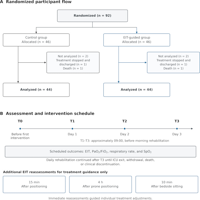
Figure 1: Participant flow and assessment schedule. (A) Randomized participant flow. (B) Assessment and intervention schedule. T0 preceded the first intervention; T1–T3 were assessed at approximately 09:00 before morning rehabilitation on days 1–3. Additional EIT reassessments were used only to guide individual treatment adjustments. Please click here to view a larger version of this figure.
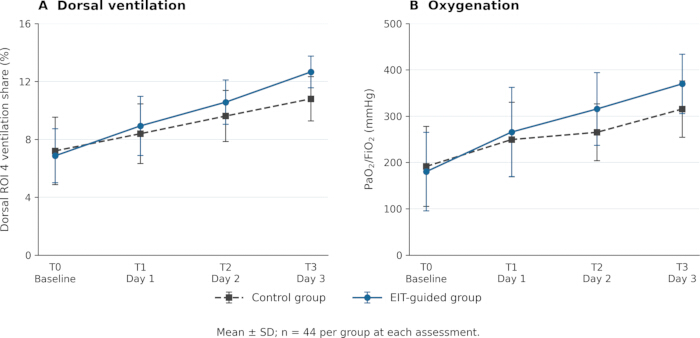
Figure 2: Changes in the main regional ventilation and oxygenation outcomes. (A) Dorsal-layer regional tidal-ventilation share (hierarchical ROI 4). (B) PaO₂/FiO₂. Points and error bars show the mean and standard deviation for 44 participants per group at each time point. Curves display the values reported in Tables 2 and 4; significance is reported in the text and tables. Please click here to view a larger version of this figure.
Table 1: Comparison of general conditions between the two groups of patients. Please click here to download this file.
Table 2: Comparison of ' hierarchical ' distribution of ROI 1–4 values at different time points between the two groups. Please click here to download this file.
Table 3: Comparison of ROI 1–4 values of ' quadrant ' distribution at different time points between the two groups. Please click here to download this file.
Table 4: Comparison of oxygenation index, pulse oxygen saturation, and respiratory rate between the two groups at different time points. Please click here to download this file.
| Characteristic | Control group | Experimental group | Statistical test value | p value |
| Length of ICU stay (days) | 7.75±3.307 | 6.55±1.976 | -2.078 | 0.041 |
| Delirium [n (%)] | 4.034 | 0.045 | ||
| Yes | 20(45.5) | 11(25) | ||
| No | 24(54.5) | 33(75) | ||
| Delirium odds ratio (experimental versus control): 0.40 (95% CI, 0.16–0.99; log-Wald). | ||||
Table 5: Comparison of ICU hospitalization time and delirium between the two groups.
Discussion
Effect of EIT-oriented pulmonary rehabilitation strategy on lung ventilation in patients with respiratory failure
The results of this study showed that there was no significant difference in the trend of ROI change over time and the difference between the two groups when the ' quadrant ' distribution was used. When the ' hierarchical ' distribution was adopted, the values of ROI 1–4 in both groups changed with time, and the ventilation of ROI 4 showed a gradual upward trend, suggesting that with the advancement of pulmonary rehabilitation intervention, the distribution of dorsolateral lung ventilation was improved. The experimental group had a higher dorsal-layer ventilation share at T2 (p = 0.008) and T3 (p < 0.001), whereas the difference at T1 was not statistically significant (p = 0.217), consistent with the overall direction of previous reports16,17. In this study, most of the patients with respiratory failure were elderly and critically ill patients, and most of them underwent tracheal intubation and invasive mechanical ventilation. In the early stage of treatment, mechanical ventilation in the semi-recumbent position under sedation is usually associated with excessive airway mucus secretion, decreased ciliary mucus clearance, and increased retention of local lung secretions18. If the patient's position is improper and the lung area with more secretions is not well drained for a long time, the accumulation of secretions can easily lead to limited local ventilation, while other areas compensate with excessive ventilation16. Through real-time dynamic monitoring, EIT can visually observe delayed ventilation, excessive expansion, or ventilation defects in the lung area, accurately locate the area of vibration expectoration, dynamically evaluate the efficacy of pulmonary physical therapy, and improve pulmonary ventilation function.
Early activity and reasonable position management are of great significance in the treatment of ICU patients, but they still face certain challenges10. Therefore, the activity plan and posture need to be individualized. Critically ill patients have poor pulmonary function reserve, and there may be risks such as increased respiratory load and hemodynamic instability during activity19. Bedside EIT monitoring can show regional ventilation changes during early activity and positioning15. In this study, changes in ROI 1–4 guided individual treatment adjustments within the combined rehabilitation program. The group-by-time effect sizes for dorsal-layer ventilation share (ηp2 = 0.200) and PaO₂/FiO₂ (ηp2 = 0.080) were consistent with the larger observed increases from T0 to T3 in the experimental group.
Effects of EIT-oriented pulmonary rehabilitation strategy on oxygenation index, respiratory rate, and pulse oxygen saturation in patients with respiratory failure
Oxygenation index can accurately reflect alveolar ventilation/blood flow matching and pulmonary ventilation function, which is more sensitive and objective than peripheral blood oxygen saturation20. The results of this study showed that the oxygenation index in the experimental group was significantly higher than in the control group at T2 and T3. EIT can monitor the distribution of ventilation in the lung area in real time at the bedside, identify alveolar collapse and uneven ventilation areas, guide position management, lung physical therapy, and lung recruitment, accurately promote collapsed alveolar recruitment, and improve local lung ventilation2. In contrast, conventional pulmonary rehabilitation measures lack quantitative evaluation of local pulmonary ventilation, and targeted improvements in local pulmonary ventilation are somewhat limited. Therefore, early application of EIT-individualized guidance for pulmonary rehabilitation can effectively improve pulmonary ventilation in patients with respiratory failure, and the intervention effect can appear early and continue to benefit in the early stage of rehabilitation, which is consistent with the study by Wang et al.21.
The results of this study showed that, compared with the control group, the EIT-oriented pulmonary rehabilitation strategy did not significantly improve respiratory rate and pulse oxygen saturation in patients with respiratory failure. The reasons for the analysis are as follows: most patients received invasive or non-invasive mechanical ventilation after admission to the ICU to relieve dyspnea, while continuously receiving sedative/analgesic drugs to maintain human-machine coordination. The respiratory rate is mainly affected by ventilator mode and sedation depth, and the spontaneous breathing drive is significantly inhibited22, making it difficult to reflect the effect of pulmonary rehabilitation on respiratory rhythm. Pulse oxygen saturation can be rapidly maintained in the target range by increasing the concentration of inhaled oxygen or increasing positive end-expiratory pressure23. Therefore, due to the mechanical ventilation intervention, the use of sedative and analgesic drugs, and adjustments to conventional oxygenation and ventilation parameters, the EIT-oriented pulmonary rehabilitation strategy failed to demonstrate additional benefits in respiratory rate and pulse oxygen saturation.
Effect of EIT-oriented pulmonary rehabilitation strategy on hospitalization time and delirium in patients with respiratory failure
EIT can enable real-time dynamic visual monitoring of lung ventilation, help accurately identify abnormal conditions such as uneven lung ventilation, local collapse, or excessive expansion, and formulate individualized pulmonary rehabilitation programs accordingly4. This precise guidance avoids the blind spots in pulmonary rehabilitation. At the same time, EIT can provide immediate feedback during the treatment process to guide the dynamic adjustment of the rehabilitation program8, thereby improving the efficiency of each treatment. In addition, EIT can monitor lung reexpansion, objectively evaluate the immediate effects of interventions such as postural changes and manual therapy, and provide an objective basis for treatment decisions. Previous studies have reported that EIT-guided interventions may shorten the duration of invasive ventilation and ICU stay24,25. In the present study, the EIT-oriented pulmonary rehabilitation strategy was associated with a shorter ICU stay in patients with respiratory failure, consistent with the findings reported by Wang et al.21, and further confirming that this strategy has important clinical value for patients' rehabilitation.
Delirium in critically ill patients is a common acute clinical syndrome in the ICU. It is a transient state of confusion caused by various factors. The main features are disturbance of consciousness and changes in cognitive function26. The causes of delirium in patients with respiratory failure are complex, mainly related to the use of sedative drugs, decreased oxygen partial pressure, use of ventilators, and other factors27. The incidence of delirium in elderly patients with respiratory failure was 31.3%28. EIT visualizes lung ventilation in real time, provides visual feedback to patients, and enables them to see the real-time improvement in their ventilation status on the screen. This immediate visual feedback helps to stimulate their cognitive engagement and active participation. Previous work has reported shorter mechanical ventilation duration and ICU stay with EIT-guided rehabilitation21. In the present study, the experimental group had a shorter ICU stay and a lower incidence of delirium; however, mechanical ventilation duration and sedation requirements were not measured. Therefore, the mechanism underlying the lower incidence of delirium cannot be determined from the present data.
Disclosures
The authors declare no conflicts of interest related to this study.
Acknowledgements
This work was supported by the Chongqing Science and Health Joint General Project (Grant No. 2023MSXM052), the Chongqing medical scientific research project (Joint Project of Chongqing Health Commission and Science and Technology Bureau, No. 2025MSXM162), and the Medical Research Foundation of Chongqing General Hospital (No. Y2023HLKYZDXM02).
Materials
| Name | Company | Catalog Number | Comments |
|---|---|---|---|
| 16-electrode band | Dräger | 8420055-8420059 (adult sizes S-XXL; size used not specified) | EIT monitoring electrode band. Placed at the 4th to 5th intercostal level of the chest. |
| Arterial blood gas testing | Standard hospital laboratory equipment | — | Collects arterial blood and yields oxygenation index. Assessed at T0 and on days 1 to 3 after intervention. |
| Balloon blowing training aid | Standard rehabilitation aid | — | Rehabilitation aid. Breathing training for alert patients. Increased gradually from 5 to 10 repetitions, twice daily. |
| Clinical Diagnosis and Treatment Guidelines - Respiratory Diseases Branch | Chinese Medical Association, Respiratory Diseases Branch | https://ctschina.cma.org.cn/ | Used as the inclusion criterion for respiratory failure. Confirmed by the ICU attending physician. |
| Confusion Assessment Method for ICU (CAM-ICU) | Vanderbilt University Medical Center | https://www.icudelirium.org/medical-professionals/delirium/monitoring-delirium-in-the-icu | Assesses delirium occurrence in ICU patients. If CAM-ICU was positive once during the ICU stay, delirium was recorded as present. |
| ECG/patient monitor | Standard hospital equipment | — | Equipment. Reads respiratory rate and pulse oxygen saturation. Used for daily synchronized assessment. |
| EIT hierarchical ROI distribution mode | Dräger | https://www.draeger.com/en_in/Products/PulmoVista-500 | Divides the lung from ventral to dorsal into ROI 1 to ROI 4 for ventilation assessment. ROI 1 ventrolateral, ROI 2 ventromedial, ROI 3 dorsomedial, ROI 4 dorsolateral. |
| EIT quadrant ROI distribution mode | Dräger | https://www.draeger.com/en_in/Products/PulmoVista-500 | Software function / analysis setting. Divides the image into four quadrants with the center as origin to detect regional ventilation deficits. |
| Electrical impedance tomography (EIT) instrument | Dräger | PulmoVista 500 | Bedside real-time monitoring of lung ventilation distribution to guide positioning, vibration sputum clearance, and early mobilization. Connected to a 16-electrode band; provides real-time state images; supports hierarchical/quadrant analysis. |
| High-flow oxygen therapy device | Standard ICU equipment | — | Treatment device/oxygen therapy mode. Recorded as patient oxygen therapy mode and baseline data. Used in Table 1 to record high-flow oxygen proportion. |
| Hospital bed/positioning support | Standard hospital equipment | — | Equipment / nursing aid. Position management and early rehabilitation activities. Head of bed raised 30 to 40 degrees; prone, seated, bedside seated, or 30 to 45 degree positioning used as needed. |
| Invasive mechanical ventilation/tracheal intubation | Standard ICU equipment | — | Treatment device/oxygen therapy mode. Recorded as patient oxygen therapy mode and baseline data. Used in Table 1 to record tracheal intubation proportion. |
| Mechanical vibration expectoration device | Standard respiratory therapy equipment | — | Equipment. Mechanical vibration sputum clearance; intensified according to EIT guidance. Routine group: twice daily, 15 min each; intervention group adjusted by EIT localization. |
| Medical records / case data form | Hospital medical records | https://en.cghhospital.org/idintro_info/ | Data source. Records demographic data, oxygen therapy, medication use, complications, and delirium. Completed by research nurses and double-checked for entry. |
| Nebulized antibiotics | Hospital Formulary | — | Drug class. Recorded nebulized medication use. |
| Nebulized bronchodilator | Hospital Formulary | — | Recorded nebulized medication use. |
| Nebulized expectorant | Hospital Formulary | — | Recorded nebulized medication use. Both groups were 44 (100%); specific drug not reported. |
| Nebulized glucocorticoid | Hospital Formulary | — | Recorded nebulized medication use. |
| Noninvasive ventilator | Standard ICU equipment | — | Recorded as patient oxygen therapy mode and baseline data. Used in Table 1 to record noninvasive ventilation proportion. |
| Random number table | N/A (noncommercial randomization method) | — | Randomization tool. Randomly assigned patients to control and test groups. |
| Respiratory failure patients | Tertiary Grade A hospital ICU in Chongqing | https://en.cghhospital.org/idintro_info/ | Study sample. Patients enrolled for control and intervention group comparison. 92 patients randomized; 44 cases completed in each group; Jul 2024 to Jan 2025. |
| Richmond Agitation-Sedation Scale (RASS) | Virginia Commonwealth University (VCU) | VCU Tech #12-063; https://techtransfer.research.vcu.edu/technologies/biomedical/richmond-agitation-sedation-scale-rass.html | Assesses sedation/agitation level to match the early mobilization plan. RASS -5 to -2, -1 to 0, and 0 correspond to different activity plans. |
| SPSS software | IBM | PID 5725-A54; version 27.0.1.0 | Statistical software. Statistical analysis including t test, chi-square/Fisher test, and repeated-measures ANOVA. P < 0.05 considered statistically significant. |
| Ultrasound diagnosis/drainage support | Standard diagnostic ultrasound equipment | — | Assists physician ultrasound diagnosis and reassessment after drainage when EIT suggests pleural effusion. Conditional use; not a primary study material. |
References
- Lagina M, Valley TS. Diagnosis and management of acute respiratory failure. Crit Care Clin. 2024;40(2):235-53.
- Standard Committee of the Critical Care Medicine Branch of China Health Information and Big Data Association, Beijing Oncology Society Critical Care Medicine Professional Committee, Chinese Critical Care Lung Electrical Impedance Working Group. Chinese expert consensus on clinical application of electrical impedance tomography in critical respiratory management. National Medical Journal of China. 2022;102(9):615-28.
- Sun G, Li Y. Advances in application of electrical impedance tomography in individualized positive end-expiratory pressure ventilation. Journal of Clinical Medicine in Practice. 2023;27(17):140-4.
- He H et al. Recommendations for lung ventilation and perfusion assessment with chest electrical impedance tomography in critically ill adult patients: an international evidence-based and expert Delphi consensus study. EClinicalMedicine. 2025;89:103575.
- Wang G et al. Characteristics and normal range analysis of electrical impedance tomography in healthy population. Journal of Clinical Medicine in Practice. 2021;25(15):14-7.
- Wang Y, Pan Y, Yang M. Clinical application of electrical impedance tomography in lung recruitment after off-pump coronary artery bypass grafting. Journal of Shanghai Jiao Tong University (Medical Science). 2018;38(6):653-7.
- Xu M et al. Study on reference range of pulmonary ventilation parameters based on electrical impedance tomography in healthy Chinese adults. Chinese Journal of Critical Care Medicine (Electronic Edition). 2023;9(4):360-7.
- Li Y et al. Application of electrical impedance tomography in respiratory rehabilitation of patients with hospital-acquired pneumonia. Chinese Journal of Respiratory and Critical Care Medicine. 2023;22(2):83-8.
- Li Z et al. Research and application of electrical impedance tomography in pulmonary ventilation function assessment. Journal of PLA Medical College. 2024;45(11):1191-6.
- Cai J et al. Interpretation of the “Guidelines for Position Management and Early Activity in ICU Patients”. Nursing Research. 2026;40(1):1-8.
- Ma F, Si M, Zhu Y. Application of precise positioning vibration sputum excretion in airway clearance in patients with acute exacerbation of chronic obstructive pulmonary disease. Tianjin Nursing. 2025;33(6):647-50.
- Gu H et al. Construction of a pulmonary physiotherapy plan for mechanically ventilated patients guided by electrical impedance tomography. Journal of Nursing Management. 2025;25(4):312-7.
- Zhou R et al. Emphasizing the application of electrical impedance tomography in pulmonary nursing of patients with severe acute respiratory distress syndrome. Chinese Journal of Emergency and Critical Care Nursing. 2020;1(6):536-9.
- Wang X et al. Study on the implementation effect of early activity multidisciplinary collaborative model in mechanically ventilated patients. Chinese Journal of Emergency and Critical Care Nursing. 2025;6(9):1029-35.
- Zhou R et al. Changes in pulmonary ventilation during early activity in patients with difficult weaning from ARDS monitored by EIT. Chinese Journal of Critical Care Medicine (Electronic Edition). 2021;7(4):319-25.
- Shen K et al. Effect of electrical impedance tomography on position management and high-frequency vibration sputum excretion in patients with severe pneumonia undergoing invasive mechanical ventilation. Chinese Journal of Antituberculosis. 2025;47(S2):96-100.
- Wang Y et al. Comparison of effects of different-position vibration sputum excretion in patients with moderate-to-severe ARDS guided by EIT technology. Journal of Precision Medicine. 2020;35(4):301-4.
- Hu M et al. Research progress on airway clearance in elderly critically ill patients. Journal of Nurses Training. 2023;38(3):233-7.
- Vignon P. Cardiopulmonary interactions during ventilator weaning. Front Physiol. 2023;14:1275100.
- Ranieri VM et al. Acute respiratory distress syndrome: the Berlin Definition. JAMA. 2012;307(23):2526-33.
- Wang Y, Pang L, Wu X. Effect of rehabilitation nursing guided by electrical impedance tomography on mechanical ventilation duration and complications in ICU patients with respiratory failure. Chinese Journal of Practical Nursing. 2024;40(27):2111-7.
- Quickfall D et al. The influence of drugs used for sedation during mechanical ventilation on respiratory pattern during unassisted breathing and assisted mechanical ventilation: a physiological systematic review and meta-analysis. EClinicalMedicine. 2024;68:102417.
- Rathi S et al. Utility of peripheral blood oxygen saturation (SpO₂)/fraction of inspired oxygen (FiO₂) ratio and arterial partial oxygen pressure (PaO₂)/FiO₂ ratio in the diagnosis of acute respiratory distress syndrome: a comparative study. Trop Doct. 2025;55(3):217-22.
- Wang H, Xi J, Jiang H. Randomized clinical study of electrical impedance tomography-guided chest physiotherapy in difficult-to-wean patients: study protocol. PeerJ. 2025;13:e19727.
- Bayat S, Guérin C, Louis B, Terzi N. Lung electrical impedance tomography during positioning, weaning and chest physiotherapy in mechanically ventilated critically ill patients: a narrative review. Ann Intensive Care. 2025;15(1):127.
- Tian J, Zhang S. Assessment and Non-pharmacological Interventions of Delirium in Adult Inpatients: A Review of Delirium Management Guidelines. Chinese Journal of Emergency and Critical Care Nursing,2020,1(03): 212-215.