This review proposes a testable oral-liver-pancreas axis in type 2 diabetes, in which periodontal dysbiosis may amplify hepatic inflammation and contribute to downstream pancreatic beta-cell stress.
Review Article
A Proposed Oral-Liver-Pancreas Axis in Type 2 Diabetes: Linking Oral Dysbiosis, Hepatic Insulin Resistance, and Pancreatic Beta-Cell Stress
In This Article
Summary
Abstract
Type 2 diabetes mellitus develops through the interaction of metabolic, inflammatory, and microbial processes that extend beyond the pancreas. Although the gut microbiota has received substantial attention, the oral microbiota may offer an additional, modifiable source of microbial and inflammatory signals. Periodontitis repeatedly exposes the circulation and gastrointestinal tract to bacteria, lipopolysaccharide, outer membrane vesicles, and host mediators. This narrative perspective asks whether oral dysbiosis may amplify pancreatic beta-cell stress through hepatic immunometabolic dysfunction, a hypothesis with direct support that requires causal testing. Human, animal, and mechanistic evidence was supplemented by targeted checks of PubMed-indexed records, PubMed Central, and publisher sources in September 2026, without a systematic search. In this hypothesis, oral dysbiosis may contribute to transient hematogenous dissemination and to an oral-gut route that can impair intestinal barrier function. The liver is proposed to act as an immunometabolic relay, converting microbial exposure into innate immune activation, hepatic insulin resistance, altered lipid handling, and circulating inflammatory or vesicular signals. Animal studies support oral-gut effects, whereas hepatic inflammatory mechanisms and beta-cell stress are supported largely by evidence not specific to oral exposure. These changes may increase the secretory, oxidative, and endoplasmic reticulum stress experienced by pancreatic beta cells. Direct evidence that oral microbes initiate beta-cell failure remains limited, and current human data are predominantly associative. The model, therefore, frames oral dysbiosis as a potential amplifier rather than a primary cause of type 2 diabetes. Longitudinal cohorts, microbial tracing studies, and intervention trials that measure periodontal, hepatic, inflammatory, and beta-cell outcomes together are needed to test this axis.
Introduction
Type 2 diabetes mellitus (T2DM) is characterized by insulin resistance and progressive failure of pancreatic beta-cell function. These processes occur within a broader inflammatory environment that is influenced by obesity, diet, tissue stress, and microbial exposure. Periodontitis is especially relevant because it has a bidirectional relationship with diabetes. Hyperglycemia promotes periodontal inflammation through advanced glycation, oxidative stress, and altered immune responses. In turn, severe periodontal inflammation is associated with poorer glycemic control and a higher systemic inflammatory burden1,2.
The oral cavity contains a dense and spatially organized microbial community. Health depends on stable biofilms, salivary flow, mucosal immunity, diet, and mechanical disruption. Diabetes can shift this ecology toward a more inflammatory community, while periodontal pockets favor proteolytic and anaerobic organisms3,4. Human studies associate oral microbial diversity with dysglycemia, while epidemiological syntheses link periodontitis with diabetes; neither establishes microbial directionality5,6. At inflamed periodontal sites, routine activities can release bacteria, lipopolysaccharide (LPS), peptidoglycan, outer membrane vesicles, and host mediators into the circulation. Swallowed oral microbes may also reshape gut communities and barrier function before their products reach the liver via the portal vein7,8,9.
These observations support a working model in which the liver connects oral dysbiosis with systemic metabolism and pancreatic stress. The liver is continuously exposed to gut-derived microbial products and can translate innate immune activation into impaired insulin signaling, increased glucose output, altered lipid handling, and circulating mediators10,11,12. Pancreatic beta cells may then face greater secretory demand along with cytokine, lipid, oxidative, and organelle stress13,14,15,16. The oral-liver-pancreas axis proposed here does not establish a new primary cause of T2DM. Instead, it organizes existing evidence around a testable hypothesis: chronic oral dysbiosis may amplify hepatic immunometabolic dysfunction and contribute to vulnerable beta cells progressing toward decompensation. The primary objective is to assess whether hepatic dysfunction plausibly mediates the association between oral dysbiosis and beta-cell stress, distinguish direct from indirect evidence, and identify experiments needed for causal testing.
The contribution is a focused synthesis and testable mediation hypothesis rather than the discovery of a new biological pathway. Harrandah's oral-gut-systemic review already connects periodontal dysbiosis, intestinal changes, and systemic inflammation7. Gut-liver frameworks examine portal exposure and hepatic metabolic responses, while gut-pancreas frameworks examine metabolite and endocrine effects on islets11,17. Marroncini and colleagues have already integrated gut-liver-pancreas crosstalk and microbiota-directed strategies17. Here, the starting exposure is periodontal dysbiosis, and the specific question is whether hepatic dysfunction mediates additional beta-cell stress beyond shared metabolic risk. The added value is to separate oral-source evidence from general gut or hepatic mechanisms and specify joint periodontal, hepatic, and beta-cell measurements that could test mediation. This framing complements those axes and does not establish liver dependence or exclude parallel oral-systemic effects.
Review and Perspective
Literature identification and selection
Targeted web-assisted literature checks were performed on 18 September 2026 using PubMed-indexed records, PubMed Central, and publisher websites; Scopus and Web of Science were not searched. Searches combined oral microbiota, periodontitis, or Porphyromonas gingivalis with type 2 diabetes, insulin resistance, or liver, and gut microbiota or microbial metabolites with gut-liver, gut-pancreas, or beta-cell concepts. Specific article titles and DOIs were used to verify relevant citations and identify comparator reviews. No publication-date filter was applied to these checks; the cited literature spans 2004-2026. English-language reports were considered. Selection was purposive and based on relevance to oral exposure, gut barrier changes, hepatic metabolism, beta-cell stress, or periodontal intervention, not on whether a report supported the proposed axis. Human observational and intervention studies, animal exposure studies, cell or tissue experiments, and relevant evidence syntheses were considered. Studies without a relevant exposure, mechanism, or outcome were not used to support the model. Pancreatic cancer and type 1 diabetes studies were retained only as indirect context, not as evidence of T2DM beta-cell injury. Evidence was interpreted by study design, oral-source specificity, temporal ordering, confounding, exposure relevance, and measured hepatic or islet outcomes. No formal risk-of-bias score, pooled estimate, or exhaustive coverage is claimed; purposive selection leaves a risk of selection bias.
Oral dysbiosis at the diabetes-periodontitis interface
Periodontitis and T2DM reinforce each other through overlapping inflammatory and metabolic mechanisms. Diabetes increases susceptibility to periodontal tissue destruction, and the severity of periodontitis generally rises with poorer glycemic control1,2. Periodontal inflammation can also release cytokines and microbial products into the circulation. This reciprocal relationship makes the mouth both a target of metabolic disease and a possible contributor to systemic inflammatory load.
Oral dysbiosis is an ecological shift rather than a single pathogen signature. Inflammation changes nutrient availability, oxygen tension, and host defenses within the periodontal pocket. These conditions favor pathobionts such as Porphyromonas gingivalis, Fusobacterium nucleatum, and other inflammation-tolerant anaerobes. Experimental transfer studies show that diabetes-associated oral communities can become more pathogenic, partly through interleukin-17-dependent changes3. Reviews of human microbiome studies nevertheless identify substantial variation in sampling, periodontal assessment, medication control, and sequencing methods4.
Human association studies, therefore, require cautious interpretation. Salivary or oral rinse profiles have been linked to diabetes, prediabetes, fasting glucose, and reduced microbial diversity5. However, oral community composition is also shaped by periodontal status, smoking, diet, obesity, medications, oral hygiene, and access to dental care. Epidemiological studies support a relationship between periodontal disease and diabetes, but they do not establish microbial causality6. The most informative future cohorts should combine repeated microbial sampling with standardized periodontal examinations and detailed metabolic phenotyping.
Routes from the oral cavity to the liver
Two nonexclusive routes can carry oral signals toward the liver. The first is hematogenous dissemination from ulcerated periodontal tissues. Recurrent bacteremia and circulating microbial products may create many small exposures rather than a single infectious event. The second is the gastrointestinal route, in which swallowed oral organisms interact with resident gut communities and the intestinal barrier7,8,9. Both routes can operate simultaneously in chronic periodontitis.
Animal studies provide the clearest support for the gastrointestinal route. Oral administration of P. gingivalis has led to gut dysbiosis, impaired barrier function, systemic inflammation, and dissemination of enteric bacteria to the liver8,9. These experiments demonstrate biological plausibility, but their exposure doses and host conditions may not reproduce human periodontal disease. They also cannot determine whether the active signal is the oral organism itself, a reorganized gut community, or a host immune response.
Population evidence increasingly links oral microbial features with liver phenotypes. A large retrospective analysis associated lower oral microbial diversity with metabolic dysfunction-associated steatotic liver disease (MASLD)18. This association does not establish an oral cause of liver disease because obesity, smoking, diet, and metabolic status can affect both sites. Even so, MASLD is a useful intermediate phenotype because hepatic fat, inflammation, insulin resistance, and T2DM are tightly connected.
Hepatic immunometabolism as a relay
The liver receives intestinal microbial products via the portal circulation, providing an anatomical rationale for considering it a candidate relay. Its control of glucose output and lipid handling also makes hepatic insulin resistance relevant to beta-cell secretory demand. This role is not exclusive: adipose tissue, muscle, gut endocrine pathways, and systemic immune responses can influence beta cells without hepatic mediation. LPS and other pathogen-associated molecular patterns activate Toll-like receptors on Kupffer cells, hepatocytes, stellate cells, and recruited leukocytes. Downstream nuclear factor kappa B and NLR family pyrin domain-containing 3 (NLRP3) inflammasome signaling can increase cytokines, reactive oxygen species, and chemokines. These pathways converge with metabolic endotoxemia and the inflammatory mechanisms already implicated in obesity and diabetes10,11.
Inflammatory signaling can disrupt insulin receptor substrate function and phosphoinositide 3-kinase/Akt signaling. Hepatic insulin then becomes less effective at suppressing glucose production. Simultaneous changes in lipid oxidation, synthesis, and export can increase hepatic fat and systemic lipid stress. Metagenomic studies of T2DM support a broader relationship between microbial ecology and host metabolism, although they do not specifically identify an oral origin12.
Microbial metabolites add another layer to this relay. Short-chain fatty acids (SCFAs) are candidate mediators of gut metabolic effects. Dietary fiber and Bacillus interventions associated with SCFA-related pathways improved glucose metabolism in diabetic mice19,20. A separate intervention study examined intestinal flora-bile acid-TGR5 signaling21, while an earlier study linked gut microbiota to fat storage22; these findings should not be treated as SCFA-specific intervention evidence. These gut-focused studies identify candidate metabolic pathways rather than direct evidence for oral therapy or oral microbial origin.
Trimethylamine N-oxide (TMAO) illustrates the same principle from a potentially harmful direction. Host-microbial processing of dietary precursors generates TMAO, which is associated with cardiometabolic risk23. Experimental work also shows that elevated TMAO levels can impair beta-cell function and glucose tolerance24. The contribution of oral organisms to this pathway in T2DM remains unresolved. TMAO should therefore be treated as a candidate mediator for testing, not as an established component of the oral-liver-pancreas axis.
Together, these mechanisms position the liver as an amplifier and translator of microbial exposure. Oral dysbiosis may increase the frequency or intensity of signals entering established hepatic inflammatory pathways. The expected outputs include cytokines, abnormal lipid flux, extracellular vesicles, and reduced insulin responsiveness. Figure 1 summarizes the proposed sequence; its pathways should be interpreted alongside the evidence distinctions below.
Liver-pancreas crosstalk and the limits of current evidence
Pancreatic beta cells compensate for insulin resistance by increasing insulin secretion. Prolonged glucotoxicity, lipotoxicity, cytokine exposure, and oxidative stress can progressively impair beta-cell identity, organelle function, and survival13,14,15,16. Hepatic inflammation may add to this burden through impaired insulin action and circulating inflammatory signals, consistent with experimental and review-level evidence10,13,14,25. A single case report in primary biliary cholangitis offers only illustrative clinical evidence and cannot establish this mechanism or its generalizability to T2DM26. In this framework, the liver does not need to infect the pancreas. It increases the metabolic cost of maintaining glycemic control.
Extracellular vesicles (EVs) are plausible long-range carriers. Small EVs enriched in saturated fatty acids can promote macrophage activation, liver inflammation, and hepatocyte insulin resistance27. Acute dietary citrate can also induce insulin resistance and hepatic inflammatory markers in mice28, illustrating how rapidly liver-centered metabolic stress can affect systemic insulin action. These studies identify hepatic or systemic metabolic responses; oral origin and downstream islet effects remain untested, as discussed below.
The direct oral-pancreas route is the least supported part of the hypothesis. Research on P. gingivalis in pancreatic cancer models indicates that oral microbial exposure can interact with the biology of pancreatic disease29. This evidence cannot be extrapolated to beta-cell failure in T2DM. Likewise, microbial interventions that inhibit NLRP3 signaling in type 1 diabetes models show that microbial metabolites can alter islet immune stress, but the disease mechanism differs from T2DM30.
Microbial effects can also be protective. The upper-gut commensal Prevotella histicola delayed the onset of diabetes in non-obese diabetic mice31. This finding cautions against classifying the microbiome as uniformly harmful. The relevant targets are ecological functions, including barrier integrity, metabolite production, and immune tone. Any oral-liver-pancreas model must therefore allow both pathogenic and protective microbial signals.
Evidence strength and causal limitations
Evidence for individual links does not establish a continuous causal chain. At the oral-diabetes interface, human observational studies support association but remain susceptible to reverse causation and shared risk factors1,2,5,6. Periodontal treatment trials provide evidence that treatment can improve glycemic control, but do not establish microbial or hepatic mediation32,33. For oral-gut-liver signaling, oral P. gingivalis exposure experiments support intestinal disruption and liver-related effects in animals8,9; the human oral-MASLD association does not establish temporal direction or microbial transfer18. Importantly, an oral P. gingivalis study in db/db mice reported aggravated hyperglycemia and hepatic gluconeogenic changes without further worsening insulin resistance or hepatic proinflammatory cytokine expression34. This supports an oral-enteric-hepatic metabolic connection but challenges an obligatory inflammatory-insulin-resistance sequence. For hepatic insulin resistance, endotoxemia and inflammation studies provide experimental plausibility, while human gut metagenomics provides association; these studies do not identify an oral source10,11,12,13,14. For liver-beta-cell communication, established insulin resistance and beta-cell stress mechanisms support increased secretory demand15,16, but the cited hepatic EV study tests hepatocyte-macrophage crosstalk rather than oral-origin EV transfer to islets27. TMAO experiments support metabolite effects on beta cells, rather than oral or hepatic mediation of periodontal exposure24. Direct oral-origin hepatic signaling to human beta-cell failure remains a hypothesis. Causal testing requires temporal tracing and an intervention that changes oral exposure and shows a corresponding hepatic-mediated beta-cell response; the current literature cannot establish that the liver is a necessary intermediary.
Therapeutic and diagnostic implications
Periodontal care is the most clinically actionable intervention because it directly reduces local inflammation and microbial burden. Systematic reviews and Cochrane evidence suggest that periodontal treatment can modestly improve glycemic control in people with diabetes, although effect estimates vary across populations, baseline disease severity, and follow-up periods32,33. These data support the use of integrated oral care, but they do not prove that microbiome modification mediates the metabolic benefit.
Probiotics, prebiotics, dietary fiber, and other microbiome-directed interventions are attractive but less mature. Most metabolic evidence comes from gut-focused animal studies or complex interventions that alter several pathways at once19,20,21,30. Oral trials should demonstrate durable ecological change in plaque or saliva, not only transient symptom improvement. Targeted antimicrobial peptides and phage approaches also require evidence on delivery, strain specificity, resistance, ecological rebound, and off-target effects.
Risk stratification may become useful before treatment applications mature. Saliva and oral rinse are convenient for repeated sampling, and microbial diversity has been associated with dysglycemia and MASLD5,18. A clinically credible model will probably combine microbial taxa with periodontal status, smoking, medications, liver measures, inflammatory markers, and glycemic indices. Single-organism biomarkers are unlikely to capture the bidirectional and confounded biology of this axis.
Research priorities for causal testing
The central problem is temporal and anatomical traceability. Prospective cohorts should determine whether oral community changes precede worsening glycemia, liver fat, insulin sensitivity, or beta-cell function. Studies should collect oral and gut samples together and use standardized periodontal phenotyping. Repeated sampling is essential because both microbial communities and metabolic control change over time.
Mechanistic experiments should track labeled oral organisms, microbial DNA, LPS, outer membrane vesicles, metabolites, and immune signals across the oral cavity, gut, portal circulation, liver, systemic blood, and pancreas. Interventions should test whether reducing oral dysbiosis changes hepatic insulin signaling and beta-cell function while accounting for food intake, body weight, and medication exposure. These designs can separate microbial translocation from host-mediated inflammatory effects.
Clinical trials should measure more than glycated hemoglobin. Useful endpoint panels include periodontal inflammation, oral and gut community structure, barrier markers, liver fat or enzymes, cytokines, SCFAs, TMAO, insulin sensitivity, and beta-cell function. This multidomain approach can explain why a glycemic outcome changes or remains stable. It can also identify the patients most likely to benefit from integrated periodontal and metabolic care.
Conclusions
The proposed oral-liver-pancreas axis links a clinically accessible inflammatory site with established mechanisms of hepatic insulin resistance and pancreatic beta-cell stress. Periodontal dysbiosis can expose the host to microbial products through the bloodstream and the gastrointestinal tract. The liver may translate these exposures into innate immune activation, altered glucose and lipid metabolism, and circulating mediators that increase beta-cell workload.
The evidence is strongest for the bidirectional relationship between periodontitis and diabetes, for oral-gut effects in animal models, and for the metabolic consequences of hepatic inflammation. Evidence for a continuous oral-to-liver-to-pancreas causal chain in humans remains incomplete. Direct oral microbial injury to pancreatic beta cells is especially uncertain and should not be presented as established.
This boundary makes the model testable. Longitudinal sampling, microbial tracing, and multidomain intervention trials can determine whether oral dysbiosis is a modifiable amplifier of T2DM progression. Until those studies are available, periodontal health should be integrated into metabolic research and routine diabetes care without overstating microbiome-directed treatment effects.
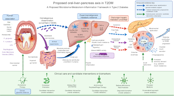
Figure 1: Proposed oral-liver-pancreas axis in type 2 diabetes mellitus. Periodontal dysbiosis may release bacteria and microbial products via transient hematogenous dissemination or swallowing into the gastrointestinal tract. The oral-gut route may alter intestinal barrier function; microbial products and metabolites, rather than swallowed oral microbes themselves, are depicted as the intestinal-to-portal signal. Portal and circulating signals can activate hepatic innate immunity, impair insulin signaling, alter lipid handling, and increase exposure to cytokines, free fatty acids, and extracellular vesicles. These mediators may add oxidative, endoplasmic reticulum, and mitochondrial stress to pancreatic beta cells, thereby reducing insulin secretion and increasing hyperglycemia. Solid colored arrows indicate mechanistic relationships supported in experimental or general metabolic settings, without establishing the full oral-origin pathway in humans. Dashed colored arrows indicate proposed oral-origin or liver-to-beta-cell links that remain unverified in T2DM. Evidence labels distinguish human associations, animal experiments, and indirect or hypothetical mechanisms. Dashed green connectors identify candidate intervention or risk-stratification points; periodontal treatment has clinical evidence for glycemic benefit, whereas the other applications require further validation. A direct oral-pancreatic route is not shown because evidence in T2DM is insufficient. Abbreviations: AMPK = AMP-activated protein kinase; ER = endoplasmic reticulum; LPS = lipopolysaccharide; NF-kappa B = nuclear factor kappa B; NLRP3 = NLR family pyrin domain containing 3; ROS = reactive oxygen species; SCFA = short-chain fatty acid; T2DM = type 2 diabetes mellitus; TLR = Toll-like receptor. Please click here to view a larger version of this figure.
Disclosures
The authors declare no conflicts of interest. Figure 1 was created using the Figure Labs platform.
References
- Preshaw PM et al. Periodontitis and diabetes: a two-way relationship. Diabetologia. 2012;55(1):21-31. doi:10.1007/s00125-011-2342-y.
- Sanz M et al. Scientific evidence on the links between periodontal diseases and diabetes: consensus report and guidelines of the joint workshop on periodontal diseases and diabetes by the International Diabetes Federation and the European Federation of Periodontology. J Clin Periodontol. 2018;45(2):138-149. doi:10.1111/jcpe.12808.
- Xiao E et al. Diabetes enhances IL-17 expression and alters the oral microbiome to increase its pathogenicity. Cell Host Microbe. 2017;22(1):120-128.e4. doi:10.1016/j.chom.2017.06.014.
- Guan H et al. Microbiomic insights into the oral microbiome's role in type 2 diabetes mellitus: standardizing approaches for future advancements. Front Endocrinol (Lausanne). 2024;15:1416611. doi:10.3389/fendo.2024.1416611.
- Saeb ATM et al. Relative reduction of biological and phylogenetic diversity of the oral microbiota of diabetes and pre-diabetes patients. Microb Pathog. 2019;128:215-229. doi:10.1016/j.micpath.2019.01.009.
- Borgnakke WS, Ylostalo PV, Taylor GW, Genco RJ. Effect of periodontal disease on diabetes: systematic review of epidemiologic observational evidence. J Periodontol. 2013;84(4 Suppl):S135-S152. doi:10.1902/jop.2013.1340013.
- Harrandah AM. The oral-gut-systemic axis: emerging insights into periodontitis, microbiota dysbiosis, and systemic disease interplay. Diagnostics (Basel). 2025;15(21):2784. doi:10.3390/diagnostics15212784.
- Arimatsu K et al. Oral pathobiont induces systemic inflammation and metabolic changes associated with alteration of gut microbiota. Sci Rep. 2014;4:4828. doi:10.1038/srep04828.
- Nakajima M et al. Oral administration of P. gingivalis induces dysbiosis of gut microbiota and impaired barrier function leading to dissemination of Enterobacteria to the liver. PLoS One. 2015;10(7):e0134234. doi:10.1371/journal.pone.0134234.
- Cani PD et al. Metabolic endotoxemia initiates obesity and insulin resistance. Diabetes. 2007;56(7):1761-1772. doi:10.2337/db06-1491.
- Tilg H, Moschen AR. Microbiota and diabetes: an evolving relationship. Gut. 2014;63(9):1513-1521. doi:10.1136/gutjnl-2014-306928.
- Qin J et al. A metagenome-wide association study of gut microbiota in type 2 diabetes. Nature. 2012;490(7418):55-60. doi:10.1038/nature11450.
- Hotamisligil GS. Inflammation, metaflammation and immunometabolic disorders. Nature. 2017;542(7640):177-185. doi:10.1038/nature21363.
- Donath MY, Shoelson SE. Type 2 diabetes as an inflammatory disease. Nat Rev Immunol. 2011;11(2):98-107. doi:10.1038/nri2925.
- Prentki M, Nolan CJ. Islet beta cell failure in type 2 diabetes. J Clin Invest. 2006;116(7):1802-1812. doi:10.1172/JCI29103.
- Weir GC, Bonner-Weir S. Five stages of evolving beta-cell dysfunction during progression to diabetes. Diabetes. 2004;53(Suppl 3):S16-S21. doi:10.2337/diabetes.53.suppl_3.s16.
- Marroncini G, Naldi L, Martinelli S, Amedei A. Gut-liver-pancreas axis crosstalk in health and disease: from the role of microbial metabolites to innovative microbiota manipulating strategies. Biomedicines. 2024;12(7):1398. doi:10.3390/biomedicines12071398.
- Liu Z et al. Association between oral microbiome alpha and beta diversity and MASLD risk: a large-scale, population-based retrospective study. Front Cell Infect Microbiol. 2026;16:1784034. doi:10.3389/fcimb.2026.1784034.
- He X et al. Dietary fiber of Tartary buckwheat bran modified by steam explosion alleviates hyperglycemia and modulates gut microbiota in db/db mice. Food Res Int. 2022;157:111386. doi:10.1016/j.foodres.2022.111386.
- Chen X et al. Bacillus amyloliquefaciens Fmb50 alleviates hyperglycemia by reshaping gut structures and mitigating metabolic disorders in type 2 diabetic mice. J Agric Food Chem. 2026;74(3):2888-2900. doi:10.1021/acs.jafc.5c15573.
- Liu R et al. Study on the mechanism of modified Gegen Qinlian decoction in regulating the intestinal flora-bile acid-TGR5 axis for the treatment of type 2 diabetes mellitus based on macro genome sequencing and targeted metabonomics integration. Phytomedicine. 2024;132:155329. doi:10.1016/j.phymed.2023.155329.
- Backhed F et al. The gut microbiota as an environmental factor that regulates fat storage. Proc Natl Acad Sci U S A. 2004;101(44):15718-15723. doi:10.1073/pnas.0407076101.
- Tang WH et al. Intestinal microbial metabolism of phosphatidylcholine and cardiovascular risk. N Engl J Med. 2013;368(17):1575-1584. doi:10.1056/NEJMoa1109400.
- Kong L et al. Trimethylamine N-oxide impairs beta-cell function and glucose tolerance. Nat Commun. 2024;15(1):2526. doi:10.1038/s41467-024-46829-0.
- Zhang Z et al. The roles of liver inflammation and the insulin signaling pathway in PM2.5 instillation-induced insulin resistance in Wistar rats. Dis Markers. 2021;2021:2821673. doi:10.1155/2021/2821673.
- Clarembeau F, Komuta M, Horsmans Y, Lanthier N. Impact of liver inflammation on whole body insulin resistance: a case report on primary biliary cholangitis. Acta Gastroenterol Belg. 2019;82(4):536-538.
- Garcia-Martinez I et al. Saturated fatty acid-enriched small extracellular vesicles mediate a crosstalk inducing liver inflammation and hepatocyte insulin resistance. JHEP Rep. 2023;5(8):100756. doi:10.1016/j.jhepr.2023.100756.
- Branco JR et al. Dietary citrate acutely induces insulin resistance and markers of liver inflammation in mice. J Nutr Biochem. 2021;98:108834. doi:10.1016/j.jnutbio.2021.108834.
- Chen SM et al. Lactobacillus attenuate the progression of pancreatic cancer promoted by Porphyromonas gingivalis in K-rasG12D transgenic mice. Cancers (Basel). 2020;12(12):3522. doi:10.3390/cancers12123522.
- Zhang Y et al. Lactobacillus plantarum NC8 and its metabolite acetate alleviate type 1 diabetes via inhibiting NLRP3. Microb Pathog. 2023;182:106237. doi:10.1016/j.micpath.2023.106237.
- Marietta E et al. Administration of human derived upper gut commensal Prevotella histicola delays the onset of type 1 diabetes in NOD mice. BMC Microbiol. 2022;22(1):8. doi:10.1186/s12866-021-02406-9.
- Teeuw WJ, Gerdes VE, Loos BG. Effect of periodontal treatment on glycemic control of diabetic patients: a systematic review and meta-analysis. Diabetes Care. 2010;33(2):421-427. doi:10.2337/dc09-1378.
- Simpson TC et al. Treatment of periodontitis for glycaemic control in people with diabetes mellitus. Cochrane Database Syst Rev. 2022;4(4):CD004714. doi:10.1002/14651858.CD004714.pub4.
- Kashiwagi Y et al. Porphyromonas gingivalis induces entero-hepatic metabolic derangements with alteration of gut microbiota in a type 2 diabetes mouse model. Sci Rep. 2021;11(1):18398. doi:10.1038/s41598-021-97868-2.