

Summary
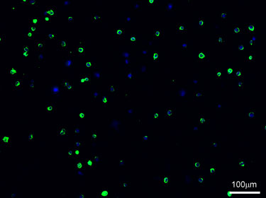
Suspension immunocytochemical staining of human pluripotent stem cells (hPSCs) for cell-surface markers (SSEA-3/SSEA-4) was achieved based on use of a self-made cytospin apparatus to create a monolayer of cells for observation and quantification.
Abstract
Human pluripotent stem cells (hPSCs) that include human embryonic stem cells (hESCs) and human induced pluripotent stem cells (hiPSCs) are exciting cell sources due to their limitless self-renewal capabilities and their potential to differentiate into multiple cell types. The pluripotent state of hPSCs is typically assessed by techniques such as qPCR, immunocytochemistry, and by other in vitro and in vivo differentiation strategies into multiple cell types. Among these, immunocytochemical techniques have been developed for routine characterization of the undifferentiated state of hPSCs based on analysis of candidate intracellular and cell-surface biomarkers. Given the fact that hPSCs grow as colonies, problems arise in quantifying the expression of these markers at the individual cell level on a routine basis. Flow cytometry analyses serve to address this issue but require cell numbers and use of reagents that are not normally conducive for routine quality control assessment of hPSC cultures. Thus, the development of practical and reproducible means of creating monolayer cell samples with preserved integrity for marker evaluation has many advantages in stem cell research. This greatly benefits immunocytochemical analysis because individual cells from the monolayer can be easily observed and quantified for the expression of specific markers. Towards this goal, a self-made cytospin apparatus was constructed and optimized for use with immunocytochemical staining. Two cell-surface markers (SSEA3/SSEA4) expression were analyzed in a variant BG01 stem cell line for the purpose of this protocol.Protocol
Part 1: Suspension Immunocytochemical Staining
- Cells are harvested into a single cell suspension.
- The cells are then transferred into 1.5-2mL microcentrifuge tubes and centrifuged at 200g for 4 minutes.
- Cells are washed by aspirating out supernatant, re-suspended in 1mL of PBS-- (without Mg2+ and Ca2+) and then centrifuged again at 200g for 4 minutes. This should be done twice.
- After washing, cells are then fixed by re-suspending them in 1mL of 4% paraformaldehyde (PFA) at room temperature for 10 minutes.
- Cells are washed again twice with 1mL of PBS--.
- After washing, block cells by re-suspending them in 1mL of Block Solution (6% serum/94% PBS--). Incubate these at room temperature for 45 minutes.
- After, centrifuge tubes at 200g for 4 minutes and aspirate out Block Solution. Re-suspend cells in 1mL of the primary antibody solution (made with Block Solution at recommended dilution). Cells can either be incubated at room temperature for 1 hour or at 4°C overnight before proceeding to the next step.
- Cells are then washed twice with 1mL of Block Solution.
- After, re-suspend cells in 1mL of secondary antibody solution (made with Block Solution at recommended dilution) and incubate at room temperature for 1 hour.
- After 1 hour, cells should then be washed again twice with 1mL of Block Solution.
- After washing with Block Solution, wash cells again with 1mL of PBS--.
- After the various washes, re-suspend cells in 1mL of DAPI nuclear stain (1:10,000 dilution of DAPI in PBS--) and incubate at room temperature for 5 minutes.
- After 5 minutes, centrifuge tubes at 200g for 4 minutes. Aspirate out the DAPI nuclear stain solution and re-suspend cells in 1mL of PBS--.
Part 2: Incorporation of hPSCs in slides generated through cytospin apparatus
- Plastic slides are prepared by making the desired amount of holes in them with a drill press. The diameter needs to be at most 1mm larger than the widest diameter of the micropipette tip(s) to be used.
- Filter paper is cut to the parameters of the plastic slide with scissors.
- Holes are then made in the filter paper. The size of the hole(s) should be large enough that the micropipette tip(s) fits snuggly through it.
- After, one of the prepared plastic slides is placed on top and a glass slide in placed at the bottom of the cut filter paper (Figure 1). The layers are secured with tape.
- A maximum of 100μL (depending on the desired monolayer density) of the stained cell suspension from Part 1 is then placed into the top of the micropipette tip(s).
- If necessary, the apparatus with cell solution can be weighed and a counter-balance of similar weight created.
- The cytospin apparatus and counter-balance can are then placed in a desktop centrifuge and centrifuged at 200g for 4 minutes.
- After centrifugation, the cytospin apparatus is carefully disassembled in a way that doesn’t dislodge the cells from the glass slide.
- If necessary, excess surrounding PBS-- on glass side in aspirated out.
- Cells can then be viewed using a fluorescence microscope to check if the desired cell monolayer has been achieved.
Part 3: Mounting slides
- A drop of the desired mounting media is placed directly in the center of each area containing cells.
- After, a cover slip is gently lowered onto the slides trying to avoid air bubbles if possible.
- Excess mounting media is then removed from slides.
- After, the sides of the cover slips are sealed with nail varnish.
- The slides can be kept in a dark storage area until results are observed and documented.
Subscription Required. Please recommend JoVE to your librarian.
Discussion
For the purposes of our lab, a 35mm dish of stem cell cultures grown on mouse embryonic fibroblasts (MEFs) is allotted for the immunocytochemical staining procedure for use with the cytospin apparatus. The cells are then collected by means of enzymatic passaging, removing as much of the MEF layer as possible, into a single-cell suspension. If more effective stem cell isolation from the MEF layer is desired, colonies can be manually picked then digested into suspension. If feeder free conditions are used in growing the stem cells cultures, the colonies can simply be scraped off and digested into suspension. While an initial single-cell suspension is ideal, any cellular clumps should effectively be broken apart throughout the numerous washing and re-suspension steps called for in the immunocytochemical staining procedure.
The video focused on the use of extracellular markers for staining hPSCs. If intracellular staining of the hPSCs is desired, an additional step before the addition of Block Solution (Step 1.6) needs to be done wherein the cells are washed in 1mL of Permeabilization Solution (50mLs of High Salt Buffer with 25μL of Tween 20) three times before re-suspending them in Block Solution. Another critical point that should be mentioned is that from Step 1.9 in the procedure, care should be taken in minimizing light exposure to the samples to prevent fluorescence bleaching of the secondary antibody and DAPI nuclear stain.
Because of the numerous washing and re-suspension steps in the procedure, an entire 35mm dish with a good amount of passage-ready stem cell colonies, estimated to have around 400k-600k cells, is recommended to be digested and used in the initial suspension. This is done in anticipation of some cell loss due to the nature of the procedure. Theoretically, a much smaller amount of cells can be used in the initial suspension but an extra amount of care must be taken in order to further minimize cell loss. When loading onto the cytospin apparatus after the immunocytochemical staining procedure, a cell density of 10k-50k is ideal. It should be mentioned that, while 100μL is the maximum amount that can be loaded onto a cytopsin apparatus that uses a 0.1μL – 10μL micropipette tip, a smaller amount (around 20μL - 30μL) containing the ideal cell density is recommended. This minimizes unnecessary overflow of liquid onto the glass slide. Finally, when using the cytospin apparatus with the centrifuge, as long as the apparatus is secure from lateral movement, the force created by the centrifuge while running has, to our knowledge, been sufficient in keeping the apparatus from flipping or falling off.
Ingredients for solutions used in the immunocytochemical procedure:
- 2% Paraformaldehyde (PFA)/2% Sucrose
- Add 75 ml distilled water, and place on heated stir plate at 56 °C.
- Weigh out 2 g PFA, and add to glass beaker.
- Weigh out 2 g sucrose, and add to PFA in glass beaker.
- Add 2 drops of 1M sodium hydroxide.
- Once all the reagents have gone into solution, add 10 ml of 10X PBS++.
- Adjust the pH of the solution to 7.2-7.4.
- Bring up volume to 100 ml with distilled water.
Store at 4 °C, and use within 1 week. Aliquots can be stored @ -20 °C for 1 month. Centrifuge briefly after thawing to remove any precipitate.
- Block Solution (10mL)
- Add 9.4 ml PBS--.
- Add 0.6 ml serum to the PBS--. (This may need to be optimized for each antibody.)
Store at 4 °C, and use within 48 hours.
Subscription Required. Please recommend JoVE to your librarian.
Disclosures
No conflicts of interest declared.
Acknowledgments
Partial funding for this work was provided by NSF-CAREER 0744556 (Rao) and a scholarship through the VCU-HHMI Science Education and Research Program (Pascual).
Materials
| Name | Company | Catalog Number | Comments |
| Lab Tek Permanox Chamber Slide | Thermo Fisher Scientific, Inc. | 117437 | Material for re-used plastic slides |
| Superfrost/Plus Microscope Slides Precleaned | Fisher Scientific | 12-550-15 | |
| 0.1-10μL Filter Tips | USA Scientific, Inc. | 1121-3810 | |
| Microscope Cover Glass | Fisher Scientific | 120542-B | |
| Cytoseal 280 | Richard-Allan Scientific | 8311-4 | Mounting Medium |
| DPBS | GIBCO, by Life Technologies | 14190 | |
| Normal Goat Serum | Invitrogen | 10000C | |
| Mouse Anti-SSEA-4 Monoclonal Antibody | EMD Millipore | MAB4304 | Primary Antibody for SSEA-4 staining |
| Rat Anti-SSEA-3 Monoclonal Antibody | EMD Millipore | MAB4303 | Primary Antibody for SSEA-3 staining |
| Alexa Fluor 488 Goat Anti-Mouse IgG | Invitrogen | A11029 | Secondary Antibody for SSEA-4 staining |
| Alexa Fluor 488 Labeled Goat Anti-Rat IgG | Invitrogen | A11006 | Secondary Antibody for SSEA-3 staining |
| DAPI, Dihydrochloride | Calbiochem | 268298 |
References
- Rao, R. R., Johnson, A. V., Stice, S. L. Cell surface markers in human embryonic stem cells. Methods in Mol. Bio. - Stem Cell Assays. 407, 51-61 (2007).
- Sisino, G., Bellavia, D., Corallo, M., Geraci, F., Barbieri, R. A homemade cytospin apparatus. Anal. Biochem. 359, 283-284 (2006).
