This video demonstrates a reverse transcriptase (RT) assay to assess the effect of a test RNA on virus-like particle (VLP) production. Cells are co-transfected with plasmids, and RT activity is measured as a quantitative indicator of VLP output. A decrease in RT activity reflects suppression of viral gene expression by the test RNA.
يتطلب اشتراك JoVE لعرض هذا المحتوى. تسجيل الدخول أو ابدأ نسخة تجريبية مجانية
مقالة منهجية
Reverse Transcriptase Activity as a Marker for Virus-Like Particle Production
195 مشاهدة
⸱
يوليو 31, 2026
في هذه المقالة
الملخص
البروتوكول
1. Viral Production Assay
- Remove the 24-well cell culture plates from the incubator and gently swirl the plates inside the biosafety level 3 (BSL3) cell culture hood. Transfer 150 µl of supernatant from each well to a corresponding well in a 96-well flat-bottom plate, which will be used to quantify HIV-1 production.
Note: Common viral quantification assays include measuring the expression of the human immunodeficiency virus (HIV)-1 capsid protein (p24) by enzyme-linked immunosorbent assay (ELISA), quantifying viral RNA by reverse transcription polymerase chain reaction (RT-PCR), and measuring the activity of the HIV-1 RT enzyme. Steps 1.2 to 1.5 explain methods to quantify HIV-1 RT activity.
- Transfer 5 µl of supernatant to the corresponding wells in a 96-well plate, containing 25 µl of a viral disruption cocktail (Table 1). Incubate the mixture for 5 min at room temperature (RT) and transfer the plate to a radioactivity workstation.
Note: Step 1.2 is only necessary if a radioactivity workstation is not available in the BSL3 laboratory. If the plates do not need to be removed from the BSL3 laboratory, proceed to step 1.3. and add 50 µl of radioactive/viral disruption cocktail (Table 1) in place of the 25 µl of radioactive cocktail.
- Prepare a radioactive cocktail (Table 1), and add 25 µl to each well of viral supernatant and disruption cocktail. Incubate the plates at 37 °C for 2 hr.
- Spot 5 µl of the reaction mixture onto corresponding squares in a glass fiber diethylaminoethyl (DEAE) filter mat paper and allow the spots to dry for 10 min. Spot the reaction mixture on every other square so that no two samples directly border one another. This helps to avoid cross-over between samples when determining counts per minute (cpm) in step 1.6.
- Wash the papers 5x for 5 min with 2x saline sodium citrate (SSC) buffer (Table 1), followed by two 1 min washes with 95% ethanol. Allow the papers to dry and seal them in sample bags.
- Clip sample bags containing the filter mat paper into a cassette and insert the cassette into a microplate scintillation counter. Set the counter to read cpm for radioactive phosphorus isotope (32P) with the reference date provided for the batch of [32P]deoxythymidine triphosphate (dTTP) used in the experiment. Select which samples to read using the plate map and start the counter.
- For any viral quantification method, divide the values obtained for each test RNA by the adjacent negative control and multiply this value by 100 to get the percent inhibition of HIV-1 production for each replicate of the test RNAs. An example of results comparing different test RNAs at different concentrations is provided in Figure 1.
Table 1: Components of non-commercial buffers and reagents. Recipes for all non-commercial buffers and reagents are provided.
| Name of Buffer / Reagent | Composition |
| Viral disruption cocktail | 60 mM Tris-HCl (from 1 M Tris-HCl, pH 7.8), 75 mM KCl, 5 mM MgCl2, 1.04 mM EDTA, 1% NP-40. |
| Radioactive cocktail | 60 mM Tris-HCl (from 1 M Tris-HCl, pH 7.8), 75 mM KCl, 5 mM MgCl2, 1.04 mM EDTA, 10 µg/ml Poly(A), 0.33 µg/ml oligo dT. Added immediately before use: 8 mM dithiothreitol (DTT, C4H10O2S2) and 5 µl [32P] dTTP (3,000 Ci/mmol) for each 500 µl of cocktail. |
| Radioactive/viral disruption cocktail | 60 mM Tris-HCl (from 1 M Tris-HCl, pH 7.8), 75 mM KCl, 5 mM MgCl2, 1.04 mM EDTA, 0.1% NP-40, 5 µg/ml Poly(A), 0.16 µg/ml oligo dT. Added immediately before use: 8 mM dithiothreitol (DTT, C4H10O2S2) and 5 µl [32P] dTTP (3,000 Ci/mmol) for each 1 ml of cocktail. |
| 2x SSC | 7.53 g NaCl and 8.82 g sodium citrate - 2H2O in 1 L H2O. |
الوصول مقيد. يرجى تسجيل الدخول أو بدء فترة تجريبية لعرض هذا المحتوى.
النتائج
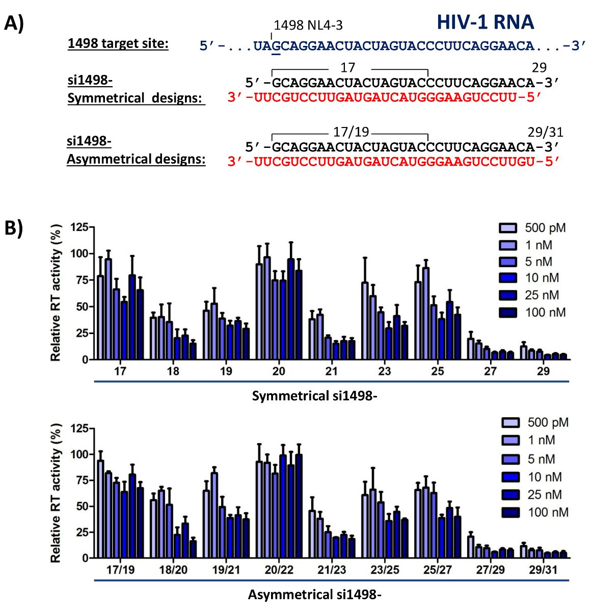
FIGURE 1: Effect of test siRNAs on HIV-1 production. (A) Design of siRNAs with different lengths and symmetries. A conserved sequence in HIV-1 RNA (1498 target site) was used to design symmetrical and asymmetrical siRNAs (si1498-) with differe...
الوصول مقيد. يرجى تسجيل الدخول أو بدء فترة تجريبية لعرض هذا المحتوى.
المواد
| الاسم | الشركة | رقم فهرسي | التعليقات |
|---|---|---|---|
| Cell culture plates, 96 well, 24 well, 6 well. | Corning | 353075, 353047, 353043 | |
| [³²P]dTTP | Perkin Elmer | BLU505H | |
| Poly(A) RNA template | Sigma-Aldrich | 10108626001 | |
| Oligo(dT)12-18 DNA primer | Thermo Fisher | 18418-012 | |
| DEAE filtermat paper | Perkin Elmer | 1450-522 | |
| Microplate scintillation counter | Perkin Elmer | 1450-024 | |
| Lysis buffer tablets | Roche | 4693159001, 4906837001 | protease and phosphatase inhibitors |
